C A S E R E P O R T
Open Access
Tonsillar Plasmacytoma: clues on magnetic
resonance imaging
İrfan Çelebi
1, Gülpembe Bozkurt
2,4*and Nedim Polat
3Abstract
Background: Malignant plasma cell proliferation may present as a disseminated disease (multiple myeloma), a solitary plasmacytoma of bone, or an extramedullary plasmacytoma of soft tissue. The latter plasmacytomas represent approximately 3% of all plasma cell proliferations, and 80% develop in the head-and-neck region. The unexpected clinical presentation of such masses may be present.
Case presentation: Here, we report a rare case of primary tonsillar plasmacytoma in a 42-year-old female. The patient presented with asymmetric tonsillar hypertrophy that was resistant to antibiotherapy. Upon further workup, we found no evidence of multiple myeloma or light-chain disease. The patient underwent surgery and, at the last follow-up, exhibited no evidence of such disease.
Conclusions: In adults presenting with asymptomatic tonsillar enlargement, the possibility of submucosal masses should be considered, thus encouraging the radiologist to evaluate crypts within the palatine tonsil on a postcontrast MRI, besides enlargement and signal change.
Keywords: Plasmacytoma, Multiple myeloma, Magnetic resonance imaging, Asymmetric tonsillar enlargement Background
Extramedullary plasmacytomas (EMPs) are rare, com-prising about 3% of all plasma cell neoplasms. Such plas-macytomas exhibit bone marrow plasma cell infiltration, < 5% nucleated cells, and no evidence of myeloma [1]. EMPs usually develop in the head-and-neck region (80%), with the nasopharynx and sinonasal cavities being the most common sites [2,3]. However, EMPs may ori-ginate in any region where lymphoid deposits are found, including submucosal tissues of the pharynx, larynx, and oral cavity, and nonmucosal regions such as the thyroid gland [1,4]. One published series found that < 5% of EMPs arose in the pharyngeal tonsils and oropharynx [5,6].
Patients typically present in the fifth to seventh decades of life with localized submucosal masses or swellings, and
symptoms reflecting compression and obstruction of local structures. In this paper, we present a rare case of a solitary tonsillar EMP presenting as a slightly asymmetric persistent enlargement of the tonsils. As far as we know, there are no reports in radiologic literature that illustrate magnetic resonance imaging (MRI) contribute to the diag-nosis of tonsillary plasmacytoma.
Case presentation
A 42-year-old female was seen by a primary care phys-ician, whose examination of the oral cavity revealed smooth bilaterally enlarged tonsils, with the right tonsil being slightly larger than the left. No surface abnormal-ity was evident. She was prescribed two courses of amoxicillin but did not improve. She was then referred to our otorhinolaryngology department. Her medical history was unremarkable. We found no unexplained cervical lymphadenopathy, no significant systemic symp-tom, no malignancy, and no immunocompromise. All
laboratory parameters were within normal limits.
Magnetic resonance (MR) images of the palatine tonsils did not reveal any obvious mass lesion. Axial noncon-trast T1 (600/8/2 [TR/TE/NEX]) and coronal STIR MR (5700/80/1) images of the palatine tonsils showed that
* Correspondence:[email protected]
The English in this document has been checked by at least two professional editors, both native speakers of English. For a certificate, please see:
http://www.textcheck.com/certificate/qd3cCC
2Department of Otolaryngology, Acıbadem University Faculty of Medicine,
Istanbul, Turkey
4Department of Otolaryngology-Head and Neck Surgery, Acıbadem Üniversitesi
Atakent Hastanesi, Halkalı Merkez Mahallesi, Halkalı Altınşehir İstanbul Cd. No:16, 34303 Küçükçekmece,İstanbul, Türkiye
Full list of author information is available at the end of the article
© The Author(s). 2018 Open Access This article is distributed under the terms of the Creative Commons Attribution 4.0 International License (http://creativecommons.org/licenses/by/4.0/), which permits unrestricted use, distribution, and reproduction in any medium, provided you give appropriate credit to the original author(s) and the source, provide a link to the Creative Commons license, and indicate if changes were made. The Creative Commons Public Domain Dedication waiver (http://creativecommons.org/publicdomain/zero/1.0/) applies to the data made available in this article, unless otherwise stated.
Çelebi et al. BMC Medical Imaging (2018) 18:19 https://doi.org/10.1186/s12880-018-0261-9
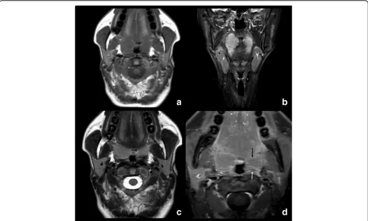
the right tonsil was larger than the left. No mass or abnormal T2 prolongation (suggestive of a tumor) was evident. An axial T2-weighted image (4400/100/2) and a postgadolinium T1-weighted image with fat saturation (550/8/1) also failed to reveal any mass in the right ton-sil, but on postcontrast MRI, the left tonsil showed mu-cosal crypts with linear enhancement, while on the right side the crypts were partly obliterated by a large mass and did not display enhancement (Fig.1).
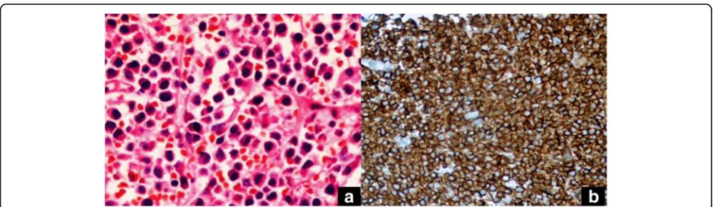
The patient underwent complete surgical resection of the right tonsil because of a possible malignancy. Histological examination of the specimen revealed diffuse sheets of monomorphous plasmacytoid cells with abun-dant, eosinophilic finely granular cytoplasm, and eccentric nuclei. Occasional binucleated and pleomorphic cells with giant nuclei and prominent nucleoli were also observed, as were mitotic figures. No amyloid deposition was appar-ent. Immunohistochemically, the plasma cells were
dif-fusely immunoreactive for the λ light chain (DAKO,
Glostrup, Denmark), IgG (DAKO), and CD43 (DAKO);
partially positive for epithelial membrane antigen
(DAKO); but negative for theκ light chain (DAKO), IgA (DAKO), IgM (DAKO), and CD20 (DAKO). Additional immunohistochemical staining showed that the cells were positive for the plasma cell markers CD138, MUM-1, and CD56. Thus, we diagnosed a plasmacytoma (Fig.2).
A metastatic workup was performed to search for multiple myeloma/light chain disease. Laboratory testing revealed the absence of anemia, and the serum calcium, albumin, total protein, and creatinine levels were nor-mal. The urine lacked Bence-Jones protein. The bone survey was negative. Bone marrow biopsy revealed < 5% plasma cells, ruling out plasmacytoma of the bone. No monoclonal gammopathy was evident, precluding an immunofixation disorder. The kappa-to-lambda ratio was normal. No adjuvant treatment was indicated, and the patient remains clinically free of disease after 6 months of follow-up.
Discussion
The 2016 World Health Organization classification of lymphoid neoplasms divided plasma cell tumors into plasma cell myeloma (multiple myeloma [MM]), solitary plasmacytoma of bone [SBP], and extraosseous plasma-cytoma [7]. EMP is rare in adults and constitutes a form
of non-Hodgkin’s lymphoma [8]. EMP involving the
tonsil is unusual and has only been sporadically reported in the literature [6,9].
It is common for tonsillar asymmetry to raise suspi-cion of malignancy, often indicating the need for a ton-sillectomy. This is especially true in adults aged 18 and above where MRI evaluations are frequently required.
Fig. 1 Axial noncontrast T1 (a), coronal STIR (b), Axial T2-weighted (c) and postgadolinium T1-weighted with fat saturation (d) MR images. Note the obliteration of the right palatine tonsillar crypts combined with linear horizontal enhancement to the left (arrow in d)
Whether tonsillar asymmetry is the only clinical finding that truly reflects a risk of EMP has been frequently dis-cussed in the otorhinolaryngologic literature. Some au-thors have suggested that all unilaterally enlarged tonsils should be excised to counter possible malignancies even when no other suspicious feature is noted at presenta-tion [10,11]. In 1999, Reiter et al. concluded that asym-metric (but otherwise normal) tonsils were suggestive of a significant risk of lymphoma, which tends to grow sub-mucosally and therefore occasionally presents with asymmetry only; histological examination was also rec-ommended [2]. Conversely, other researchers recommend watchful waiting when asymmetry is the only abnormal clinical finding because of the low incidence of malignancy in unilaterally enlarged tonsils [12, 13]. Several authors have reported that unilateral tonsillar enlargement may be attributable to mucosal asymmetry in the tonsillar pillars or differences in the depths of the tonsillar fossae. When considering management options in such patients, the risk associated with unnecessary tonsillectomy should be bal-anced against that of delayed diagnosis of a tonsillar ma-lignancy. As far as we know, there are no radiologic contributions reported in radiologic literature to diagnose submucosal tonsillar masses with the exception of tonsil-lary enlargement and signal change.
MM is a known sequela of EMP. The rate of conver-sion of EMP to MM is lower than that of other plasma cell neoplasms such as SPB; the reported rates range from 11 to 33% over 10 years [14]. The risk of conver-sion is highest in the first 2 years, but converconver-sion has been recorded up to 15 years after diagnosis. Therefore, a lack of clinical suspicion in our patient was associated with a risk of delayed diagnosis and the possible unavail-ability of optimal therapy.
Today, imaging modalities such as computed tomog-raphy and MR imaging are used to stage and assess the responses of malignant lymphomas, rather than for
lymphoma diagnosis per se [15]. No report has yet
described how to image plasmacytomas optimally. The
only striking finding in our patient was obliteration of the right palatine tonsillar crypts which did not show enhancement, as compared to linear enhancement in the crypts seen in the normal left tonsil. This case report emphasizes that disappearing of tonsillar crypts on post-contrast MRI is the most effective diagnostic sign for extramucosal intratonsillar masses which have no signal
differentiation on non-contrast MR examination.
However, this feature is not exclusive of EMP —but
helped decide that such a tonsil needed further investiga-tion due to strong possibility of neoplasia. And that fur-ther corroboration of this feature is needed in more cases.
The differential diagnoses should include malignant lymphoma (which is more common). Negativity for leukocyte common antigen (LCA) and other lymphoid markers (CD3, CD20, CD7, CD4, CD79a, CD30, CD68, PAX5, ALK1, and TdT) rules out malignant lymphoma. Positivity for vimentin, CD138, MUM-1, and CD56 strongly supports a plasma cell origin, as does monoclo-nal kappa light-chain positivity [16].
Plasma cell tumors are well known to be radiation-sen-sitive, and radiotherapy represents the treatment of choice for EMP. Complete surgical excision is appropriate only when a lesion is small and localized; any role for chemo-therapy remains unclear [17]. Surgery is optimal when the disease is localized and amenable to complete resection. In our case, the lesion was easily accessible, and complete surgical removal was achieved.
Conclusion
EMP is rare, and no controlled or cohort study has exam-ined the roles played by clinical manifestations in EMP diagnosis in adults. In adults presenting with asymptom-atic tonsillar enlargement, the possibility of submucosal masses should be considered. The radiologist is encour-aged to study the crypts within the palatine tonsil on post-contrast MRI, besides enlargement and signal change on T2W images. The pathologist must also carefully examine and preserve tissue for immunological characterization.
Fig. 2 Plasma cells with eccentric nucleus and abundant cytoplasm (hematoxylin eosin × 40) (a), Diffuse cytoplasmic, focal membranous CD138 staining on the plasma cells (streptavidinebiotin peroxidase × 400) (b)
Availability of data and materials
The datasets generated and/or analysed during the current study are not publicly available due to the patients’ privacy but are available from the corresponding author on reasonable request.
Authors’ contributions
Concept -İÇ, GB, NP; Design - İÇ, GB; Supervision - İÇ, GB, NP; Resource - İÇ, GB, NP; Materials - GB, NP; Data Collection &/or Processing - GB, NP; Analysis &/or Interpretation -İÇ, GB, NP; Literature Search - GB, NP; Writing - İÇ, GB, NP; Critical Reviews -İÇ, GB, NP. All authors have read and approved the final version of the article.
Ethics approval and consent to participate Not applicable.
Consent for publication
Written informed consent was obtained from the patient for publication of this case report and any accompanying images.
Competing interests
There authors declare that they have no competing interests.
Publisher’s Note
Springer Nature remains neutral with regard to jurisdictional claims in published maps and institutional affiliations.
Author details
1
Department of Radiology, Sisli Hamidiye Etfal Education and Research Hospital, Istanbul, Turkey.2Department of Otolaryngology, Acıbadem
University Faculty of Medicine, Istanbul, Turkey.3Department of Pathology,
Beykent University Medical Faculty, Istanbul, Turkey.4Department of
Otolaryngology-Head and Neck Surgery, Acıbadem Üniversitesi Atakent Hastanesi, Halkalı Merkez Mahallesi, Halkalı Altınşehir İstanbul Cd. No:16, 34303 Küçükçekmece,İstanbul, Türkiye.
Received: 30 August 2017 Accepted: 11 June 2018
References
1. Susnerwala SS, Shanks JH, Banerjee SS, Scarffe JH, Farrington WT, Slevin NJ. Extramedullar plasmacytoma of the head and neck region:
clinicopathological correlation in 25 cases. Br J Cancer. 1997;75:921–7. 2. Reiter ER, Randolph GW, Pilch BZ. Microscopic detection of occult
malignancy in the adult tonsil. Otolaryngol Head Neck Surg. 1999;120:190–4. 3. Miller FR, Lavertu P, Wanamaker JR, Bonafede J, Wood BG. Plasmacytomas of
the head and neck. Otolaryngology—head and neck. Surgery. 1998;119:614–8. 4. Wiltshaw E. The natural history of extramedullary plasmacytoma and its
relation to solitary myeloma of bone and myelomatosis. Medicine (Baltimore). 1976;55:217–38.
5. Galienia P, Cavi M, Pulsoni S, Avvisati G, Bigazzi C, Neri S, et al. Clinical outcome of extramedullary plasmacytoma. Haemtologica. 2000;85:47–51. 6. Tokatli F, Puyan FO, Alas RC, Tuncbilek N, Uzal C. Extramedullary
plasmacytoma: clinicopathology, immunohistochemistry and therapeutic approach to a case with a tonsillar site. Hematology/Oncology and Stem Cell Therapy. 2008;1:241–5.
7. Swerdlow SH, Campo E, Pileri SA, Harris NL, Stein H, Siebert R, et al. The 2016 revision of the World Health Organization classification of lymphoid neoplasms. Blood. 2016;127:2375–90.
8. Alexiou C, Kau RJ, Dietztelbinger H, Kremer M, Spiess JC, et al.
Extramedullary plasmacytoma: tumor occurrence and therapeutic concepts. Cancer. 1999;85:2305–14.
9. Sakai Y, Ikeya J, Morita I, Sato K. Extramedullary plasmacytoma of the tonsil diagnosed by fine-needle aspiration cytology. Ann Diagn Pathol. 2008;12:41–3.
10. Ridgway D, Wolff LJ, Neerhout RC, Tilford DL. Unsuspected nonHodgkin’s lymphoma of the tonsils and adenoids in children. Pediatrics. 1987;79:399–402.
11. Cortez EA, Maltox DE, Holt GR, Gates GA. Unilateral tonsil enlargement. Otolaryngol Head Neck Surg. 1979;87:707–16.
12. Cinar F. Significance of asymptomatic tonsil asymmetry. Otolaryngol Head Neck Surg. 2004;131:101–3.
13. Spinou C, Kubba H, Konstantinidis I, Johnston A. Role of tonsillectomy in histology for adults with unilateral tonsillar enlargement. Br J Oral Maxillofac Surg. 2005;43:144–7.
14. D’Aguillo C, Soni RS, Gordhan C, Liu JK, Baredes S, Eloy JA. Sinonasal extramedullary plasmacytoma: a systematic review of 175 patients. Int Forum Allergy Rhinol. 2014;4:156–63.
15. Kwee TC, Kwee RM, Nievelstein RA. Imaging in staging of malignant lymphoma: a systematic review. Blood. 2008;111:504–16.
16. Terada T. Primary extramedullary malignant plasmacytoma mimicking carcinoma of rectum. Case Reports in Clinical Pathology. 2016;3:6–10. 17. Soutar R, Lucraft H, Jackson G, Reece A, Bird J, Low E, et al. Guidelines on
the diagnosis and management of solitary plasmacytoma of bone and solitary extramedullary plasmacytoma. Br J Haematol. 2004;124:717–26.