Abstract
Legionnaires’ disease (LD) is a systemic infectious disease caused by Legionella species. It mainly presents with lung involvement. Herein, we present a case with suspected myocarditis associated with LD and review of the relevant literature. An 81-year-old male tourist patient with high fever, cough, imbalance while walking, and confusion presented to the emergency department. The patient was diagnosed with LD based on increased density in the left lower zone on chest x-ray and a positive Legionella urine antigen test. He was administered a combination of claritromycin and levofloxacine on the day of admission. The diagnosis of acute myocarditis was made after worsening of the cardiac functions, ST elevation and troponin I positivity. The patient’s symptoms regressed with antibiotic therapy and the patient was transferred to his home country by ambulance plane ten days after admission. A search of PubMed and Web of Science using the keywords “Legionella and myocarditis” revealed 15 case reports, nine of which were in English and were reviewed. There were three female and six male patients with a mean age of 44 years (range: 32-56 years). Seven were diagnosed with LD by urine antigen testing, one by serological testing and culture, and one by direct fluorescent-antibody staining and culture. Myocarditis was diagnosed by biopsy in two patients and by clinical and laboratory findings in the rest. Myocarditis without existing pneumonia was detected in one case. Electrocardiography abnormalities such as atrial flutter, atrioventricular block, torsade de pointes, sinus tachycardia, QT prolongation, ST elevation, and T wave inversion were detected in seven patients. Ventricle dysfunction on echocardiography and cardiac marker abnormality were detected in all but one of the patients (not tested in one patient). Antimicrobial monotherapy was chosen for three of the cases. One patient died due to myocarditis. In conclusion, myocarditis may develop rarely during the course of LD. Clinical suspicion is essential for the diagnosis. Early diagnosis and appropriate treatment may be life-saving.
Keywords: Pneumonia, Legionella pneumophila, Legionnaires’ disease, myocarditis, travel medicine
Lejyoner hastalığı (LH), Legionella bakterisinin etkeni olduğu ve ön planda akciğerlerin tutulduğu sistemik bir enfeksiyon hastalığıdır. Burada LH tanısı alan ve miyokardit şüphesi olan bir olgu sunuldu ve ilgili literatür gözden geçirildi. Seksen bir yaşında turist erkek hasta yüksek ateş, öksürük, yürümede bozukluk ve bilinç değişikliği şikayeti ile acil servise başvurdu. Akciğer grafisinde sol alt zonda dansite artışı ve idrarda Legionella antijeni olumlu saptanması üzerine LH tanısı aldı. Klaritromisin ve levofloksasin tedavisine yattığı gün başlandı. Kardiyak fonksiyonların kötüleşmesi, ST elavasyonu ve troponin I değerlerinde yükseklik akut miyokardit ile uyumlu bulundu. Antibiyotik tedavisi ile şikayetleri gerileyen hasta kendi isteği üzerine yatışının onuncu günü uçak ambulansla ülkesine gönderildi. PubMed ve Web of Sciences’ta yapılan “Legionella ve miyokardit” taramasında 15 makale bulundu ve İngilizce bildirilen dokuz olgu irdelendi. Hastaların üçü kadın, altısı erkek olup yaş ortalaması 44 yıl (minimum: 32-maksimum: 56 yıl) idi. Yedi tanesi idrarda Legionella antijeni, bir tanesi seroloji ve kültür, bir tanesi direkt floresan antikor boyama ve kültür ile LH tanısı almıştır. Miyokardit tanısı iki olguda biyopsi, diğer olgularda ise klinik ve laboratuvar bulgularına göre konmuştur. Bir olguda pnömoni olmadan miyokardit saptanmıştır. Hastaların yedisinde elektrokardiyografide (atrial flatter, atriyoventriküler blok, torsade de pointes, sinüs taşikardisi, QT uzaması, ST elavasyonu, T dalgası inversiyonu) bozukluk saptanmış. Olguların hepsinde ekokardiyografide ventrikül disfonksiyonu ve kardiyak biyomarkırlarda (bir olguda bakılmamış) bozukluk bildirilmiştir. Üç olguda LH’ye karşı antimikrobiyal tedavide monoterapi tercih edilmiştir. Bir hasta hayatını kaybetmiştir. Sonuç olarak LH seyri
Öz
Published: 21 April 2017 Address for Correspondence/Yazışma Adresi: Haluk Erdoğan MD, Başkent University Alanya Medical and
Research Center, Deparment of Infectious Diseases and Clinical Microbiology, Alanya, Turkey E-mail: [email protected]
Received/Geliş Tarihi: 27.12.2016 Accepted/Kabul Tarihi: 24.03.2017 ©Copyright 2017 by the Infectious Diseases and Clinical Microbiology Specialty Society of Turkey Mediterranean Journal of Infection, Microbes and Antimicrobials published by Galenos Yayınevi.
1Başkent University Alanya Medical and Research Center, Department of Infectious Diseases and Clinical Microbiology, Alanya, Turkey 2Başkent University Alanya Medical and Research Center, Department of Cardiology, Alanya, Turkey
Haluk ERDOĞAN1, Halil Olcay ELDEM2
Lejyoner Hastası Bir Hastada Miyokardit Şüphesi: Olgu Sunumu ve Literatürün Gözden
Geçirilmesi
A Patient with Suspected Myocarditis Associated with
Legionnaires’ Disease: A Case Report and Review of the Literature
Mediterr J Infect Microb Antimicrob 2017;6:2 Erişim: http://dx.doi.org/10.4274/mjima.2017.2
Presented in: As a poster in Antalya EKMUD 2016.
Introduction
Legionnaires’ disease (LD) is a systemic infectious disease caused by Legionella bacteria which primarily affects the lungs. More than 90% of LD is caused by Legionella pneumophila. Legionella bacteria accounts for approximately 2-15% of community-acquired pneumonia[1,2]. In LD, involvement of extrapulmonary
organs such as heart, liver, kidneys is rare and results from the direct or indirect influence of bacteria (toxins, etc.)[1,3,4]. Cardiac
involvement in LD may lead to the development of endocarditis, myocarditis, and pericarditis, as well as arrhythmias such as sinus bradycardia, second-and third-degree atrioventricular (AV) block, atrial fibrillation, and premature ventricular contractions. There have also been reports of cases with cardiac involvement in the absence of pulmonary involvement[5,6]. In this paper, we present
our management and follow-up of a case of LD with signs of cardiac involvement and review the related literature.
Case Report and Literature Review
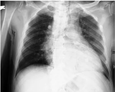
An 81-year-old male patient presented to our emergency department on September 25, 2015 with high fever and cough for four days. The patient exhibited fever with rigors, malaise, gait disorder, and later, altered level of consciousness. According to the patient’s history, he was retired, did not smoke, rarely used alcohol, and had a history of coronary artery disease and arterial fibrillation. He was in Turkey as a tourist and was using the water and air conditioning facilities in the hotel. Physical examination revealed moderate general condition with confusion. His vital signs were as follows: body temperature: 39.3 ˚C, pulse: 102/ min, blood pressure: 110/80 mmHg, respiratory rate: 24/minute, and SpO2 at room temperature: 88%. There were no signs of meningeal irritation. Respiratory system examination revealed rough bilateral inspiratory and expiratory sounds; rales and rhonchi were not observed. The patient’s laboratory results at admission were as follows: white blood cell count: 13800/µL (92% PNL), hemoglobin: 12.6 g/dL (13.5-17.5), hematocrit: 36.8% (40-52), platelet count: 139000/µL (140000-440000), C-reactive protein: 295 mg/L (0-5), glucose: 149 mg/L (80-110), blood urea nitrogen: 20 mg/dL (6-26), creatinine: 1 mg/dL (0.7-1.3), sodium: 127 meq/L (132-136), aspartate aminotransferase (AST): 58 U/L (5-35), alanine aminotransferase (ALT): 41 U/L (0-55), lactate dehydrogenase: 271 U/L, creatinine kinase (CK): 253 U/L (<200), CK-MB: 20 U/L (0-24), and troponin I: 40.6 pg/mL (0-34). Eight hours later, troponin I level was 15.3 pg/mL (0-34). Increased
density was observed in the lower left zone on posteroanterior chest radiography (Figure 1, 2, 3). Legionella urine antigen test was positive (Binax NOW®, Legionella urine antigen card, Alero
Co, USA) and Streptococcus pneumoniae urine antigen was negative. No growth was observed in the sputum, blood, and urine cultures. Polymerase chain reaction for viruses potentially causing myocarditis was not performed. No pathological findings were observed on brain magnetic resonance (MR). Treatment with intravenous (IV) levofloxacin 750 mg/day and IV clarithromycin 1000 mg/day was initiated. The patient suffered increased respiratory distress (respiratory rate 32/minute) after seven hours of hospitalization and was transferred to the intensive care unit for oxygen support. During follow-up, he was given nasal oxygen support at two liters/minute. His respiratory distress improved with treatment, the need for oxygen support decreased, and he became agitated in the intensive care unit. As a result, on the following day, he was transferred to inpatient wards.
During consultation with the cardiology department while the patient was in the emergency unit, electrocardiography (ECG) showed sinus tachycardia without ischemic changes. Echocardiography (ECHO) revealed an ejection fraction of 60%, normal wall appearance, and biatrial dilatation. On the 4th day
of hospitalization, anterior ST-elevation myocardial infarction pattern (Figure 4) was observed, and akinesia in the apical segments was evident on ECHO. Laboratory values were as follows: ALT: 89 U/L, AST 99 U/L, CK: 288 U/L, and troponin 1: 0.054 ng/mL (0-0.023). Coronary angiography could not be performed because the patient and his relative did not consent to the procedure. The patient’s agitation resolved on the 5th day of hospitalization
and oral antibiotic treatment was initiated on the 7th day.
The patient’s overall condition improved and his symptoms diminished; on the 10th day of hospitalization, the patient
voluntarily returned to his home country by air medical transport. The patient’s spouse and doctor reported that the patient continued to be treated for LD in a hospital in his home country and was discharged on October 27, 2015. The patient recovered after undergoing physical therapy rehabilitation and lived in good health for over two years.
Review of Literature
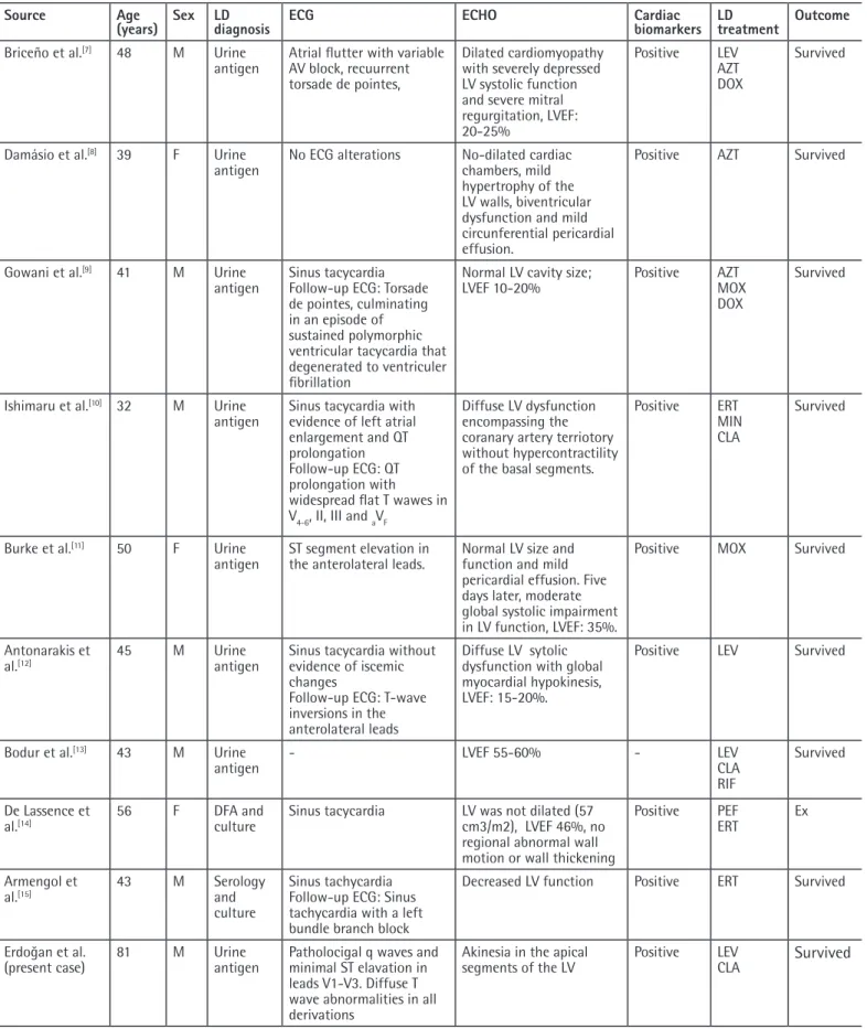
A literature search of PubMed and Web of Sciences on 1 December 2016 using the keywords “Legionella and myocarditis” yielded 15 articles. The nine cases published in English were analysed and are summarized in Table 1.
sırasında nadiren de olsa miyokardit gelişebileceği akılda tutulmalıdır. Klinik şüphe tanıda esastır. Lejyoner hastalığının erken tanısı ve uygun tedavisi hayat kurtarıcı olabilir.
Discussion
The incidence of LD depends on the degree of water reservoir colonization, individual sensitivity, the intensity of contact, and the availability of diagnostic methods able to identify the infection. Cases may be sporadic, or can occur in clusters or epidemics. Legionnaires’ disease should be considered in cases of pneumonia with underlying chronic disease, advanced age, smoking habit, history of travel, or extrapulmonary involvement. Elevated liver enzymes, diarrhea, abdominal pain, nausea, relative bradycardia, headache, confusion/lethargy, microscopic hematuria, and elevated CK are more common in LD than in other forms of pneumonia[1,2]. In our case, the patient had risk
factors, such as advanced age and travel history, as well as extrapulmonary symptoms (gait disorder, altered mental status). Laboratory analyses demonstrated abnormal liver function tests, elevated CK, and hyponatremia. Due to the presence of high fever and altered level of consciousness, the differential diagnosis
Figure 1. Chest radiograph showing no infiltrates on the day of
admission
Figure 2, 3. Chest radiograph, two and four days later, showing
infiltrates in left lower zone.
Figure 4. Pathological q waves and minimal ST elevation in leads
Table 1. Demographic, clinical, and laboratory data of ten patients with Legionnaire’s disease and myocarditis
Source Age
(years) Sex LD diagnosis ECG ECHO Cardiac biomarkers LD treatment Outcome
Briceño et al.[7] 48 M Urine
antigen Atrial flutter with variable AV block, recuurrent torsade de pointes,
Dilated cardiomyopathy with severely depressed LV systolic function and severe mitral regurgitation, LVEF: 20-25% Positive LEV AZT DOX Survived
Damásio et al.[8] 39 F Urine
antigen No ECG alterations No-dilated cardiac chambers, mild hypertrophy of the LV walls, biventricular dysfunction and mild circunferential pericardial effusion.
Positive AZT Survived
Gowani et al.[9] 41 M Urine
antigen Sinus tacycardia Follow-up ECG: Torsade de pointes, culminating in an episode of sustained polymorphic ventricular tacycardia that degenerated to ventriculer fibrillation
Normal LV cavity size;
LVEF 10-20% Positive AZTMOX
DOX
Survived
Ishimaru et al.[10] 32 M Urine
antigen Sinus tacycardia with evidence of left atrial enlargement and QT prolongation Follow-up ECG: QT prolongation with widespread flat T wawes in V4-6, II, III and aVF
Diffuse LV dysfunction encompassing the coranary artery terriotory without hypercontractility of the basal segments.
Positive ERT
MIN CLA
Survived
Burke et al.[11] 50 F Urine
antigen ST segment elevation in the anterolateral leads. Normal LV size and function and mild pericardial effusion. Five days later, moderate global systolic impairment in LV function, LVEF: 35%.
Positive MOX Survived
Antonarakis et
al.[12] 45 M Urine antigen Sinus tacycardia without evidence of iscemic
changes
Follow-up ECG: T-wave inversions in the anterolateral leads
Diffuse LV sytolic dysfunction with global myocardial hypokinesis, LVEF: 15-20%.
Positive LEV Survived
Bodur et al.[13] 43 M Urine
antigen - LVEF 55-60% - LEVCLA
RIF
Survived De Lassence et
al.[14] 56 F DFA and culture Sinus tacycardia LV was not dilated (57 cm3/m2), LVEF 46%, no
regional abnormal wall motion or wall thickening
Positive PEF
ERT Ex
Armengol et
al.[15] 43 M Serology and
culture
Sinus tachycardia Follow-up ECG: Sinus tachycardia with a left bundle branch block
Decreased LV function Positive ERT Survived
Erdoğan et al.
(present case) 81 M Urine antigen Patholocigal q waves and minimal ST elavation in leads V1-V3. Diffuse T wave abnormalities in all derivations
Akinesia in the apical
segments of the LV Positive LEVCLA Survived
LD: Legionnaire’s disease, ECG: Electrocardiography, ECHO: Echocardiography, AV: Atrioventricular, DFA: Direct fluorescent antibody staining, F: Female, M: Male, AZT: Azithromycin, CLA: Clarithromycin, ERT: Erythromycin, DOX: Doxycycline, LEV: Levofloxacin, MIN: Minocycline, MOX: Moxifloxacin, PEF: Pefloxacin, RIF: Rifampicin LV: Left ventricular, LVEF: Left ventricular ejection fraction
included acute central nervous system infections and central vascular events. However, lack of any findings of meningeal irritation or signs of pathology on cranial MR were unsupportive of these prediagnosis.
Criteria for definitive diagnosis of LD are isolation of Legionella bacteria in cultures, positive Legionella urine antigen test, and observing at least a 4-fold increase in antibody titer against L.
pneumophila serogroup 1 in paired serology. The Legionella urine
antigen test is valuable in LD diagnosis because patients become positive within the first few days of infection; it is not affected by antibiotic use, is easily performed, and provides rapid results. The test has a sensitivity of 70-80% and specificity close to 100%; the sensitivity and specificity rates of the urinary card test are comparable to those of ELISA. The biggest disadvantage of the test is the lack of reliability for species and serogroups other than L. pneumophila serogroup 1[1,2]. Definitive LD diagnosis was
established for our patient upon obtaining a positive Legionella urine antigen result. Legionella bacteria could not be isolated in the sputum culture obtained when the patient was receiving antibiotic treatment. Serologic tests were not used in our case because of their limited benefit in early diagnosis.
Myocarditis is an inflammatory disease of the cardiac muscle which may exhibit different clinical presentations depending on the severity of cardiac involvement (focal, diffused, etc.). It may show wide clinical variability, ranging from mild symptoms to heart failure, cardiogenic shock, arrhythmia, and sudden death. ECHO findings can range from diffused wall motion abnormalities to regional motion abnormalities. Clinical suspicion is essential for the diagnosis of myocarditis. Elevated cardiac biomarkers (such as troponin, CK-MB), ECG changes indicating acute myocardial damage, arrhythmia, and new and unexplained disruptions of cardiac function are also suggestive of myocarditis[16]. Endomyocardial biopsy is the gold standard in
diagnosis, but it must be noted that this technique is applicable only in selected cases due to the difficulty and the risk involved. Of the nine cases identified in our literature review, the male:female ratio was 2:1 and the mean age was 44 years (range: 32-56 years). Seven of the cases exhibited abnormalities on ECG (atrial flutter, AV block, torsade de pointes, sinus tachycardia, prolonged QT, ST elevation, T-wave inversion), and all cases showed ventricular dysfunction on ECHO. Cardiac biomarker abnormalities were reported in eight of the cases. In only two cases, the myocarditis diagnosis was based on cardiac biopsy. In our case, clinical findings were accompanied by elevated troponin I values, newly emergent ST elevation, and akinesia in apical segments on ECHO. Cardiac biopsy was not performed. When observed only in the apical myocardial segments, as in our case, wall motion abnormalities may be indicative of focal myocarditis. ECG abnormalities associated with myocarditis may
initially mimic ST-elevation or non-ST-elevation acute myocardial infarction[17]. However, considering how ST-T abnormalities evolve
in the course of time, the ECG changes associated with acute myocarditis evolve much slower than those seen with acute myocardial infarction[18]. As in our case, persistence for even a few
days without forming pathological Q wave is indicative of acute myocarditis. In cases with ECG and ECHO findings and elevated cardiac biomarkers, stress cardiomyopathy or neurogenic heart syndrome should also be considered in the differential diagnosis. Stress cardiomyopathy is a condition that occurs in patients under severe physical or psychic stress. Findings include apical aneurysm, as well as ECG changes mimicking acute myocardial infarction and cardiac enzyme abnormalities[19]. In our case, the
confirmed presence of an etiology (LD) that may cause may cause myocarditis, increased the probability of myocarditis. However, the likelihood of the two conditions co-occurring is even higher. Combination therapy is usually preferred in cases with severe LD and extrapulmonary organ involvement. Macrolides are administered with rifampicin or a quinolone drug as combination therapy. The duration of treatment is 7-10 days. Treatment should be extended to 14-21 days for cases with severe course, extrapulmonary organ involvement, and immunosuppression[1,2].
It should be kept in mind that macrolides and quinolones used in the specific treatment of LD may have an arrhythmogenic effect or exacerbate an underlying condition. We preferred a combination of quinolone and macrolide for our patient. Six of the nine cases found in our literature review were also treated with combination antibiotic therapy. Even under appropriate antibiotic therapy, a patient’s clinical condition may deteriorate or increased density may be observed on chest radiography, as in our case. In a case reported by Briceño et al.[7], the patient developed severe left
ventricular failure and cardiogenic shock due to repolarization anomaly despite receiving appropriate antibiotic therapy. A mechanical assist device (TandemHeart pVAD) was used briefly to treat cardiogenic shock. Damásio et al.[8] used intra-aortic
balloon pump and extracorporeal membrane oxygenation in a patient who developed cardiogenic shock. Only one of the nine cases did not respond to treatment and died.
In conclusion, LD should be suspected in cases of severe community-acquired pneumonia with prominent extrapulmonary symptoms. It is important to be aware that symptoms and findings related to cardiac involvement may be encountered during the clinical course of LD. Clinical suspicion is essential for diagnosis. Early diagnosis and proper treatment of LD may be life-saving.
Ethics
Informed Consent: Consent form was filled out by patient
Peer-review: Externally and internally peer-reviewed. Authorship Contributions
Surgical and Medical Practices: H.E., H.O.E, Concept: H.E., H.O.E, Design: H.E., H.O.E, Data Collection or Processing: H.E., H.O.E, Analysis or Interpretation: H.E., H.O.E, Literature Search: H.E., H.O.E, Writing: H.E.
Conflict of Interest: No conflict of interest was declared by the authors.
Financial Disclosure: The authors declared that this study received no financial support.
References
1. Phin N, Parry-Ford F, Harrison T, Stagg HR, Zhang N, Kumar K, Lortholary O, Zumla A, Abubakar I. Epidemiology and clinical management of Legionnaires’ disease. Lancet Infect Dis. 2014;14:1011-21.
2. Erdogan H, Erdogan A, Lakamdayali H, Yilmaz A, Arslan H. Travel-associated Legionnaires disease: clinical features of 17 cases and a review of the literature. Diagn Microbiol Infect Dis. 2010;68:297-303.
3. Rihs JD, Yu VL, Zuravleff JJ, Goetz A, Muder RR. Isolation of Legionella
pneumophila from blood with the BACTEC system: a prospective study
yielding positive results. J Clin Microbiol. 1985;22:422-4.
4. Evans CP, Winn WC Jr. Extrathoracic localization of Legionella pneumophila in Legionnaires’ pneumonia. Am J Clin Pathol. 1981;76:813-5.
5. Gross D, Willens H, Zeldis SM. Myocarditis in legionnaires’ disease. Chest. 1981;79:232-4.
6. Castellani Pastoris M, Nigro G, Midulla M. Arrhythmia or myocarditis: a novel clinical form of Legionella pneumophila infection in children without pneumonia. Eur J Pediatr. 1985;144:157-9.
7. Briceño DF, Fernando RR, Nathan S, Loyalka P, Kar B, Gregoric ID. Tandem heart as a bridge to recovery in Legionella myocarditis. Tex Heart Inst J. 2015;42:357-61.
8. Damásio AF, Rodrigues L, Miranda L, Coelho P, Banazol N, Colaço J, Fragata J. Fulminant myocarditis caused by Legionella pneumophila: case report. Rev Port Cardiol. 2014;33:185.e1-5.
9. Gowani SA, Kumar A, Arora S, Lahiri B. Legionella pneumonia complicated by myocarditis and torsades de pointes: A case report and review of literature. Conn Med. 2013;77:331-4.
10. Ishimaru N, Suzuki H, Tokuda Y, Takano T. Severe Legionnaires’ disease with pneumonia and biopsy-confirmed myocarditis most likely caused by
Legionella pneumophila serogroup 6. Intern Med. 2012;51:3207-12.
11. Burke PT, Shah R, Thabolingam R, Saba S. Suspected Legionella-induced perimyocarditis in an adult in the absence of pneumonia: a rare clinical entity. Tex Heart Inst J. 2009;36:601-3.
12. Antonarakis ES, Wung PK, Durand DJ, Leyngold I, Meyerson DA. An atypical complication of atypical pneumonia. Am J Med. 2006;119:824-7.
13. Bodur H, Savran Y, Koca U, Kilinç O, Albayrak S, Itil O, Akoglu S. Legionella pneumonia with acute respiratory distress syndrome, myocarditis and septic shock successfully treated with Drotrecogin Alpha (activated). Eur J Anaesthesiol. 2006;23:808-10.
14. De Lassence A, Matsiota-Bernard P, Valtier B, Franc B, Jardin F, Nauciel C. A case of myocarditis associated with Legionnaires’ disease. Clin Infect Dis. 1994;18:120-1.
15. Armengol S, Domingo C, Mesalles E. Myocarditis: a rare complication during
Legionella infection. Int J Cardiol. 1992;37:418-20.
16. Sinagra G, Anzini M, Pereira NL, Bussani R, Finocchiaro G, Bartunek J, Merlo M. Myocarditis in Clinical Practice. Mayo Clin Proc. 2016;91:1256-66. 17. Nisbet BC, Breyer M. Acute myopericarditis with focal ECG findings mimicking
acute myocardial infarction. J Emerg Med. 2010;39:e153-8.
18. Nable JV, Brady W. The evolution of electrocardiographic changes in ST-segment elevation myocardial infarction. Am J Emerg Med. 2009;27:734-46. 19. Tsuchihashi K, Ueshima K, Uchida T, Oh-mura N, Kimura K, Owa M, Yoshiyama M, Miyazaki S, Haze K, Ogawa H, Honda T, Hase M, Kai R, Morii I; Angina pectoris-myocardial infarction investigations in Japan. Transient left ventricular apical ballooning without coronary artery stenosis: a novel heart syndrome mimicking acute myocardial infarction. Angina Pectoris-Myocardial Infarction Investigations in Japan. J Am Coll Cardiol. 2001;38:11-8.