©BEYKENT UNIVERSITY
CELLULAR NERURAL NETWORK APPROACH
FOR ENHANCEMENT OF DENTAL IMAGE
WITH LOW DOSED X-RAY
Niyazi KILIC
1, Osman N. UCAN
1, Fatih OZKARSLI
2,
Bulent YILMAZ
2and Seckin DINDAR
2Istanbul University, Engineering Faculty, Electrical-Electronics Dept. 34320 Avcilar, Istanbul, Turkey. [email protected]
2Istanbul University, Dentistry Faculty, Capa, Istanbul , Turkey
ABSTRACT
Despite radiography's great importance in dental treatment, they caused serious health problems when were used repeatedly with high amount of radiation. In this paper, we have presented an algorithm for image enhancement in dental image with low dosed x-ray (radiation). We aimed to get high quality images (like images with high dosed x-ray) while we have reduced the amount of radiation in Radio VisioGraphy (RVG), which were used for those patients, by use of Cellular Neural Network (CNN). When compared with input images and CNN output images, CNN outputs better clear and quality than inputs from various angle have been seen.
Keywords: Image enhancement, Cellular neural networks, RadioVisioGraphy,
X-ray
ÖZET
Radyografların diş tedavisinde vazgeçilemez önemine karşın, tekrarlanan ve aşırı dozda kullanılan radyasyon ciddi sağlık sorunlarına neden olmaktadır. Bu çalışmada, düşük dozlu dişsel görüntülerin iyileştirilmesi için bir algoritma sunduk. Hücresel yapay sinir ağları (HYSA) kullanılarak hasta ağızlarına uygulanacak RVG cihazının x- ışını miktarı düşürülülüp yüksek kalitede görüntü (yüksek dozlu x-ışınlı görüntü) elde edebilmeyi amaçladık. HYSA sonuçları ile giriş verileri karşılaştırıldığında HYSA çıktılarının, girişlere göre birçok açıdan daha net ve kaliteli görüntüye sahip olduğu görülmüştür.
Anahtar Kelimeler: Görüntü iyileştirme, Hücresel yapay sinir ağları,
Radyovizyografi, X-ışını
1. INTRODUCTION
Since their inventions in 1895, Radiographies had been an indispensable diagnosis tools. Radiographies were used at different intervals of dental treatments. They were used for the purpose of diagnosis before the treatment, but generally periodically used for the assessment of the success of the treatment in the end. [1]
Despite their great importance in dental treatment, they caused serious health problems when were used repeatedly with high amount of radiation. Among
these health problems, loss of taste sense, mount dryness, destruction in hard
dental tissues (like teeth decay) and delayed teeth growth can be cited [2]. In addition to these, when high amount of radiation is exposed to, especially
during intervals 18th and 45th days of pregnancy period, it may lead to weight
loss in infant baby, tumors and vulnerability to childhood leukemia [3]. RadioVisioGraphy (RVG) System invented by Mouyen and et al. in 1989 so as to decrease high amount of radiation generated by conventional radiographies and make use of the software system advantages, has been manufactured by Trophy Radiologic. It has been found out that digital imaging systems have reduced the rate of radiation 80%, when compared with conventional radiographies.[4]
Cellular Neural Networks (CNNs) model has been defined firstly in 1988 by Chua [5-6]. The basic circuit unit of cellular neural networks is called a cell. It contains linear and non-linear circuit elements. There are many well-known applications of CNN like image processing, motion detection, pattern recognition.
In this paper, we aimed to get high quality images while we have reduced the amount of radiation in RVG, which were used for those patients, by use of Cellular Neural Network (CNN) techniques which have been explained in the second section. In the third section, we have applied CNN techniques on dental images and shown the results of the study.
2. MATERIAL AND METHOD
2.1. CELLULAR NEURAL NETWORKS
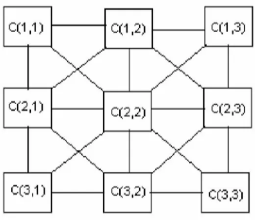
Most neural Networks fall into two main classes: (1) memoryless neural Networks and (2) dynamical neural Networks. As in Hopfield Networks and CNN, dynamical neural Networks have usually been designed as dynamical systems where the inputs are set to some constant values and the path approach to a stable equilibrium point depends upon the initial state. A Cellular Neural Network is composed of large-scale nonlinear analog circuits, which processes signals in real time [5]. Like cellular automata, the CNN is made of a massive aggregate of regularly spaced identical circuits, called cells, which communicate with each other directly only through their nearest neighbors (Figs. 1 and 2). Adjacent cells can, therefore, interact directly with each other. Cells not directly connected together affect each other indirectly because of the propagation effects of the continuous-time dynamics of CNN. An example of a two-dimensional (2-D) CNN is shown in Figure 1.
C(1,1) C(1,2) 0(1,3) C(1,1) C(1,2) 0(1,3)
X X
C(2,1) 0(2,3) C(2,1) 0(2,3)X X
0(3,1) 0(3,2) 0(3,3) 0(3,1) 0(3,2) 0(3,3)Figure 1: Sample 3-D cellular neural network.
A cellular neural network, which consists of M line and N column (MxN) is
considered. In this structure ith line and jth column are named (i,j) cell and
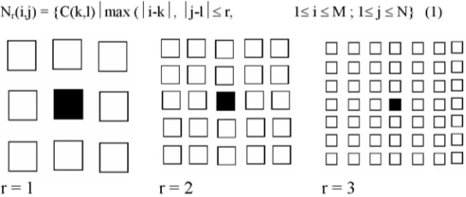
explained as C(i,j). Figure 2 shows the neighborhoods of the C(i,j) cell (located at the center and shaded) for neighborhood of first second and third (r=1,2,3). In addition, the neighborhood has the property of symmetry (if
C(k,l) e Nr(i,j, C(i,j) e Nr(k,l) ). The r-neighborhood of a cell C(i,j) in a
cellular neural network is defined by: Nr(i,j) = {C(k,l) 1 max
•
n
•
•
r = 1
n
1< i < M ; 1< j < N} (1)• • • • •
r = 2
•
•
•
•
•
•
•
•
•
•
•
•
•
•
• •
• •
• •
• •
• •
• •
• •
• •
• •
• •
• •
• •
• •
• •
r = 3
Figure 2: Neighborhood in a cellular neural network.
Cells are multiple-input single-output nonlinear processors described by one, or one among several different, parametric functional. A cell is characterized by a state variable, which is generally not observable as such outside the cell itself. It contains linear and nonlinear circuit elements, such as linear resistors, capacitors and nonlinear controlled sources (Fig. 3). Every cell is connected to other cells within its neighborhood. In this scheme, information is only exchanged between neighboring neurons and this local information characteristic does not prevent the capability of obtaining global processing. The CNN is a dynamical system operating in continuous or discrete time. Cells can be characterized by a functional block diagram that is typical of neural network theory: Figure 3 depicts a two-stage functional block diagram of a cell, composed of a generalized weighted sum (in general nonlinear with memory) integration, output nonlinear function / functional [7-8]. Data can be fed to the CNN through two different ports: initial conditions of the state and input u. Bias value / may be used as a third port.
•4
I „(UM
I (¡JM xyA general form of the cell dynamical equations may be stated as follows:
dxn(t)
= -Xj(t) + 2 A(i' j;
k'')y *(t) + S
B(i' j;ki)
uki(t) +
1d t C(kl)eNr (i,j) C(k,l)eNr (i,j)
(2) y.j(t) = f [x j(t)]= 1(\xj(t) + i\-\xtj(t) -1\) (3) a-1,-1 a-1,0 a-1,1 b-i,-i b-1,0 b-i, i
A =
a0,-1 a0,0 a0,1, B =
b0,-1 b0,0 b0,1,
I ai , - 1 ai , 0 ai , 1_
A-1 b1,0 bi,i_
(4)where; x, y, u, I denote respectively cell state, output, input, bias and j and k are cell indices. CNN parameter values are assumed to be space-invariant and nonlinear function is chosen as piece-wise linear (Fig. 3). A, B and I, the cloning matrices, are identically repeated in the neighborhood of every neuron. The network behavior of a CNN depends on the initial state of the cells, namely the bias I, and the weights values of A and B matrices, which are associated with the connections inside the well-defined neighborhood of each cell. CNNs are arrays of locally and regularly interconnected neurons, or, cells, whose global functionality are defined by a small number of parameters (A, B,
I) that specify the operation of the component cells as well as the connection
weights between them. CNN can also be considered as a nonlinear convolution with the template.
2.2. CNN APPLICATIONS
For the cellular neural networks, there is not an analytically developed learning algorithm to perform any kind of processing. In this study, Recurrent Perceptron Learning Algorithm (RPLA) is used in the applications. Information in detail about RPLA algorithm is in [9].
RPLA is a learning algorithm. The aim in using this learning algorithm is to obtain the weighting coefficients and the threshold value that will perform the desired image processing procedure. For this reason, the w vector, which contains all of these values is defined and used.
The error function E[w] is a measure of the difference between the original output (steady-state output) value and the desired output value. The aim in using the RPLA algorithm is to obtain the w weighting coefficients that is supposed to minimize this error function.
A, B templates that are obtained from the learning process and the threshold
level, I are shown below:
A =
1.6402 1.6335 1.6281
1.6460 0.8026 1.6460
1.6281 1.6335 1.6402
(6)B =
1.3287 1.3287 1.3287
1.3287 1.3287 1.3287
1.3287 1.3287 1.3287
(7) I = 2.850Using these values, w may be calculated:
(8)
w = [1.6402 1.6335 1.6281 1.6460 0.8026 1.3287 1.3287 1.3287
1.3287 2.85]
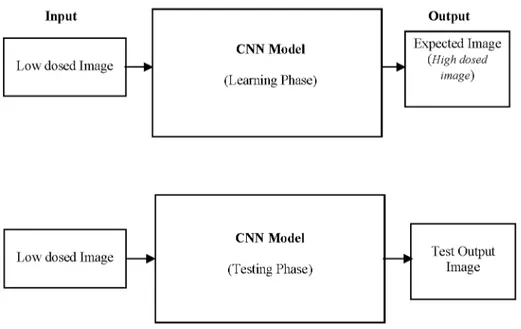
CNN image processing involves two stages. The first stage involves the learning of the low levelled x-ray image by using the RPLA algorithm. At this stage, the input object is the low levelled radiation image whereas the output object is the target (high levelled x-ray) image. This stage is where the weighting coefficients are determined.
After the learning stage is completed, next stage involves the testing where another low levelled x-ray image is used as the input value to the system. At this stage, the weighting coefficients (determined at the previous stage) are used to get the output image. This image is the desired target image.
In the upper part of Figure 4, input and output parts for the learning stage can be seen. The w values are determined by using the relationship between the input and output values. In the testing phase, only the input value is given to the system to obtain the desired image by using the algorithm.
The images obtained by RVG are enhanced by CNN and the results are shown in section 3.
Figure 4. The block scheme of CNN training/test phases.
3. RESULTS
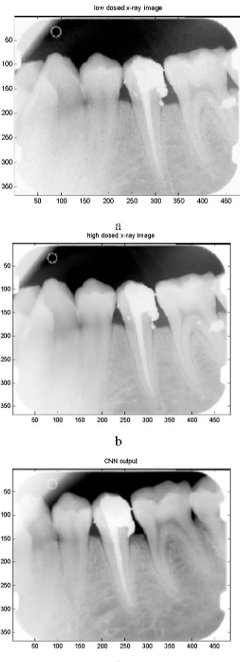
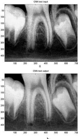
The Cellular Neural Networks (CNN) introduced in [5] have well-suited structures for image processing. Various applications of CNN have been considered and the performance of the proposed approach has been evaluated. CNN with templates obtained was applied to images obtained from the RVG imaging system. Figure 5 shows the results obtained during the learning phase of CNN. Data obtained from this figure are taken as initial data and applied to CNN as input as well as initial state. Similar results are obtained for another image (test phase), which are shown in Figure 6.
When compared results, CNN outputs clearly show the difference and development in terms of the clear differences between various phases, which is the target of this study. On the other hand, CNN outputs better than the inputs and we have reached high quality images while we have reduced the amount of radiation in RVG
low dosed x-ray image 50 100 150 200 250 300 350 50 100 150 200 250 300 350 400 450 50 100 150 200 250 300 350 50 100 150 200 250 300 350 400 450 50 100 150 200 250 300 350 50 100 150 200 250 300 350 400 450
c
Figure 5. a) CNN input (low dosed x-ray image) b) Target (high dosed x-ray image b) CNN output
*
I
>
100 200 300 400 500 600 700
a
CNN test output
100 200 300 400 500 600 700
Figure 6. a) CNN input , b) CNN output
50
00
50
4. CONCLUSSION
We presented an algorithm for image enhancement in dental image with low dosed x-ray. We used Cellular neural network image enhancer. The CNN found by Chua in 1988 has attracted a lot of attention. Not only from a theoretical point of view these systems have a number of attractive properties, but also furthermore, there are many well-known applications like image processing, motion detection, pattern recognition, simulation. In this study, we have used them to enhance the dental images with very low dosed radiation using CNN approach. We can also conclude that CNN may be one of the compromising methods in the image enhancement.
REFERANCES
[1] Frederiksen N.L.; Specialized radiographic techniques. In: Pharoah MJ, White SC, editors. Oral Radiology Principles and Interpretation. 4th Edition. St. Louis, Mosby, (2000).
[2] Brocklebank L.; Dental Radiology—Understanding the X-Ray Image, Oxford University Press, Oxford, U.K, (1997).
[3] Curry T.S., Dowdey J.E., and Muny R.C.; Christensen's Physics of Diagnostic
Radiology, Lea & Febiger, Philadelphia, (1990).
[4] Mouyen F., Benz C., Sonnabend E., and Lodter J.P.; Presentation and physical evaluation of RadioVisioGraphy, Oral Surg Oral Med Oral Pathol. (1989), 68(2):238-42.
[5] Chua L.O., and Yang L.; Cellular Neural Networks: Theory. IEEE Trans. On
Circuit and Systems (1988); 35: 1257-1272.
[6] Chua L.O., and Roska T.; Cellular neural networks and visual computing. Cambridge University press, (2002)
[7] Cimagalli V.; Cellular Neural Networks A Review, Proceedings of sixth Italian workshop on parallel architectures and Neural Networks, Vietri Sul Mare, Italy, (1993) [8] Albora, A. M., Ucan, O. N., Ozmen A., and Ozkan, T.; Separation of Bouguer anomaly map using cellular neural network, Journal of Applied Geophysics, (2001), 46(2):129-142
[9] Kilic, N.; Biyomedikal işaretlerin Markov Rastgele Alanları Kullanılarak İşlenmesi, İstanbul Üniversitesi Fen Bilimleri Enstitüsü Yüksek Lisans Tezi.