Summary
The aim of this study was to determine clinical, haematological, biochemical and pathological findings in Sakız crossbred lambs with Peste des Petits Ruminants, using a competitive enzyme linked immunosorbet assay (c-ELISA). In the study, 12 lambs infected with PPR 4 to 6 months age and diff erent sexes were used. Furthermore, 6 clinically healthy lambs were also used as control group. High fever, anorexia, salivation, pneumonia, diarrhea, erosive or necrotic stomatitis on the mucosae of lips, cheek and tongue, purulent ocular and nasal discharges were observed in infected lambs. In the haematological examination, White blood cell (WBCs), mean corpuscular volume (MCV), mean corpuscular hemoglobin (MCH) and mean corpuscular hemoglobin concentration (MCHC) parameters were significantly increased, but haemoglobin (Hb), red blood cell (RBCs), peak cell volume (PCV) and platelets (PLT) parameters were decreased in infected group. According to the biochemical examination, serum gamma glutamyl transferase (GGT), alanin aminotransferaz (ALT) and aspartate transaminase (AST) concentrations were significantly higher in infected group compared to those of control group. In the histopathological examinations, bronchitis, bronchiolitis, and syncytial cell formation in the lumen of alveoles, in lung and also erosive stomatitis with syncytial cells were seen. Furthermore, nuclear or cytoplasmic virus-associated inclusions were detected both epithelial and syncytial cells. In immunohistochemical examination, virus-associated antigens were found most frequently in the cytoplasm and rarely in the nucleus. Results of the present study indicated that infection with PPR in lambs provide valuable datas about clinical, haematological, biochemical and histopathological findings
Keywords: Lamb, Peste des Petits Ruminants, Immunohistochemistry, Enzyme, ELISA
Peste des Petits Ruminants’lı Kuzularda Klinik, Hematolojik,
Biyokimyasal ve Patolojik Bulgular
Özet
Bu çalışmada competitive enzyme linked immunosorbet assay (c-ELISA) testi kullanılarak Peste des petits ruminants (PPR) tespit edilen Sakız melezi kuzularda, hastalığın klinik, hematolojik, biyokimyasal ve patolojik bulgularının belirlenmesi amaçlandı. Çalışmada 4-6 aylık yaşlarda ve farklı cinsiyetlerde peste des petits ruminants (PPR) ile enfekte 12 kuzu kullanıldı. Bununla birlikte, klinik olarak sağlıklı olan 6 kuzu da kontrol grubunu oluşturdu. Enfekte kuzularda yüksek ateş, anorexia, salivasyon, pnömoni, ishal, dudak, dil ve yanakta nekroz, eroziv ve nekrotik stomatit, purulent gözyaşı ve burun akıntısı gözlendi. Hematolojik muayenede infekte grupta, lökosit (WBCs), ortalama korpusküler volüm (MCV), ortalama korpusküler hemoglobin (MCH) ve ortalama korpusküler hemoglobin konsantrasyonu (MCHC) parametreleri anlamlı olarak arttı, fakat hemoglobin (Hb), eritrosit (RBCs), trombosit (PLT) ve hematokrit (PCV) parametreleri azaldı. Biyokimyasal muayene sonuçlarına göre, enfekte grupta serum gamma glutamil transferaz (GGT), alanin aminotransferaz (ALT) ve aspartat transaminaz (AST) konsantrasyonları, kontrol grubuyla karşılaştırıldığında anlamlı olarak yüksekti. Histopatolojik incelemede, akciğerde bronşitis, bronşiolitis ve alveol lümenlerinde sinsitiyal hücre oluşumları ve ayrıca, sinsitiyal hücreleri de içeren eroziv stomatitis gözlendi. Bunun yanısıra, nüklear ya da sitoplazmik yerleşimli, hem epitel hem de sinsitial yerleşimli viral antijenler belirlendi. İmmunohistokimyasal incelemede, virus ilişkili antijenler sıklıkla sitoplazmada ve nadiren de çekirdekte bulundu. PPR ile enfekte kuzularda yapılan bu çalışmanın sonuçları; hastalığın klinik, hematolojik, biyokimyasal ve histopatolojik bulguları hakkında önemli bilgiler sağladı.
Anahtar sözcükler: Kuzu, Küçük ruminant vebası, İmmunohistokimya, Enzim, ELISA
Clinical, Haematological, Biochemical and Pathological Findings
in Lambs with Peste des Petits Ruminants
İsmail AYTEKİN * Nuri MAMAK ** Aykut ULUCAN *** Aslan KALINBACAK ****
* ** *** ****
Department of Internal Medicine, Faculty of Veterinary Medicine, Mustafa Kemal University, TR-31040 Hatay - TURKEY Department of Internal Medicine, Faculty of Veterinary Medicine, Mehmet Akif Ersoy University, TR-15100 Burdur - TURKEY Department of Pathology, Faculty of Veterinary Medicine, Afyon Kocatepe University, TR-03200 Afyonkarahisar - TURKEY Department of Internal Medicine, Faculty of Veterinary Medicine, Ankara University, TR-06110 Ankara - TURKEY
Makale Kodu (Article Code): KVFD-2010-3386
İleti şim (Correspondence)
+90 326 2455840/1547INTRODUCTION
Peste des petits ruminants (PPR), is an acute or sub-acute and highly contagious viral disease of goats, sheep and small wildlife ruminants. The common clinical findings of the disease are including high fever, depression, salivation, pneumonia, anorexia, erosive or necrotic stomatitis, purulent ocular and nasal discharges, and ulceration of the mucous membranes and infl ammation of the digestive system associated with severe diarrhea 1-3.
Peste des petits ruminants virus (PPRV) infection has been determined in many countries including Middle East, the Arabian Peninsula, Southern Asia and West, Central and East African countries. It is more commonly seen in North Africa, the Middle East and Turkey 1-7 and its
mortality is considerably high in these countries.
Morbidity and mortality rates of PPR vary in small ruminants. Its morbidity and mortality have been reported to range between 10% to 80% and 0% to 90% in sheep and goats, respectively 8,9. The disease was shown to
be more severe among young animals, and, therefore, causes high economical losses in these animals. It is also more severely aff ects goats than sheep 8,9. On the other
hand, its morbidite and mortalite rates rise with presence of secondary infections such as pox, ectima, E. coli, pasteurellosis, coccidiosis and cryptosporidiosis infections. The virus spreads by droplet infection. But it may also happen by feeding of contaminated foods or water and direct contact with infected animals or their excretions or secretions 4,6.
The mainly histopathologic findings of PPR are observed in the oral cavity, digestive, respiratory and lymphoid systems. The digestive system lesions involve erosive and ulcerative stomatitis and fibrinohaemorrhagic enteritis in the digestive system and haemorrhages in the abomasum mucosa. The pulmoner lesions involve bronchitis, bronchiolitis, interstitial pneumonia, syncytial cells and intracytoplasmic and intranuclear inclusion bodies in bronchiolar and alveoler epithelium 1,4,10,11.
Peste des petits ruminants virus infection occurs most commonly in Asia and African countries however it may spread through other parts of the world 1-7.
Attention should be given to this disease because it has very high morbidity and mortality rate and causes heavy economic loss. For these reasons, this severe disease must be investigated carefully with multiple parameters. To describe the disease better, to make an accurate and fast diagnosis and for appropriate treatment (symptomatic) and protection against PPRV infection; clinical, hematologic, biochemical and histopathologic findings should be examined carefully.
Histopathological findigs in PPRV infected sheep have been well-studied 1,4,6,10-15. However, clinical, biochemical
and hematological parameters in PPRV infected sheep have not been well-documented. Therefore these para-meters in sheep infected with PPRV need to be studied to clarify the patogenez of the PPRV infection.
The aim of this study was to determine clinical, haematological, biochemical and pathological findings in Sakız crossbred lambs with Peste des Petits Ruminants, using a competitive enzyme linked immunosorbet assay (c-ELISA).
MATERIAL and METHODS
Animals
A PPR outbreaks were observed in the Şuhut-Afyonkarahisar, aff ecting a lamb fl ock in August 2009. The outbreak occurred in a fl ock of 86 lambs. In the study, 12 infected and 6 clinically healthy lambs (control group) were used. In both groups, lambs age ranged between 4-6 months and they included diff erent sex. Control group was located in a seperate place and their contact with infected animals were prevented. There was no sign of PPR infection in healthy group and they were not vaccinate against PPRV.
Lambs with clinical signs of PPRV infection were develope fever, anorexia, dehydration, dullness, muco-purulent oculonasal discharge, lacrimation, conjunctivitis, oral lesions, halitosis, coughing, bronchopneumonia, dyspnea and diarrhea. Most clinical cases of mouth lesions were characterized by swelling around mouth, erosion, ulceration and necrosis on the lips, gingiva, buccal cavity, tongue and palate.
Treatment and Vaccination
All infected animals was monitored and symptomatic therapy was administered to them. For this aim; amoxicillin/ clavulanic acid (Synulox®-Pfizer, 8.75 mg/kg IM, every 24 h for 5 days), vitamin A (Ademin®-Dif, 0.5 ml IM, single dose), vitamin C (Vitce®-Sanovel, 1.5 ml/10kg SC, 24 h for 5 days), Sulphadimidine sodium (Sulphamezatine®-Dif-Sanofi, 16%, initial dose of 3 ml/25 kg body mass orally. Maintenance dose of 1.5 ml/25 kg for a further 2 days) and Neomycin Sulphate/Bismuth subcarbonate (Cesamolin®-Topkim, 1 gr PO, every 12 h for 3 days) were administered. Dehydrated animals were treated with iso-tonic sodium chloride, 5% dextrose monohydrate (Dextro-Flex 5%®; Eczacıbaşı-Baxter, 25 ml/kg IV) and 8.4% sodium bicarbonate (Bikarvil Enjektabl®-Vilsan) in proportion to dehydration degree. Lamb barns were disinfected mouth lesions were cleaned with antiseptic (Gliserin iode 200®-Ülkem). Besides, lamb barns were disinfected with benzalkonium chloride (Dezen %20®- Vetaş). Further-more, after describe infection, the fl ock was quarantined and the disease was informed the Animal Health Department of the Provincial Directorate of the Ministery
of Agriculture and Rural Aff airs located in Afyonkarahisar. Şuhut district directorate of agriculture was output in the disease. All of the fl ocks in Şuhut region were vaccinated aganist infection.
Serology
Serum samples were collected from both infected and control lambs and used to determine antibodies against PPR virus, using a competitive enzyme linked immunosorbet assay (c-ELISA). The tests were performed according to the instructions in the manual included with Office International des Epizooties Manual of Standards 16.
Clinical Examination
Routine clinical examination was performed for all the animals and heart rates, body temperature, respiration and pulsation rate were recorded. All the clinical parameters were done one set of measurement.
Haematological Analysis
Blood was collected from jugular vein into EDTA-containing tubes for hematological and plane tubes for biochemical analysis. Blood samples with EDTA were used to determine white blood cell (WBCs), haemoglobin (Hb), red blood cell (RBCs), peak cell volume (PCV), mean corpuscular volume (MCV), mean corpuscular hemoglobin (MCH), mean corpuscular hemoglobin concentration (MCHC) and platelets (PLT) parametres using a blood cell counter (Beckman Coulter Gen-S Hematology Analyzer, USA).
Biochemical Analysis
Collected serum samples were used to determine bio-chemical parameters including phosphorus (P), chlorine (Cl), potassium (K), calcium (Ca), sodium (Na), iron binding capacity, iron (Fe), total bilirubin, direct bilirubin (conjugated), uric acid, urea, gamma glutamyl transferase (GGT), alanin aminotransferaz (ALT) and aspartate trans-aminase (AST) were analyzed by an automatic analyzer (ILAB Tm 900/1800, Instrumentation Laboratory, Italy).
Pathological Analysis
Necropsy was performed to dead lambs (n=4) showing stomatitis, purulent ocular and nasal discharges, diarrhae
and pneumonia. Gross lesions in the mouth, lungs, rumen, reticulum, small and large intestine, mesenteric lymph nodes, spleen, liver and kidneys were recorded and tissue samples taken from these organs. These samples were fixed in 10% neutral buff ered formalin, processed in paraffin wax and sectioned at 5 μm for histopatological examination. Sections were stained by routine methods with haematoxylin and eosin (H&E) and examined under a light microscope.
Immunohistochemical Analysis
To detect the presence and distribution of PPR associated-viral antigen in the tissue samples, Avidin-Biotin Complex Peroxidase (ABC-P) technique was applied according to the staning procedure of commercial immuno-peroxidase kits (CadenzaTags immuno-peroxidase kit with AEC, Shandon Inc. Pittsburgh, PA, USA). For these purposes, sections that were separated from same tissues prepared for the histopathological examination were passed xylene, alcohols series and digested with 0.1% trypsin solution. To quench endogenous peroxidase activity and reduce background staining, the sections were treated with 3% hydrogen peroxide in 70% methanol for 5 min at room temperature and then washed in buff er for 2 min. All slides were treated with Protein Blocking Agent to reduce nonspecific binding of antibodies. Every section was applied with polyclonal rabbit-anti-rinderpest virus (RPV) hyperimmun serum (the anti-RPV serum was kindly supplied from Etlik-Ankara Central Veterinary Control and Research Institute) diluted 1:100 as a primary anti-body, and incubated 30 min in room temperature in a humidified chamber. The sections were incubated with biotinylated commercial secondary antibody (OmniTags Biotinylated Secondary “Anti Rb, Gt, Mo” 55757- 11/2007) and streptavidin-peroxidase reagent. As a chromogen, 3-amino-9-ethylcarbazole (AEC) was applied to the sections. All sections were counterstained with Mayer’s haematoxylin, washed in tap water, covered with lamella using gelatine and observed for signs of virus-associated antigen under a light microscope. Instead of the primary antibodies, phosphate buff ered saline solution (PBS) was used to every section control slide. Immunoperoxidase staining procedure was performed with manual immuno-staining equipment (Sequenza Immunoimmuno-staining -Shandon).
Statistical Analysis
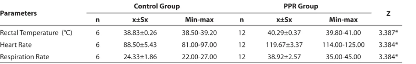
Table 1. Clinical parameters of control lambs and PPR groups Tablo 1. PPR ve kontrol grubundaki kuzularda klinik parametreler
Parameters
Control Group PPR Group
Z
n x±Sx Min-max n x±Sx Min-max
Rectal Temperature (°C) 6 38.83±0.26 38.50-39.20 12 40.29±0.37 39.80-41.00 3.387* Heart Rate 6 88.50±5.43 81.00-97.00 12 119.67±3.37 114.00-125.00 3.384* Respiration Rate 6 24.33±1.86 22.00-27.00 12 38.92±2.57 35.00-45.00 3.384*
Statistical analyses were conducted using Microsoft Exel (Microsoft Excel, 2007) and SPSS 12.0 for Windows® (SPSS 12.0 for Windows, Version 2.0. Champaign, IL: Lead Technologies, Inc.). The diff erences of the values between infected and control group were accepted as statistically significant when P value was lower than 0.05 (P<0.05).
RESULTS
Clinical Findings
Lambs with clinical signs of PPRV infection were develope fever, anorexia, dehydration, dullness, muco-purulent oculonasal discharge, lacrimation, conjunctivitis, oral lesions, halitosis, coughing, bronchopneumonia, dyspnea and diarrhea. Most clinical cases of mouth lesions were characterized by swelling around mouth, erosion, ulceration and necrosis on the lips, gums, buccal
cavity, tongue and palate. The outbreak occurred in a fl ock of 86 lambs. All infected animals was administered to symptomatic therapy. But the infection was so severe that some of animals died before and after treatment. Mortality rate, especially among kids, was high that of the 25 lambs died, 21 (84%) were kids and 4 (16%) were adult. Rectal temperature, heart rate and respiration rates of the infected animals were statistically high compared to those of control group (P<0.01; Table 1).
Serological Findings
All the serum samples collected from infected lambs were tested positive for antibodies specific to PPRV. Contrastingly, there was no detectible antibody against PPRV in samples collected from control lambs.
Haematological Findings
Hematological values obtained from both groups were
Table 2. Haematological parameters of control lambs and PPR groups Tablo 2. PPR ve kontrol grubundaki kuzularda hematolojik parametreler Parameters
Control Group PPR Group
Z n x±Sx Min-max n x±Sx Min-max WBC (x103/ml) 6 9.52±3.37 5.50-13.50 12 36.30±17.25 14.90-62.30 3.380a HGB (mg/dL) 6 11.48±3.22 7.10-16.50 12 6.13±1.42 4.60-8.70 3.193a RBC (x106/ml) 6 12.43±4.17 7.60-18.50 12 2.85±0.55 2.05-3.74 3.380a PCV (%) 6 26.70±7.57 19.20-38.70 12 10.76±2.34 7.60-14.00 3.380a MCV (fl ) 6 26.95±8.57 17.70-38.20 12 37.86±1.73 36.60-41.50 2.452b MCH (fl ) 6 6.10±1.41 3.90-7.80 12 22.00±5.64 14.20-31.70 3.380a MCHC (g/dl) 6 32.77±3.01 28.20-36.20 12 59.00±16.55 34.30-86.10 3.005a PLT (x103/ml) 6 473.50±93.08 350-567 12 321.08±81.98 195-420 2.441b
Diff erent superscripts a.b in the same line indicate significant diff erences between the 2 groups, a P<0.01; b P<0.05
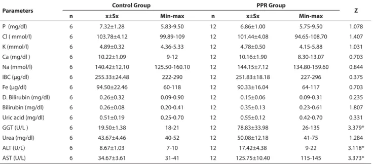
Table 3. Biochemical parameters of control lambs and PPR groups Tablo 3. PPR ve kontrol grubundaki kuzularda biyokimyasal parametreler
Parameters Control Group PPR Group Z
n x±Sx Min-max n x±Sx Min-max P (mg/dl) 6 7.32±1.28 5.83-9.50 12 6.86±1.00 5.75-9.50 1.078 Cl ( mmol/l) 6 103.78±4.12 99.89-109 12 101.44±4.08 94.65-108.70 1.407 K (mmol/l) 6 4.89±0.32 4.36-5.33 12 4.78±0.50 4.15-5.88 1.031 Ca (mg/dl ) 6 10.22±1.09 9-12 12 10.16±1.90 8.30-13.07 0.703 Na (mmol/l) 6 140.42±12.10 125.50-160.10 12 144.15±7.12 134.80-159.60 0.844 IBC (μg/dl) 6 255.33±24.48 222-290 12 251.83±18.18 227-296 0.375 Fe (μg/dl) 6 94.50±22.46 60-118 12 90.33±16.04 64-117 0.703 D. Bilirubin (mg/dl) 6 0.26±0.32 0.09-0.90 12 0.15±0.06 0.09-0.31 0.235 Bilirubin (mg/dl) 6 0.26±0.08 0.20-0.41 12 0.35±0.13 0.23-0.61 1.807 Uric acid (mg/dl) 6 0.51±0.19 0.25-0.70 12 0.55±0.12 0.42-0.70 0.331 GGT (U/L ) 6 19.50±1.38 18-21 12 78.83±33.98 26-135 3.379* Urea (mg/dl) 6 43.67±4.46 40-52 12 50.08±12.18 41-75 1.284 ALT (U/L) 6 8.67±1.03 7-10 12 17.42±4.38 9-22 3.118* AST (U/L) 6 34.67±3.61 31-41 12 125.75±10.40 115-145 3.373*
given as mean and mean of the standard division (mean ± SD) (Table 2). Significant differences in all of the para-meters were obtained between infected and control groups. Hemoglobin concentrations, percentage of PCV and RBCs and PLT counts were low in infected group compared to that of control group, but WBCs, MCV, MCH and MCHC values were higher in infected group than those of control group (Table 2).
Biochemical Findings
Biochemical parameters of control and infected groups were presented in Table III. According to the biochemical analysis, there were statistically significant increases in the serum activity of GGT, ALT and AST (P<0.01) in samples obtained from infected lambs compared to those of control group. But there was no statisticly significant diff erences in remaining tested parameters between infected and control groups (Table 3).
Macroscopic Findings
The carcasses (n=4) were observed to be dehydrated. There were apparent swelling around the mouths. Necrotic spots and erosive areas and in some cases haemorrhages were observed on the lips. Oral mucosa had occasional haemorrhagica. Severely congested tracheal mucous membranes and consolidation in the lungs were observed. The gastrointestinal tracts were mildly to moderately oedematous in some cases. Haemorrhages were seen in rumen, reticulum, rarely omasum and abomasum. Lymph nodes were usually oedematous, hyperemic and enlarged.
Histopathological Findings
Result of the histopathological findings indicated that
the lesions were mainly located on the respiratory system and upper digestive systems. In the lung, interstitial mono-nuclear cells, bronchitis, bronchiolitis, intracytoplasmic settled virus-associated inclusion bodies in the bronchioler epithelium (Fig. 1A) and syncytial cell formation in the
lumen of alveoles (Fig. 1B) were seen. The histopathology
of sore mouth was exhibited stomatitis with syncytial cell formations and intranuclear placed virus-associated inclusion bodies were generally seen in the syncytial cells (Fig. 1C). In immunohistochemical examination,
virus-associated antigens were found most frequently in the cytoplasm and rarely in the nucleus of the oral mucosal epithelium (Fig. 1D).
DISCUSSION
Common clinical signs of PPRV infection such as high fever, dehydration, oculonasal discharge, lacrimation, erosive-ulcerative oral lesions, coughing, broncho-pneumonia, diarrhea and death were observed in the present study which were also reported elsevere 2,3,6.
In the present study, clinical findings including body temperature, heart and respiratory rates were significantly higher in infected group than in control group (P<0.01) which were also reported by Yarım et al.17.
In this study, mortality of the PPR virus infection, especially among kids were high. Similarly, it has been informed in many studies 18 and it depends upon such
as be application or not symptomatic therapy, secondary infections, sheltering type and the various factors. It is higher in young animals. Morbillivirus infections have immune suppressor eff ect on the animals and increase susceptibility of PPRV infected animals to secondary pathogens 19. Therefore increased mortality rate obtained
Fig 1. A. Lung, virus-associated inclusion bodies in the bronchioler epithelium (arrows), H&E x 80 B. Lung, syncitial cells in the alveoles (arrows), H&E x 80 C. Oral mucosa, virus-associated inclusion bodies in the syncitial cells (arrows), H&E x 80 D. Oral mucosa, virus-associated anti-gens in the epithelial cells (arrows), ABC-P with Mayer’s haematoxylin x 160
Şekil 1. A. Akciğer, bronşioler epi-telde virus-ilişkili inklüzyon cisimcik-leri (oklar), H&E x 80 B. Akciğer, alveollerde sinsitial hücreler (oklar), H&E x 80 C. Ağız mukozası, sinsitial hücrelerde virus-ilişkili inklüzyon cisimcikleri (oklar), H&E x 80 D. Ağız mukozası, epitelyal hücrelerde virus-ilişkili antijenler (oklar), ABC-P, zemin boya Mayer’s haematoxylin x 160
in this study may occurs due to secondary infections reported elsevere 20.
Clinical and macroscopic findings may be helpfull for a quick diagnosis of PPR in endemic areas, but histo-pathological and serological examination are need to be applied to definitive diagnosis of PPRV infection in outbreaks 21,22. In PPR, the mucosal lesions in the oral
cavity and intestinal lesions are characterized by erosive and ulcerative stomatitis and enteritis, and the presence of inclusion bodies are pathognomic 12,13. In the PPR
infections, the pulmonary lesions were characterized by bronchiolitis, bronchitis, necrotic bronchitis or broncho-interstitial pneumonia, viral inclusion bodies and the presence of syncytial cells formations 1,12,13,15,21-23. Yener et
al.11 suggested that the absence of viral inclusion bodies
could be due to the animals in acute phase of disease or recovering from the PPR. İt was reported that the syncytial cells were observed during the terminal stage of infection 24. Toplu et al.11 in the some of
broncho-pneumonia cases was reported that eosinophilic intra-cytoplasmic inclusion bodies and syncytial cells were detected.
In this study, microscopic findings were observed mainly in the mouth and lung. Interstitial mononuclear cells, bronchitis, bronchiolitis, and syncytial cell formations in the alveoles, and intranuclear inclusion bodies were detected particularly in the cytoplasma of alveolar, bronchial and bronchiolar epithelial cells. The antigen was also detected in the syncytial cells of mouth tissues. The present findings have also been reported in previous experimental and naturally studies 1,12,14,25.
In this study; there were statistically significant differences between all parameters. White blood cells (WBCs), MCV, MCH and MCHC parameters were significantly high, but HGB, RBCs, PCV and PLT parameters were low in infected group compared to those of control group (Table 2). Olaleye et al.25 there was initial neutrophilic
leucocytosis during the phase of fever followed by marked lymphopaenic leucopaenia which progressed terminally in most of the infected goats. Leucocytosis obtained in the present study may be occurs due to development of neutrophilia during the febrile period reported by Olaleye et al.25 and İssi et al.26. Conversely, Kataria et al.20
and Yarım et al.17 determined low WBC value in infected
lambs and sheep. Moreover, Kataria et al.20 determined
high RBC, HGB, PCV values. The diff erences between these hematological parameters may be due to the diff erent phase of the disease 25, presence secondary infections 1,
and nutrition or dehydration degree 19,20,27.
In present study; according to the biochemical test results, there was statistically significant differences between control and infected group of GGT, ALT and AST (P<0.01, Tablo 3). Similarly, results were also reported by Yarım et al.17 they also reported that total bilirubin and
direct bilirubin were high. Conversely, in this study, these parameters were not statistically significant. Kataria et al.20 determined increased serum sodium and potassium
values, but in this study, serum sodium and potassium were not statistically significant in infected lambs. These diff erences may result from infection severity, stage of the infection, presence of secondary infections, age and species of animals.
In conclusion, in the present study, PPRV infection cause considerable erosive-ulcerative lesions on various organs and alter haematological and biochemical para-meters in sheep. Therefore, it is suggestive that the infection impaire the function of many organs or systems such as hearth, lung, liver, skin, oral cavity and digestive and respiratory systems. These valuable data obtained in the present study may helpfull in the diagnosis and treatment (symptomatic) of the disease. In addition, these obtained clinical, haematological, biochemical and histopathological parameters should be kept in mind by the veterinary practitioners for the possible occurance of new outbreaks in the region and for its diagnosis.
A
CKNOWLEDGEMENTSWe would like to thanks Prof.Dr. Tolga GÜVENÇ (Department of Pathology, Veterinary Faculty, Ondokuz Mayıs University) and Assoc. Prof. Dr. Oğuz KUL (Department of Pathology, Veterinary Faculty, Kırıkkale University) for their kind help.
REFERENCES
1. Aruni AW, Lalitha PS, Mohan AC, Chitravelu P, Anbumani SP: Histo-pathological study of a natural outbreak of peste des petits ruminants in goats of Tamilnadu. Small Rumin Res, 28, 233-240, 1998.
2. Aytekin İ: A subclinic Peste des Petits Ruminants case in a lamb.
Atatürk Üniv Vet Bil Derg, 3, 8-10, 2008.
3. Çam Y, Gençay A, Beyaz L, Atalay O, Atasever A, Özkul A, Kibar M: Peste des petits ruminants in a sheep and goat fl ock in Kayseri province, Turkey. Vet Rec, 157, 523-524, 2005.
4. Al-Dubaib MA: Peste des petitis ruminants morbillivirus infection in lambs and young goats at Qassim region, Saudi Arabia. Trop Anim Health
Prod, 41, 217-220, 2009.
5. Albayrak H, Gür S: A serologic investigation for Peste des petits ruminants infection in sheep, cattle and camels (Camelus dromedarius) in Aydın province, West Anatolia. Trop Anim Health Prod, 42, 151-153, 2010. 6. Kwiatek O, Minet C, Grillet C, Hurardy C, Carlsson E, Karimov B, Albina E, Diallo A, Libeau G: Peste des Petits Ruminants (PPR) outbreak in Tajikistan. J Comp Pathol, 136, 111-119, 2007.
7. Saeed IK, Yahia HA, Khalafalla AI, Rahman-Mahasin EA: Current situation of Peste des petits ruminants (PPR) in the Sudan. Trop Anim
Health Prod, 42, 89-93, 2010.
8. Diallo A, Minet C, Le Goff C, Berhe G, Albina E, Libeau G, Barrett T: The threat of peste des petits ruminants: Progress in vaccine development for disease control. Vaccine, 25, 5591-5597, 2007.
9. Lefevre PC, Diallo A, Schenkel F, Hussein S, Staak G: Serological evidence of peste des petits ruminants in Jordan. Vet Rec, 128, 110, 1991. 10. Kul O, Kabakcı N, Özkul A, Kalender H, Atmaca HT: Concurrent Peste des Petits Ruminants virus and Pestivirus infection in stillborn twin
lambs. Vet Pathol, 45, 191-196, 2008.
11. Toplu N: Characteristic and non-characteristic pathological findings in Peste des Petits Ruminants (PPR) of sheep in the Ege District of Turkey.
J Comp Pathol, 131, 135-141, 2004.
12. Kumar P, Tripathi BN, Sharma AK, Kumar R, Sreenivasa BP, Singh RP, Dhar P, Bandyopadhyay SK: Pathological and immunohistochemical study of experimental Peste des Petits ruminants virus infection in goats.
J Vet Med, 51, 153-159, 2004.
13. Ozmen O, Kale M, Haligur M, Yavru S: Pathological, serological, and virological findings in sheep infected simultaneously with Bluetongue, Peste-des-Petits-Ruminants, and Sheeppox viruses. Trop Anim Health
Prod, 41, 951-958, 2009.
14. Rowland AC, Scott GR, Hill DH: The pathology of an erosive stomatitis and enteritis in West-African dwarf goats. J Pathol, 98, 83-87, 1969. 15. Alçığır G, Atalay VS, Toplu N: Türkiye’de kuzularda Peste des Petits Ruminants virus enfeksiyonunun patomorfolojik ve immunohistolojik ilk tanımı. Ankara Univ Vet Fak Derg, 43, 181-189, 1996.
16. Anon: Office International des Epizooties Manual of Standards. Peste des Petits Ruminants. Chapter 2.1.5., Manual of Diagnostic Testes and Vaccine for Terrestrial Animals. 5th ed., Paris. 2004.
17. Yarım GF, Nisbet C, Yazıcı Z, Gumusova SO: Elevated serum total sialic acid concentrations in sheep with Peste des Petits Ruminants.
Medcyna Wet, 62, 1375-1377, 2006.
18. Albayrak H, Alkan F: PPR virus infection on sheep in blacksea region of Turkey: Epidemiology and diagnosis by RT-PCR and virus isolation. Vet
Res Commun, 33, 241-249, 2009.
19. Heaney J, Barrett T, Cosby SL: Inhibition of in vitro leukocyte proliferation by morbilliviruses. J Virol, 76, 3579-84, 2002.
20. Kataria AK, Kataria N, Gahlot AK: Large scale outbreaks of Peste des Petits Ruminants in sheep and goats in Thar Desert of India. Slov Vet Res, 44, 123-32, 2007.
21. Yener Z, Saglam YS, Temur A, Keles H: Immunohistochemical detection of peste des petits ruminants viral antigens in tissues from cases of naturally occurring pneumonia in goats. Small Rumin Res, 51, 273-277, 2004.
22. Brown CC, Mariner JC, Olander HJ: An immunohistochemical study of the pneumonia caused by peste des petits ruminants virus. Vet Pathol, 28, 166-170, 1991.
23. Sağlam YS, Temur A: Immunohistochemical detection of Peste des
Petits Ruminants (PPR) viral antigen from the cases of naturally occurring pneumonia in sheep. Kafkas Univ Vet Fak Derg, 15, 423-428, 2009.
24. Gulyaz V, Ozkul A: Pathogenicity of a local peste des petits ruminants virus isolate in sheep in Turkey. Trop Anim Health Prod, 37, 541-547, 2005. 25. Olaleye OD, Ikede BO, Durojaiye OA: Fluid, electrolyte and haematological changes in experimental peste des petits ruminants virus infection in West African dwarf goats. Cytobios, 58, 39-51, 1989.
26. İssi M, Gül Y, Dabak M: Küçük ruminant vebası saptanan keçilerde serum vitamin C düzeyeleri. Turk J Vet Anim Sci, 25, 539-544, 2001. 27. Aikhuomobhogbe PU, Orheruata AM: Haematological and blood biochemical indices of West African dwarf goats vaccinated against Pestes des Petit Ruminants (PPR). Afr J Biotechnol, 5, 743-748, 2006.