DOI: 10.2478/acve-2019-0020
Case report
*Corresponding author: e-mail: [email protected]
EXTRAMEDULLARY PULMONARY PLASMACYTOMA IN A DOG
IPEK Volkan1*, ACAR Hilal2, KOCATURKMeric3, SALCI Hakan2, SONMEZ Gursel4 1Department of Pathology, Faculty of Veterinary Medicine, Burdur Mehmet Akif Ersoy University,Burdur, Turkey; 2Department of Surgery, Faculty of Veterinary Medicine, Bursa Uludag University, Bursa,
Turkey; 3Department of Internal Medicine, Faculty of Veterinary Medicine, Bursa Uludag University,
Bursa, Turkey; 4Department of Pathology, Faculty of Veterinary Medicine, Bursa Uludag University,
Bursa, Turkey
(Received 21 January, Accepted 28 March 2019)
In this case report we are presenting a rare case of primary pulmonary plasmacytoma in a dog in the context of clinical and pathological fi ndings. A six-years-old, female Rottweiler was brought to the clinic with respiratory complaints. The patient was dyspneic and tachypneic, and there were friction sounds on auscultation of the lungs. Laryngeal and tracheal palpation induced severe cough. Lateral and ventrodorsal radiographs of the thorax showed increased opacity in the lungs and loss of cardiac silhouette. Based on clinical and radiological fi ndings, diagnosis of a lung mass was made and surgery recommended. Under general anesthesia, bilobectomy of the right lung lobes by medial sternotomy was performed. Upon cytological and histopathological fi ndings, plasmacytoma was diagnosed.
Key words: dog, histopathology, lung, plasmacytoma
INTRODUCTION
Plasma cells originate from B lymphocytes and can give rise to a group of neoplastic conditions [1]. Multiple myeloma as a systemic disease is the clinically most important form of plasma cell tumor in canines and is often associated with irregular immunoglobulin secretion from neoplastic plasma cells in the medullary cavity of the bone [1,2]. According to WHO, extramedullary plasmacytoma (EMP) is “a localized tumor composed of atypical neoplastic plasma cells” [3]. EMP refers to tumors that form outside the bone marrow and can spread to local or regional lymph nodes or metastasize to distant areas [4]. In a study in which 751 cases were examined, it was reported that extramedullary plasmacytoma was present in the skin (86%), mucous membranes (9%), rectum and colon (4%) and 1% in the stomach, spleen, genital regions, eyes, uterus and liver [5]. Wright et al. reported that EMP was present in 16
of 302 (5.2%) oral tumors [6]. In addition, EMP was found on other sites of the body such as the brain and larynx [7-9].
In humans, EMP is rarely formed in the lungs. Pulmonary EMP may be the fi rst predictor of multiple myeloma or may be confi ned to the lungs with or without spread to local lymph nodes [4]. Primary pulmonary plasmacytomas are rare in humans [10,11]. Canine pulmonary plasmacytoma is also very rare and according to the authors’ knowledge, it has only been reported in one previous case [2]. Here, we presented a case of extramedullary pulmonary plasmacytoma in a dog in the context of clinical and pathological fi ndings.
CASE PRESENTATION
A six-years-old, female Rottweiler was brought to the university polyclinics with respiratory complaints. There was friction noise on auscultation of the lungs. Severe coughing was observed during laryngotracheal palpation. Abdominal palpation revealed distention and ondulation due to ascites. Cardiac auscultation was non-specifi c and other clinical fi ndings were normal. Hematological evaluation revealed neutrophilic leukocytosis (white blood cells: 19.98 ×10⁹/L, reference: 6-17 ×109/L; neutrophiles: 17.01× 10⁹/L, reference: 3-12 ×109/L) and normocytic and normochromic anemia (red blood cells: 4.16 ×10¹²/L, reference: 5.5-8.5 ×10⁹/L; mean corpuscular volume: 64 fl , reference: 60-77 fl ; mean corpuscular hemoglobin: 20.4 pg, reference: 19.5-24.5 pg). In blood serum biochemistry analysis, aspartate aminotransferase (AST) was increased (124 U/l) compared to the reference (21-44 U/I). In the radiographic examination, lateral and ventrodorsal radiographs of the thorax revealed increased opacity and loss of cardiac silhouette. In the abdominal radiography, generalized opacity was observed due to ascites. There was no radiographic abnormality on the skeletal tissue or other visceral organs. According to clinical and radiological fi ndings, a pulmonary mass was diagnosed. Echocardiographic evaluation of the heart was performed and parameters of the patient were within the reference ranges. Sinus tachycardia, small complex QRS (R: 0,3 mV, 2nd derivation, P:168 bpm) and notching on R waves were seen on electrocardiographic evaluation of the patient due to pleural effusion in the pre-operative period (50 mm/sn, 10 mm/1m). Preoperatively, furosemide (2 mg/ kg, po., tid) to reduce pleural effusion, enalapril maleate (0.5 mg/kg po., bid) and cefi xime (20 mg/kg po., bid) were used for three days. For sedation and induction, xylazine HCl (1.5 mg/kg, im.) and ketamine HCl (6 mg/kg, im.) were applied. General anesthesia and maintenance were provided with isofl urane at 2% concentration with mechanical ventilation. After surgical preparation, the region was approached with median sternotomy, and then bilobectomy of the cranial and middle lobe of the right lung where the mass was located was performed. The thorax tube was placed and the tissues were closed according to the appropriate technique. On the 3rd day after the operation normal sinus rhythm with normal R waves (2,1 mV, 2nd derivation, P: 118
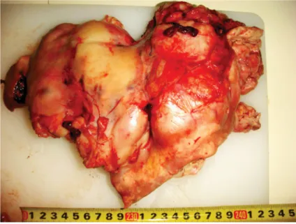
and necropsy could not be done because of owner rejection. The removed mass was 23x20x6 cm in size, soft and yellowish in color (Fig. 1). Cytologically, numerous plasma cells which had mild basophilic cytoplasm, eccentric nucleus and perinuclear clear areas were seen. Occasionally cells with two or more nuclei were present (Fig. 2). Histopathologically, numerous neoplastic plasma cells with large eosinophilic cytoplasm showing marked anisocytosis and anisonucleosis with nucleus-cytoplasm asynchrony were noted. Some of these cells had notched nuclei. Multiple giant cells with up to 4-5 nuclei were seen. There were mitotic fi gures in some tumor cells (Fig. 3). Upon these fi ndings, plasmacytoma was diagnosed.
The diagnosis of extramedullary plasmacytomas usually requires fi ne needle aspiration or tissue biopsy. Various immunohistochemical studies can be performed to differentiate the non-differentiated solitary plasmacytic tumors from other round cell tumors [12,13]. In our case, the diagnosis was made based on cytological and histopathological fi ndings. Cytology is a useful tool for diagnosis of round cell tumors including plasmacytomas. Cytological differential diagnosis includes lymphoma, histiocytoma, amelanotic melanoma, neuroendocrine (Merkel cell) tumor and peripheral nerve tumor. Characteristic morphology usually reveals perinuclear clear areas, binucleated cells and eccentrically placed nuclei [14]. In our case, giant cells with up to 3-4 nuclei and neoplastic cells with eccentric nuclei, perinuclear clear area, mild basophilic cytoplasm, prominent anisocytosis and anisonucleosis were found on cytological examination. Some of the cells contained distinct vacuoles.
Histologically EMP is classifi ed into fi ve subtypes [15,16]. Platz et al. reported a study that included histological classifi cation of EMP and prognostic importance of this classifi cation [15]. In this study, 117 EMPs in the skin and digestive system were discussed. EMPs have been classifi ed after histopathological examination as hyaline Figure 3. Numerous neoplastic plasma cells with large eosinophilic cytoplasm, some
multinucleated cells (arrow heads) and a high number of mitotic fi gures (arrows), x200, H&E.
Figure 2. Numerous plasma cells with perinuclear clear areas (arrows), and a multinucleated
the hyaline type is recognized to have sickle-shaped nuclei and a small number of binucleated giant cells. In the mature type, tumor cells are similar to mature plasma cells with vacuolated or eosinophilic fi ne granular cytoplasm and binucleated cells may be observed similar to the hyaline type. The cleaved type has been indicated as the most often observed type and is characterized by cleaved nucleus structure. Also, many giant cells containing up to four nuclei may be observed. In most cases of asynchronous type, cleaved type transition is observed, and the dominant feature is the nucleus-cytoplasm asynchrony in maturation. They also contain a large number of giant cells, similar to the cleaved type, and the tumor cells have a vacuolar eosinophilic cytoplasm and a blastic nucleus with a central nucleolus. The main feature of the polymorphous-blastic type is anisocytosis in tumor cells and a large number of giant cells with up to nine polymorphous nuclei. Tumor cells usually have eosinophilic and vacuolated cytoplasm without a perinuclear halo [15]. In a previous study [15] transitions between two or more types of tumors have been reported. According to this classifi cation, fi ndings in our case are compatible with polymorphous blastic type with transition to both asynchronous and cleaved types.
In dogs, multiple myeloma and extramedullary plasmacytoma observed in the internal organs more easily metastasized than mucocutaneous plasmacytomas [17]. In our case, no distant metastasis was observed in the radiographic examinations. However, neither serum electrophoresis of urine or bone marrow aspirate could be done to rule out multiple myeloma but there was no skeletal abnormality on radiography.
In the study of Platz et al, local recurrence was observed in nine cases but only two of them were confi rmed by histopathological examination. Both tumors were classifi ed as polymorphous-blastic type of EMP [15]. It has been seen that this detailed histological classifi cation based on cell morphology does not have a strong relationship with the biological behavior of the tumor [18].
A standard treatment protocol for human and veterinary medicine has not been established due to the rarity of the disease but complete surgical resection may be curative [2,19,20]. Surgery, chemotherapy and radiation combinations did not reveal any differences in survival [4]. Adelman et al, reported a long survival time (1.5 year) in a dog with extramedullary pulmonary plasmacytoma. In our case, it was not possible to determine the prognosis since the patient died fi ve days after the surgery.
As a co nclusion, extramedullary pulmonary plasmacytoma should be included in the differential diagnosis list in cases of a lung mass. Further studies are needed on the classifi cation and prognosis of extramedullary pulmonary plasmacytomas in canine patients.
Authors’ contributions
MK made clinical examinations and performed echocardiographic evaluation. HA and HS performed radiographic examination and surgery. GS and VI made cytological and histopathological examinations. All authors read and approved the fi nal manuscript.
Declaration of confl icting interests
The author(s) declared no potential confl icts of interest with respect to the research, authorship, and/or publication of this article.
REFERE NCES
1. Morris J, Dobson J: Skin. In: Small Anim Oncology. Oxford, United Kingdom: Blackwell Science; 2001, 65.
2. Adelman L, Larson V, Sissener T, Spotswood T: Extramedullary plasmacytoma in the lung of a Doberman pinscher dog. Can Vet J = La Rev Vet Can Canadian Veterinary Medical Association; 2014, 55:1237–1240.
3. Mathé G, Rappaport H, O’Connor G, Torloni H: Histological and cytological typing of neoplastic diseases of haematopoietic and lymphoid tissues. Int Histol Classif Tumours No 14 Geneva: World Health Organization; 1976.
4. Montero C, Souto A, Vidal I, Fernández M del M, Blanco M, Verea H: Plasmocitoma pulmonar primario: aportación de 3 casos. Arch Bronconeumol 2009, 45:564–566. 5. Kupanoff PA, Popovitch CA, Goldschmidt MH: Colorectal Plasmacytomas: A Retrospective
Study of Nine Dogs. J Am Anim Hosp Assoc 2006, 42:37–43.
6. Wright ZM, Rogers KS, Mansell J: Survival Data for Canine Oral Extramedullary Plasmacytomas: A Retrospective Analysis (1996–2006). J Am Anim Hosp Assoc 2008, 44:75–81.
7. Hayes AM, Gregory SP, Murphy S, McConnell JF, Patterson-Kane JC: Solitary extramedullary plasmacytoma of the canine larynx. J Small Anim Pract 2007, 48:288–291.
8. Witham A, French A, Hill K: Extramedullary laryngeal plasmacytoma in a dog. N Z Vet J 2012, 60:61–64.
9. Wettere AJ Van, Linder KE, Suter SE, Olby NJ: Solitary Intracerebral Plasmacytoma in a Dog: Microscopic, Immunohistochemical, and Molecular Features. Vet Pathol 2009, 46:949-951.
10. Lu R, Medina KL, Lancki DW, Singh H: IRF-4,8 orchestrate the pre-B-to-B transition in lymphocyte development. Genes Dev 2003, 17:1703–1708.
11. Wiltshaw E: The natural history of extramedullary plasmacytoma and its relation to solitary myeloma of bone and myelomatosis. Medicine (Baltimore) 1976, 55:217–238.
12. Withrow S, Vail D: Withrow &MacEwen’s Small Animal Clinical Oncology. 4th ed. St. Louis, Missouri: Saunders Elsevier; 2001.
13. Cangul IT, Wijnen M, Van Garderen E, van den Ingh TSGAM: Clinico-pathological aspects of canine cutaneous and mucocutaneous plasmacytomas. J Vet Med A Physiol Pathol Clin
14. Raskin R: Skin and subcutaneous tissues. In: Canine and feline cytology: a color atlas and interpretation guide 2nd ed. St. Louis, Missouri, USA: Saunders; 2010, 26–76.
15. Platz SJ, Breuer W, Pfl eghaar S, Minkus G, Hermanns W: Prognostic Value of Histopathological Grading in Canine Extramedullary Plasmacytomas. Vet Pathol 1999, 36:23–27.
16. Gross T, Ihrke P, Walder E, Affolter V: Skin diseases of the dog and cat: clinical and histopathologic diagnosis 2nd ed. Oxford, United Kingdom: Blackwell; 2005.
17. Trevor PB, Saunders GK, Waldron DR, Leib MS: Metastatic extramedullary plasmacytoma of the colon and rectum in a dog. J Am Vet Med Assoc 1993, 203:406–409.
18. Albanese F: Cytology of Skin Tumours. In: Canine and Feline Skin Cytology Switzerland: Springer International Publishing Switzerland; 2017, 321–328.
19. Wang J, Pandha HS, Treleaven J, Powles R: Metastatic Extramedullary Plasmacytoma of the Lung. Leuk Lymphoma Taylor & Francis; 1999, 35:423–425.
20. Hayes-Lattin B, Blanke CD, Deloughery TG: Pulmonary and intracerebral plasmacytomas in a patient without multiple myeloma: A case report. Am J Hematol 2003, 73:131–134.
EKSTRAMEDULARNI PLAZMOCITOM PLUĆA KOD PSA
IPEK Volkan, ACAR Hilal, KOCATURK Meric, SALCI Hakan, SONMEZ Gursel U ovom prikazu slučaja, predstavili smo redak slučaj primarnog plazmocitoma pluća kod psa u konteksu kliničkih simptoma i patološkog nalaza. Ženka rotvajler rase, stara 6 godina, primljena je na kliniku sa simptomima respiratornih smetnji. Kod pacijenta, bili su prisutni dispneja i tahipneja kao i zvuci frikcije pri auskultaciji pluća. Palpacijom larinksa i traheje, mogao je da se izazove intenzivan kašalj. Lateralna i ventrodorzalna radiografi ja toraksa, pokazala je intenzivno zasenčenje u plućima kao i gubitak srčane siluete. Na osnovu kliničkog i radiografskog nalaza, dijagnostikovane su promene na plućnom parenhimu pri čemu je preporučena hirurška intervencija. Operacija je obav-ljena u opštoj anesteziji, a primenom sternotomije, urađena je bilobektomija desnih lobusa pluća. Citološkim i histopatološkim pregledom, ustanovljen je plazmocitom.