Araştırma/Research
Investigation of the positive effects of silymarin on valproic acid-induced liver damage in rats
İbrahim AKTAŞ1*, İlkay ARMAĞAN2
1School of Health Services, Departmentof Pharmacology, Adiyaman University, Adiyaman, Turkey.
2Faculty of Medicine, Department of Histology and Embryology, Süleyman Demirel University, Isparta, Turkey.
Abstract
Purpose: In this study; We aimed to investigate the possible hepatoprotective effects of silymarin
against hepatic damage in valproic acid-induced rats using histological and biochemical evaluations.
Method: Experimental procedures were performed on 21 male Sprague Dawley rats. Rats were
seperated into three groups: group 1, control; group 2, valproic acid; group 3, valproic acid + silymarin. The groups were administered 500 mg/kg/day valproic acidand 100 mg/kg/daysilymarin for 14 days, except control group.
Results: Silymarin treatment decreased the levels of serumgamma glutamyl transferase, alanine
amino transferase, aspartate aminotransferase and increreased serum albumin levelssignificantly (p<0.05). In addition, increased amount of malondialdehyde and decreased levels of glutathione with valproic acid were significantly suppressed by silymarin in liver tissue (p<0.05).The combination of silymarinwith valproic acid reduced loss of body weight in the present study. Histologically, the extent of liver damage was remarkably lower in the valproic acid+silymarin group (p<0.005). When the valproic acid + silymarin group compared to the valproic acid group; it was determined that antioxidant activity was increased, oxidative stress.
Conclusion: This study revealed that the liver injury induced by valproic acid was attenuated with
silymarin administration. Silymarincan protect rat liver against valproic acid induced injury by its anti-oxidative effect, and might be useful for reducing the severity of liver injury.
Key Words: Valproic acid; Silymarin; Liver injury; Rat.
Doi: 10.30569.adiyamansaglik. 568226
YazışmadanSorumluYazar İbrahim AKTAŞ
1School of Health Services, Departmentof
Pharmacology, Adiyaman University, Adiyaman, Turkey
Tel : +90 0506 594 27 25
Email: [email protected],
GelişTarihi: 21.05.2019 Kabul Tarihi: 19.06.2019
Sayfa1446 Silimarinin, ratlarda valproik asitin indüklediği karaciğer hasarı üzerine olumlu
etkilerinin araştırılması ÖZET
Amaç:Bu çalışmada; valproik asit kaynaklı ratlarda karaciğer hasarına karşı silimarin'in olası hepatoprotektif
etkilerini histolojik ve biyokimyasal değerlendirmeler kullanarak araştırmayı amaçladık.
Gereç ve yöntemler:Deneysel işlemler, 21 adet Sprague Dawley sıçanı üzerinde gerçekleştirildi. Sıçanlar üç
gruba ayrıldı: grup 1, kontrol; grup 2, valproik asit; grup 3, valproik asit + silimarin. Gruplara, kontrol grubu hariç 500 mg/kg/gün valproik asit ve 100 mg/kg/gün silimarin 14 gün uygulandı.
Bulgular:Valproik asitile artan serum gama glutamil transferaz, alanin amino transferaz, aspartat
aminotransferaz ve azalan serum albumin seviyesi silimarin tedavisi ile tersine çevrildi (p<0.05). Ek olarak, karaciğer dokusunda valproik asit ile malondialdehit seviyesinin artması ve glutatyon seviyesinin azalması, silimarin tarafından önemli ölçüde baskılandı (p<0.05). Silimarin'in,valproik asit ile kombinasyonu bu çalışmada vücut ağırlığı kaybını azaltmıştır. Histolojik olarak, valproik asit + silimarin grubunda karaciğer hasarının derecesi anlamlı olarak dah düşük bulundu (p<0.005). Valproik asit + silimarin grubunda; valproik asit grubuna göre oksidatif streste azalma, antioksidan aktivitede artışve histopatolojik değişikliklerde azalma kaydedildi.
Sonuç:Bu çalışma davalproik asit'in indüklediği karaciğer hasarının silimarin uygulaması ile azaldığı ortaya
konuldu. Silimarin, sıçan karaciğerini anti-oksidatif etkisi ile valproik asit kaynaklı hasara karşı koruyabilir ve karaciğer hasarının boyutunu azaltmak için faydalı olabilir.
Anahtar Kelimeler:Valproik asit; Silimarin;Karaciğer hasarı; Sıçan.
Introduction
Valproic acid (VPA) are used to treat cases worldwide such as brain neoplasm (1), generalized epilepsy (2), migraine, bipolar mania (3) and psychiatric diseases. It inhibits Gamma aminobutyric acid (GABA) recovery from the presynaptic terminals by inhibiting GABA transaminase and increasing GABA in the synaptic cleft(4). VPA improves lipid peroxidation and serum liver enzymes from the start of chronic liver therapy. Moreover, oxidative stress, apoptosis and necrosis were caused by histopathological and biochemical studies(5). During the production and disposal of reactive oxygen species (ROS), an inconsistency of oxidative stress occurs (6). Large xenobiotic metabolism in the liver is considered the source of ROS production, especially in this tissue. Excessive ROS production causes damage to cellular molecules such as lipid, DNA and protein(3). High liver enzymes and lipid peroxidation were observed in 44% of chronic VPA patients(7). VPA strongly binds to plasma proteins at therapeutic levels or at high concentrations. This predisposes patients to
Sayfa1447 fluctuations in therapeutic effects and, more importantly, to unpredictable toxicity or drug interactions(8). Previous studies, there is a raise in lipid peroxidation and free radical products, and a reduce in antioxidant enzymes(9).
Recently,silymarin (SLY) have received attention for treatment of liver disease and dysfunction, and to promote liver regeneration(10). SLY increases the detoxification capacity of the liver by elevating the amount of glutathione, a strong antioxidant that is exposed to oxidative stress in the liver. It shows membrane stabilizer property by inhibiting lipid peroxidation. It promotes hepatocyte regeneration by stimulating RNA and DNA polymerase synthesis (11). The binding to the receptors in the membranes prevents the binding of toxins in these regions thereby reducing drug-induced hepatocellular damage(12). It is used in the treatment of diseases such as alcohol, toxin and viral hepatitis (13).
However, the effect of SLY on oxidative stress as a result of VPA-induced liver damage has not been investigated.Consequently, we aimed to investigate the potential hepatoprotective effect of SLY against VPA-related hepatocellular injury in rats.
Materials and Methods Chemicals
VPA (as Convulex 500 mg capsules) from Liba Co. (İstanbul, Turkey). Silymarin was obtained as Legalon fort (100 mg/kg capsules)from Madaus Co.(İstanbul, Turkey).VPA and SLY doses were determined based on previous studies, respectively(5,12).
Hydrochloric acid (HCL), thiochleroacetic acid, thiobarbuturic acid and paraffin were obtained from, Sigma-Aldrich (Germany). 5.5 '-dithio-bis-2-nitrobenzoic acid and formalin obtained from Chem-Impex (USA) and Tekkim (Turkey), respectively. Xylene, hematoxylin-eosinand ethanol were obtained from Merck (Germany).
Animals
In this study, 21 male Sprague-Dawley rats (210-240 g for 8 weeks) were used.The study was carried out according to the protocol approved by the Experimental Animal Ethics Committee of Fırat University (Protocol # 2017/92).The rats were maintained with a 12 h dark: 12 h light cycle at 21 °C with free access to water and food.
Sayfa1448 Treatment protocol
Animals were randomised into three groups, with seven rats in each group, as follows: Control; VPA; VPA + SLY. The control group received 1mL 0.9 % NaCl orally for 14 days.
The VPA group was given VPA peros 500 mg / kg / day for 14thdays (5).The VPA + SLY group
was received 500 mg / kg VPA and 100 mg / kg / day SLY peros for 14th days(12).Body weights
were recorded at the beginning and the end of the study. Liver weights were recorded at the end of the experiment. The rats were sacrificed by cervical dislocation under anesthesia with ethyl ether at the end of 14th day. Blood samples were harvested on from the jugular vein, centrifuged for 5 minutes at 5,000 xg and the serum was separated and stored at -86 °C for biochemical analysis. The entire liver was excised and preserved at -86 °C until analysis.
Body weight and liver index
Since the body weight was decreased by VPA, it was evaluated in order to effect of SLY. Body weights of rats were measured at the beginning and the end of the experiment. The ratio of liver weight to body weight (g/100 g BW) were calculated.
Biochemical evaluation
Liver serum biomarkers including Gamma glutamyl transferase (GGT) U / L, aspartate aminotransferase (AST) U / L, alanine aminotransferase (ALT) U / L and albumin g / dL were analyzed with the Olympus 2700 analyzer (Olympus Diagnostica GmbH, Germany). In addition, AST and ALT activities were evaluated according to Reitman–Frankel colorimetric transaminase procedure (14).
Oxidative stress biomarkers
Malondialdehyde (MDA)measurements were conducted in paw tissue(15). The amount of lipid peroxidation was measured according to the concentration of thiobarbituric acid
reactive species MDA was reacted with TBA at pH 2–3 and 95 °C for 15 min. The residue was
centrifuged at 2500 xg for 10 min. The test samples were then read at 532 nm with spectrophotometer(16).
Glutathione (GSH) levels in paw tissues were measured according to Sedlak and Lindsay method (17). The sample was precipitated with 50% TCA and centrifuged at 1000 ×g for 5 minutes. 2 mL Tris-EDTA buffer (0.2 M, PH = 8.9) and 0.1 mL 0.01M 5.5 '-dithio-bis-2
Sayfa1449 by taking 0.5 mL of the supernatant from the supernatant. -nitrobenzoic acid was added. The mixture was allowed to stand at room temperature for 5 min and the absorbance at 412 nm wavelength with spectrophotometry.
Histopathological examinations
During the necropsy, all rats in the control and experimental groups were removed and their livers were macroscoped. 10% neutral formalin was used for fixation. After the tissues were fixed, the formalin was removed by washing in the stream. It was passed through a series of graded alcohols for dehydration and kept in xylene for transparency. It was then buried in paraffin. From the obtained paraffin blocks, 3-4 μm sections were taken with rotary microtome (RM2125RTS, Leica, Germany). 3-4 μm sections were stained with Hematoxylin-Eosin for histopathological evaluation. Modified semi-quantitative scale were used for the evaluation of histopathological changes; [(0): none, (1): mild, (2): moderate, (3): severe grade]. Samples were evaluated and visualized with imaging-assisted binocular light microscopy (ECLIPSE Ni-U, Nikon, and Tokyo, Japan).
Statistical analysis
SPSS software, version 20.0, was used for statistical analyses. Data were means ± SEM. Body weight data were analyzed by the paired-samples t-test. The groups were compared with the paired-samples t-test at the beginning and end of the study. The Shapiro–Wilk test was performed to assess normality. Inter-group and intra-group comparisons were made using one-way ANOVA post hoc LSD for parametric values; for nonparametric values, the Kruskal– Wallis test was used for biochemical parameters. To assess semiqualitative evaluation of histopathological scores, the Kruskal-Wallis test was used. Differences in the parameters measured among the groups were analyzed by Kruskal-Wallis test. A Mann-Whitney U test was used to compare dual groups. Values for p ≤ 0.05 were considered statistically significant.
Results
Measure of body weight
Total body weight was measured at the beginning and at the end of the experiment. In relation to body weight, a significant increase was observed between the initial and final weight for the control group (p< 0.021). On the other hand, VPA and VPA + SLY groups showed a
Sayfa1450 significant decrease when comparing their initial and final weights. The final body weight of the VPA + SLY group was higher than VPA group. The body weight of theVPA group was significantly less than control and VPA + SLY groups (p<0.01 for both). The mean body weights of all groups are shown in Table 1 and Figure 1.
There was a significantly increased total relative liver weight (g/100g body weight) gain in the control group, and a significantly decreased weight in the VPA, VPA+SLY groups. In addition, treatment with SLY resulted in a significant increase in VPA-reduced liver weight (p=0,021,Table 1).
Table 1. Comparison of changes in body weight of the experimental rats and liver organ weight parameters.
Stage of study Control VPA VPA + SLY
Initial weight (g)
223.57 ± 2.1 217.57 ± 2.10 224.71 ± 2.90 Final weight (g)
242.85 ± 6.28 a,c 201.28 ± 2.88 a,b 212.28 ± 1.98 a,d
Relative difference (%)
8 ± 3 c 7 ± 1 b 5 ± 1 d
Relative liver weight (g/100g body weight)
6.53 ± 0.14 b,c 3.96 ± 0.02 a,c 5.08 ± 0.22 a,b
Data are means ± SEM, n = 7. Body weight is expressed in grams. VPA, valproic acid; SLY, slymarin. a Means in
the same group are significantly different at p<0.05, b significantly different from control at p<0.05, c significantly
different from VPA treated rats at p<0.05, d significantly different from VPA treated rats at p<0.02.
Figure 1. Changes in the body weight of experimental rats. Data are means ± SEM (n = 7). VPA, valproic acid; SLY, silymarin. aMeans in the same group are significantly different at p<0.05. bSignificantly different from
control at p<0.05. cSignificantly different from VPA treated rats at p<0.05. dSignificantly different from VPA
treated rats at p <0.02. a,d a,b a,c a,c a,b
Sayfa1451 Biochemical evaluation
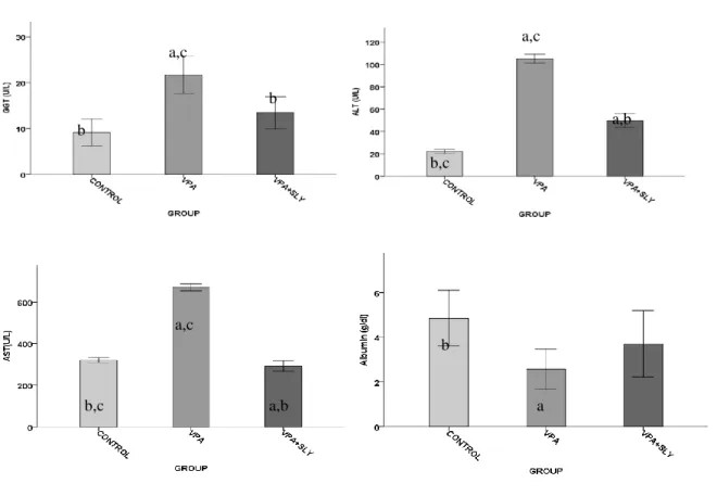
ALT, ASTand GGT levels were significantly increased in VPA group compared to control group and VPA + SLY groups. However, in the case of albumin, this was the opposite. (Table 2 and Figure 2). SLY therapies resulted in a significant decrease in VPA-induced ALT, AST and GGT levels. In addition, treatment with SLY resulted in a significant increase in VPA-reduced albumin level.
Oxidative stress biomarkers
The results in Table 2, Figure 3 showed a significantly higher level of MDA and significantly lower levels of GSH in the VPA group.SLY therapies resulted in a significant decrease in VPA-induced MDA level and a significant increase in VPA-reduced GSH level. Table 2. Serum biochemical and liver tissue oxidative stress biomarkers of the experimental groups.
Control VPA VPA + SLY
Serum biochemical biomarkers
GGT(U/L) 9.14±1.20 b 21.71±1.67 a,c 13.43±1.42 b
ALT (U/L) 22.14±0.80 b,c 105.29±1.56 a,c 49.86±2.48 a,b
AST U/L 320.71±4.77 b,c 669.86±6.97 a,c 292.00±10.32 a,b
Albumin g/dl 4.86±0.50 b 2.57±0.36 a 3.71±0.60
Liver tissue oxidative stress biomarkers
GSH (µmole/g) 20.71±2.24 b,c 67.43±2.36 a,c 27.57±1.52 a,b
MDA
(nmole/g tissue)
0.314 ± 0.010b 0.360 ± 0.012a,c 0.313 ± 0.015b
Data are means ± SEM, n = 7. a Significantly different from control, b significantly different from VPA, c
significantly different from VPA + SLY. VPA, valproic acid; SLY, slymarin; GSH, glutathione; MDA, malondialdehyde; GGT, gamma glutamyl transferase; AST, aspartate aminotransferase; ALT, alanin aminotransferaz and albumin.
Table 3. Histopathological scoring of liver sections of experimental groups.
Parameters/scores Control VPA VPA + SLY
Vacuolar degeneration in hepatocytes – +++a +b
Pycnotic nucleus in hepatocytes – ++a +b
Vascular hemorrhage / congestion – +++a +b
Sinusoidal dilatation – ++a +b
Scoring as described in the Methods section. n = 7. VPA, valproic acid; SLY, slymarin; a: VPA increased liver damage, p <0.05 vs. control group.
Sayfa1452
Figure 2.Effects of VPA and VPA + SLY on serum biochemical parameters. Values are means ± SEM (n = 7). ap
< 0.05 vs Control, bp < 0.05 vs. VPA treated rats, c p<0.05 vs. VPA + SLY treated rats. GGT, Gamma
glutamyl transferase;AST,aspartate aminotransferase;ALT,alanin aminotransferaz; and albumin.
Figure 3.Effects of VPA, VPA + SLY on hepatic lipid oxidation and antioxidant profile of rats after fourteen days. Values are means ± SEM (n = 7). ap<0.05 vs. control, bp< 0.05 vs. VPA treated rats, c p<0.05 vs. VPA +
SLY treated rats. MDA, malondialdehyde; GSH, glutathione. b a,c b b,c a,c a,b b,c a,b a,c b a b a,c b b,c a,c a,b
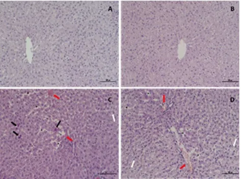
Sayfa1453 Histopathological results
Normal liver histology was observed in the control group (Fig. 4A). In VPA group, vacuolar degeneration and vascular haemorrhage/congestion are severe in hepatocytes. In the hepatocytes, picnotic nucleus and sinusoidal dilatation were moderately observed (Figure 4C-D). These histopathological changes were much less common in the SLY group (Figure 4B).
Figure 4. Rat liver tissue section. A) Normal liver histology is observed in the control group. B) In the SLY group, mild histopathological changes are observed. C, D) Valproic acid group showed significant histopathological changes including vacuolar degeneration (white arrow) in hepatocytes, pycnotic nucleus (black arrow), sinusoidal dilatation (asteric), vascular hemorrhage/congestion (red arrow) in hepatocytes. H.E, scale bar 100 barm, x200.
Discussion
In current study, one of the reasons for VPA application in SLY group was done in order to minimize the damage and the possible protective effects of oxidative stress. Oxidative stress was accepted as one of the factors causing impaired liver function. VPA administration can cause hepatocellular damage by promoting ROS and oxidative damage (18). Previous studies have shown that the liver plays a significant role in the excretion and detoxification of toxic materials (19). Our study confirms previous studies showing that VPA causes damage to the liver (20). According to recent studies, SLY is an antioxidant and a ROS cleaner (21). According to numerous studies, different antioxidant agents have been used to improve the toxic effect of VPA(18). Additional protective effects of SLY can lead to more successful
Sayfa1454 management of toxicity. However, there is little data on the protective effect of SLY on VPA-induced liver injury.
The mechanism of VPA-induced liver injury has been reported to be a consequence of the destructive effect of oxidative stress. ROS production is a process that follows events such as lipid peroxidation and reduction of GSH(22). In this study, a significant increase in MDA level and a decrease in GSH level were reported with VPA administration. High MDA levels suggest that increases of lipid peroxidation and decreases of antioxidant protection mechanisms.
Liver damage may be caused by the reactive intermediates of VPA. According to previous studies, treatment with SLY improved liver enzyme activity, which is an indicator of liver function characteristics (23). In a study, SLY reduced ROS production as a result of CCl4 administration and thus prevented liver damage (24). Accordingly, in our study, these biochemical changes significantly improved the toxicity of VPA after SLY treatment. In addition, increased antioxidant enzyme activity is a result of possible organ damage. Therefore, SLY therapy may have a preventive effect against VPA-induced liver damage by inhibiting lipid peroxide formation and blocking the oxidative chain reaction. This study shows that SLY can lead to hepatoprotection by decreasing oxidative stress. We evaluated the serum concentrations of GGT, ALT and AST to examine liver function. In addition, increased serum levels may be caused by toxic compounds affecting liver tissues (13). The results of the study showed that VPA administration caused liver damage in rats. Subsequently, with this treatment, ALT, AST, and GGT, which are indicators of liver injury, increased significantly. In addition, a significant improvement in liver biomarker levels was detected with SLY treatment (25, 26). Hard conditions may have increased the activity of ALT, AST, and GGT.These conditions may have been caused by accumulation of VPA with toxic activity in the liver, which may have led to cellular destruction or elevation in the permeability of hepatic cells. It has been found that SLY treatment results in a significant reduction in liver enzymes compared to VPA administration in rats. Increasing liver enzyme levels can be interpreted as the stabilizing effect of SLY on the hepatocyte cell membrane. Decreased levels of liver biomarkers can be considered as an indication of the regeneration capabilities of damaged hepatocytes (12). Our results demonstrate that SLY therapy reduced serum marker enzyme activities to normal levels. Therefore, it can be said that SLY protects the structural integrity of the hepatocellular
Sayfa1455 components and protects the liver against the destructive effects of VPA. These data suggest that SLY may be useful in reversing liver damage caused by VPA.
In addition, our results demonstratethe level of serum albumine was significantlyreduced in the VPA group. Although the relevant mechanisms are not well described, basic antimicrobials suggest that they can mediate hepatotoxicity by inhibiting mitochondrial oxidation (27). Low albumin level was successfully restored with SLY therapy.
In our study, VPA caused a significant reduction in body weight of rats. According to previous studies, VPA-induced oxidative stress can be effective on the gastrointestinal tract and may reduce food intake that may cause weight gain. Reduction in body weight of VPA-administered rats may be due to gastrointestinal toxicity-induced dysfunction. Furthermore, the anorexic effect of this drug; VPA may be due to an increased metabolic rate thought to be a side effect(28). On the other hand, this reduction in VPA-induced body weight; it may be due to dysfunction caused by tissue damage. In contrast, co-administration of SLY with VPA reduced body weight loss in this study. Similarly, in a study of Malekinejad, it was reported that treatment with SLY after cisplatin injection significantly reduced body weight loss(23). These results show that the mechanism of action of SLY is achieved by reducing energy consumption, increasing energy storage and increasing energy intake.
The histological findings in this study confirmed the biochemical results and showed that VPA had significant histological changes in the liver. In the histological examination of the VPA group; increased vacuolar degeneration, psychnotic nucleus, sinusoidal dilatation, vascular hemorrhage/congestion in the hepatocytes. Therefore, VPA-induced liver damage. It causes an increase in ROS formation followed by toxic events. These degenerative findings were also found in the SLY-treated group, but were less severe than the VPA group. In studies with various toxic substances, histological findings showing hepatic damage in SLY groups were close to control groups (29). The histopathological findings in our study supported the hypothesis that SLY effectively protects the histological structure and that the endogenous antioxidant defense system is effective against VPA-induced injuries. SLY plays an important role in the maintenance of cellular damage as a result of oxidative stres (26). Therefore, SLY may be the best option for side effects caused by VPA.
Sayfa1456 In conclusion, the results of our study revealed that SLY improved biochemical, histological and structural changes of VPA-induced liver damage in rats. Furthermore, the mechanisms of these effects may include the prevention of lipid peroxidation and the preservation of antioxidant enzymes. SLY may be due to its antioxidant activity and other unknown mechanisms to protect against VPA-induced liver damage. This suggests that SLY may be effective in the treatment of liver damage. Further studies are needed to examine the exact mechanism underlying the therapeutic effects of SLY.
References
1. Semmler A, Frisch C, Bleul C, Smith D, Bigler L, Prost J-C, et al. Intrauterinevalproate exposure is associated with alterations in hippocampal cell numbers and folate metabolism in a rat model of valproate teratogenicity. Seizure [Internet]. 2017 Mar [cited 2019 May 21];46:7–12. Available from: http://www.ncbi.nlm.nih.gov/pubmed/28212902
2. Kudin AP, Mawasi H, Eisenkraft A, Elger CE, Bialer M, Kunz WS. Mitochondrial Liver Toxicity of Valproic Acid and Its Acid Derivatives Is Related to Inhibition of α-Lipoamide Dehydrogenase. Int J Mol Sci [Internet]. 2017 Sep 6 [cited 2019 May 21];18(9). Available from: http://www.ncbi.nlm.nih.gov/pubmed/28878165
3. Ahangar N, Naderi M, Noroozi A, Ghasemi M, Zamani E, Shaki F. Zinc Deficiency and Oxidative Stress Involved in Valproic Acid Induced Hepatotoxicity: Protection by Zinc and Selenium Supplementation. Biol Trace Elem Res. 2017 Sep;179(1):102–9.
4. Holland KD. Efficacy, pharmacology, and adverse effects of antiepileptic drugs. Neurol Clin. 2001 May;19(2):313–45.
5. Tong V, Teng XW, Chang TKH, Abbott FS. Valproic Acid I: Time Course of Lipid Peroxidation Biomarkers, Liver Toxicity, and Valproic Acid Metabolite Levels in Rats. Toxicol Sci. 2005 Aug;86(2):427–35.
6. Graf W, Oleinik O, Glauser T, Maertens P, Eder D, Pippenger C. Altered Antioxidant Enzyme Activities in Children with a Serious Adverse Experience Related to Valproic Acid Therapy. Neuropediatrics. 1998 Aug;29(04):195–201.
7. El-Mowafy AM, Katary MM, Pye C, Ibrahim AS, Elmarakby AA. Novel molecular triggers underlie valproate-induced liver injury and its alleviation by the omega-3 fatty acid DHA: role of inflammation and apoptosis. Heliyon. 2016 Jul;2(7):e00130.
8. Gynther M, Peura L, Vernerová M, Leppänen J, Kärkkäinen J, Lehtonen M, et al. Amino Acid Promoieties Alter Valproic Acid Pharmacokinetics and Enable Extended Brain Exposure. Neurochem Res. 2016 Oct;41(10):2797–809.
Sayfa1457
10. Sokar SS, El-Sayad ME-S, Ghoneim ME-S, Shebl AM. Combination of Sitagliptin and Silymarin ameliorates liver fibrosis induced by carbon tetrachloride in rats. Biomed Pharmacother. 2017 May;89:98–107.
11. Sayın FK, Kübra F. Silybum marianum ekstresinin yüksek yağlı diyetle beslenen ratlarda insülin rezistansı, karaciğer fonksiyonları, lipit düzeyleri ve leptin seviyesi üzerine etkilerinin araştırılması. 2012;
12. Beydilli H, Yilmaz N, Cetin ES, Topal Y, Celik OI, Sahin C, et al. Evaluation of the protective effect of silibinin against diazinon induced hepatotoxicity and free-radical damage in rat liver. Iran Red Crescent Med J. 2015 Apr;17(4):e25310.
13. Abdel-Dayem MA, Elmarakby AA, Abdel-Aziz AA, Pye C, Said SA, El-Mowafy AM. Valproate-induced liver injury: modulation by the omega-3 fatty acid DHA proposes a novel anticonvulsant regimen. Drugs R D. 2014 Jun;14(2):85–94.
14. Crowley L V. The Reitman-Frankel Colorimetric Transaminase Procedure in Suspected Myocardial Infarction. Clin Chem. 1967;13(6).
15. Parlar A, Arslan SO, Doğan MF, Çam SA, Yalçin A, Elibol E, et al. The exogenous administration of CB2 specific agonist, GW405833, inhibits inflammation by reducing cytokine production and oxidative stress. Exp Ther Med [Internet]. 2018 Dec [cited 2019 May 18];16(6):4900–8. Available from: http://www.ncbi.nlm.nih.gov/pubmed/30542446.
16. Placer ZA, Cushman LL, Johnson BC. Estimation of product of lipid peroxidation (malonyl dialdehyde) in biochemical systems. Anal Biochem. 1966 Aug;16(2):359–64.
17. Sedlak J, Lindsay RH. Estimation of total, protein-bound, and nonprotein sulfhydryl groups in tissue with Ellman’s reagent. Anal Biochem. 1968 Jan;25:192–205.
18. Hepatoprotective Herbal Drug, Silymarin From Experimental Pharmacology to Clinical Medicine - Redorbit. 19. Lee M-H, Hong I, Kim M, Lee BH, Kim J-H, Kang K-S, et al. Gene expression profiles of murine fatty liver
induced by the administration of valproic acid. Toxicol Appl Pharmacol. 2007 Apr;220(1):45–59.
20. Powell-Jackson PR, Tredger JM, Williams R. Hepatotoxicity to sodium valproate: a review. Gut. 1984 Jun;25(6):673–81.
21. de Avelar CR, Pereira EM, de Farias Costa PR, de Jesus RP, de Oliveira LPM. Effect of silymarin on biochemical indicators in patients with liver disease: Systematic review with meta-analysis. World J Gastroenterol. 2017 Jul;23(27):5004–17.
22. Pourahmad J, Eskandari MR, Kaghazi A, Shaki F, Shahraki J, Fard JK. A new approach on valproic acid induced hepatotoxicity: Involvement of lysosomal membrane leakiness and cellular proteolysis. Toxicol Vitr. 2012 Jun;26(4):545–51.
23. Malekinejad H, Rokhsartalab-Azar S, Hassani-Dizaj S, Alizadeh-Fanalou S, Rezabakhsh A, Amniattalab A. Effects of silymarin on the pharmacokinetics of atorvastatin in diabetic rats. Eur J Drug Metab Pharmacokinet. 2014 Dec;39(4):311–20.
24. Younis T, Khan MR, Sajid M. Protective effects of Fraxinus xanthoxyloides (Wall.) leaves against CCl4 induced hepatic toxicity in rat. BMC Complement Altern Med. 2016 Oct;16(1):407.
25. Kidd P, Head K. A review of the bioavailability and clinical efficacy of milk thistle phytosome: a silybin-phosphatidylcholine complex (Siliphos). Altern Med Rev. 2005 Sep;10(3):193–203.
Sayfa1458
26. Kandimalla R, Dash S, Bhowal AC, Kalita S, Talukdar NC, Kundu S, et al. Glycogen-gold nanohybrid escalates the potency of silymarin. Int J Nanomedicine. 2017;12:7025–38.
27. El-Mowafy AM, Abdel-Dayem MA, Abdel-Aziz A, El-Azab MF, Said SA. Eicosapentaenoic acid ablates valproate-induced liver oxidative stress and cellular derangement without altering its clearance rate: Dynamic synergy and therapeutic utility. Biochim Biophys Acta - Mol Cell Biol Lipids. 2011 Jul;1811(7–8):460–7. 28. Goda K, Saito K, Muta K, Kobayashi A, Saito Y, Sugai S. Ether-phosphatidylcholine characterized by
consolidated plasma and liver lipidomics is a predictive biomarker for valproic acid-induced hepatic steatosis. J Toxicol Sci. 2018;43(6):395–405.
29. Altınok-Yipel F, Ozan Tekeli İ, Özsoy ŞY, Güvenç M, Kaya A, Yipel M. Hepatoprotective Activity of Linalool in Rats Against Liver Injury Induced by Carbon Tetrachloride. Int J Vitam Nutr Res. 2019 Apr;1-7.