The Effect of Triclosan on In vitro Embryonic Development in Rat
[1] [2]Dicle ÇAYAN
1,a Erdoğan UNUR
2,bMehtap NİSARİ
2,cDilara PATAT
3,dErtuğrul DAĞLI
4,eHilal AKALIN
5,f[1] The project was supported by Erciyes University Scientific Research Projects Commission (TDK-2017-7083)
[2] This study was presented “Effects of Triclosan on in vitro Embryonic Rat Development” as oral presentation in 1st
International Mediterranean Anatomy Congress, 6-9 September 2018, Konya, Turkey
1 Niğde Ömer Halisdemir University, Niğde Zübeyde Hanım School of Health, Department of Nursing Management, TR-51100 Niğde - TURKEY
2 Erciyes University, Faculty of Medicine, Department of Anatomy, TR-38280 Kayseri - TURKEY
3 Lokman Hekim University, Faculty of Medicine, Department of Anatomy, TR-06510 Ankara - TURKEY 4 Ahi Evran University, Faculty of Medicine, Research Hospital, TR-40200 Kırşehir - TURKEY
5 Erciyes University, Faculty of Medicine, Department of Medical Genetics,TR- 38280 Kayseri - TURKEY
ORCIDs: a 0000-0001-7233-063X; b 0000-0003-2033-4350; c 0000-0002-1126-7478; d 0000-0001-5237-4846; e 0000-0002-5331-1289 f 0000-0002-2580-836X
Article ID: KVFD-2020-23873 Received: 07.01.2020 Accepted: 23.07.2020 Published Online: 24.07.2020
How to Cite This Article
Çayan D, Unur E, Nisari M, Patat D, Dağlı E, Akalın H: The effect of triclosan on in vitro embryonic development in rat. Kafkas Univ Vet Fak Derg, 26
(5): 595-602, 2020. DOI: 10.9775/kvfd.2020.23873
Abstract
Triclosan (TCS) is a broad spectrum antimicrobial agent showing its effect by deactivating the fatty acid synthesis of bacteria. The aim of this study was to investigate the effects of TCS on in vitro embryonic development in rats and to determine the levels of caspases 2, 7, 8, and 9 inducing cells to apoptosis through gene expression. According to the TCS dose added to the culture whole rat serum, 3 experimental groups and a control group were formed with each including 10 embryos. After 48 h culturing period, embryos were subjected to morphological scoring for developmental evaluation. The levels of caspases 2, 7, 8, and 9 were measured by performing gene expression on 40 embryos. Significant decreases were obtained in all parameters of morphological scoring in the experimental groups as the dose of TCS increased. While the caspase-2 level showed a significant decrease among the groups and was inversely proportional to the level of TCS, the caspase-9 level showed a significant increase among the groups and was directly proportional to the level of TCS. In conclusion, TCS was determined to cause apoptosis in the cells via the intrinsic pathway during pregnancy period and lead to embryonic growth retardation, which increased with increased dose of TCS.
Keywords: Triclosan, Rat embryo culture, Apoptosis, Gene expression, Caspase
Triklosanın In vitro Embriyonik Rat Gelişimi Üzerine Etkisi
Öz
Triklosan, bakterilerin yağ asidi sentezini bozarak etkisini gösteren geniş spektrumlu bir antimikrobiyal ajandır. Bu çalışmanın amacı, triklosanın ratlarda in vitro embriyonik gelişim üzerindeki etkisini araştırmak ve gen ekspresyon yöntemi ile hücreleri apoptoza sürükleyen kaspaz 2, 7, 8 ve 9 değerlerini belirlemektir. Kültür şişesine eklenen triklosan dozuna göre, kontrol ve 3 deney grubu oluşturuldu. 48 saatlik kültür periyodundan sonra embriyolar gelişimsel değerlendirme amacıyla morfolojik skorlamaya tabi tutuldu. Ardından 40 embriyoya gen ekspresyonu yapılarak kaspaz-2, kaspaz-7, kaspaz-8 ve kaspaz-9 değerleri ölçüldü. Morfolojik skorlamaya ait tüm parametrelerde istatistiksel olarak anlamlı bir gerileme tespit edildi. Kaspaz-2 değerlerinin triklosan miktarı ile ters orantılı bir şekilde, gruplar arasında istatistiksel olarak anlamlı bir azalma gösterdiği, kaspaz-9 değerlerinin ise triklosan miktarıyla orantılı bir biçimde istatistiksel olarak anlamlı bir artış gösterdiği tespit edildi. Bu verilere göre, gebelik döneminde triklosan kullanımının, hücrelerde intrinsik yol aracılığıyla apoptoza yol açarak, embriyonik gelişme geriliğine neden olduğu, bu durumun doza bağlı olarak arttığı belirlendi.
Anahtar sözcükler: Triklosan, Rat embriyo kültürü, Apoptoz, Gen ekspresyonu, Kaspaz
INTRODUCTON
Triclosan (TCS) is a fat-soluble, broad-spectrum anti-microbial agent produced in laboratory in 1966. The area
of its usage was first expanded into antibacterial soaps and then into products such as toothpastes, cosmetics, and deodorants in the following years [1]. TCS can pass the biological barriers because of its lipophilic property
Correspondence
+90 533 4507645 Fax: +90 388 2112813and can aggregate in living organisms [2]. The target organs have been reported to be the liver, spleen, brain, heart, reproductive system, and the immune system. Compared with other well-identified target organs, high concentrations of TCS up to 7 ng/mL can be detected in the placenta, cord blood, and amniotic fluid, which directly indicate a high risk of adverse effects on the embryo in an utero [3]. TCS is a relatively small compound with a molecular weight of 289.5 g/mole [4]. Owing to its molecular weight, TCS can pass the placenta and affect the embryo. TCS metabolism mainly occurs in the liver; however, it can also be metabolized in the skin in small amounts [5]. TCS is metabolized to glucuronide and sulfate conjugates (phase II metabolism) and primarily excreted by urine [6]. As a result of metabolism, it passes to body fluids, breast milk, and urine [7,8] and then merges into sewerage system via wastes, which causes wide contamination in the environment, such as in drinking water and surface waters [1,9]. Therefore, people using TCS-containing products are directly at risk and individuals drinking water contaminated with TCS are indirectly at risk. Many cell culture studies have shown that TCS is cytotoxic to human hepatocytes and cardiotoxic to cardiomyocytes, has neurodegenerative effects on rat neural stem cells, and shows strong embryotoxic effects on bone development [3,10-12]. However, only a few studies have reported its embryotoxic effects on embryo culture [4,13,14]. Moreover, the exact mechanism of the effects of TCS on early embryonic development is still not well understood. In a study with zebrafish embryos, growth retardation has been detected in the embryos exposed to 300 µg/L of TCS [13]. Another study has also demonstrated embryotoxic effects of TCS at high doses (250 µg/L) in zebrafish embryos, such as abnormal phenotypes, short tails, heart edema, and decreased hatching rate [4]. Developmental parameters are proved to be excellent indicators of TCS toxicity [4].
Developmental delay in babies has made the reliability of drugs and chemicals used in pregnancy period a current issue. The European Center for the Validation of Alternative Methods, established in 1991, has accepted the Rat Whole-Embryo Culture Test as one of the methods used to determine the teratogenic and toxic effects of chemical substances [15]. Therefore, this test is a preferred method in toxicology studies [16-19]. Previously, it was suggested that toxic substances triggered pregnancy loss, embryonic death or structural abnormalities by affecting the mechanisms that regulated normal embryonic development. However, today, it is known that apoptosis plays an important role in embryogenesis with cell proliferation and cell differentiation and that toxic sub-stances disrupt the apoptosis pathway in the embryonic organs and cause malformations [20]. Apoptotic stimulus leads to intracellular activation of caspases. Pathways that activate initiator caspases vary with an apoptotic stimulus and there are two different pathways; extrinsic and intrinsic pathways. Extrinsic pathway is driven by caspase-8 and
caspase-7. Intrinsic pathway, which is also called classical or mitochondrial pathway, is the major route to apoptotic death in mammalian cells. This step triggers caspase-9 and caspase-2. These caspases are not specifically present in the tissue [21,22].
In the present study, we aimed to investigate the direct toxic effects of TCS on embryonic growth and development in cultured rat embryos and to evaluate its possible genotoxic effects by determining the potential role of apoptosis on the toxic effects of TCS using the gene expression method. Real-time PCR is the most sensitive molecular method with high sensitivity (105-106), which has the risk of contamination and has a short lead time. The activations of caspase-2, caspase-7, caspase-8, and caspase-9, which may adversely affect cell survival, were measured to explore the mechanisms underlying the effects of TCS in embryo.
MATERIAL and METHODS
The study was approved by the Animal Care and Use Committee (Ethics Committee) of Erciyes University (date: 13.01.2016 and No: 16/012). All procedures throughout the study were carried out in accordance with the ethical issues. Wistar rats were obtained from the Clinical and Experimental Research Center in the Medical Faculty of Erciyes University.
Chemicals
TCS used in the study was provided from Sigma-Aldrich (CAS No 3380-34-5) (Lot# LRAA9502) and GAPDH (Lot# 0000054846), caspase-2 (Lot# 0000054850), caspase-7 (Lot# 0000054845), caspase-8 (Lot# 0000054847), and caspase-9 (Lot# 90017681) were obtained from Roche. Embryo Culture and Morphological Scoring
Female rats approximately 4-10 months of age and weighing 150-250 g were paired with their male partners in cages at about 5.00 pm and left overnight. On the next day morning around 8 am, the female rats were examined regarding the presence of vaginal plugs as an indication of mating and thereby fertilization. The female rats with vaginal plug were considered as 0.5 day pregnant at noon. The conceptuses were dissected from the uteri, decidua and Reichert’s membranes by general anesthesia on day 9.5 of gestation after the blood samples of the females were collected from the abdominal aorta. The conceptuses were explanted into whole embryo culture by the method of New [23].
In order to assess the toxic effect of TCS on embryonic growth, the embryos were divided into four groups (each consisting 10 embryos); three experimental groups and one control group. In the literature, no study of TCS dose to be applied in embryo culture was found. Therefore, the
administration dosages of TCS were determined according to the data gained from previous studies and the reference dose was accepted as 300 ng/mL [4,13,24]. The control group embryos were cultured in whole rat serum (WRS) and the experimental groups were cultured in WRS containing 100 ng/mL, 200 ng/mL, and 300 ng/mL of TCS, in accordance with the technique developed by New [23]. Using this method, the effects of TCS on in vitro embryonic development during the early organogenesis period (between 9.5 and 11.5 days) were evaluated. Moreover, the embryonic development was also compared between the embryos of control and experimental groups morphologically on day 11.5 (after 48 h of culturing period) via morphologic scoring system [14]. In this scoring system, taking the growth and differentiation of different embryological features into consideration, 11.5 day embryos were evaluated by 17 para- meters; each parameter was divided into 6 stages; and each stage was scored with a numerical value between 0 and 5. As morphological parameters, mean yolk sac diameter; yolk sac vessel development; allantois development; embryonic flexion; heart and caudal neural tube development; hind-brain, midbrain and forebrain development; development of eye, ear, nose and pharynx; maxillary and mandibular processes; differentiation fore and hind limbs; crown-rump length; and somite number were evaluated.
Gene Expression Stages
Tissue specific RNA isolation was not possible in immature embryos due to TCS toxicity. Therefore, we used the whole embryo to perform RNA isolation of caspases that are not specific to any tissue.
RNA isolation and cDNA synthesis: Rat embryos were placed into 500 μL of TriPure Isolation Reagent (Roche Applied Science, Basel, Switzerland) for RNA isolation. Total RNA isolation was performed using the protocol for High Pure RNA Tissue Isolation Kit (Roche Applied Science, Mannheim, Germany). Qualification and quantification of RNA samples was performed using the Nanodrop 1000 Spectrophotometer (Thermo Fisher Scientific, Dreieich, Germany). RNA concentrations were assessed by optical density measurement at 260 nm and 280 nm and purity was determined by the ratio of 260/280 nm. Synthesis of cDNA from the total RNA (100 ng) was performed using random hexamer as a primer via the Transcriptor High Fidelity cDNA Synthesis Kit (Roche Applied Science, Mannheim, Germany) according to the manufacturer’s protocol.
Quantitative real-time polymerase chain reaction (PCR) analysis: From the obtained cDNA samples, mRNA expression levels of caspase-2, caspase-7, caspase-8, and caspase-9 genes, which have roles in apoptotic pathways, were investigated using quantifier real-time PCR via LightCycler® 480 II device (Roche Diagnostics, Mannheim, Germany). The primer sequences specific to the cDNAs of the investigated genes and the Universal Probe Library (UPL) probe numbers are presented in Table 1. Amplifications were performed in the total reaction volume of 20 μL using cDNA, mRNA-specific primers, UPL probe, LightCycler® probe master mixture (Roche Diagnostics, Mannheim, Germany), and distilled water according to the PCR cycling program: 95°C for 10 min, then 45 cycles of 95°C for 10 sec, 60°C for 30 sec, followed by 72°C for 10 sec. The level of mRNA expression of glyceraldehyde-3-phosphate dehydrogenase (GAPDH) was accepted as reference for normalizing the amounts of caspase-2, caspase-7, caspase-8, and caspase-9 gene expression. The procedure was repeated three times for each concentration of TCS. The expression levels of target genes were calculated using the relative quantitation method using the software program of the LightCycler® 480 II device (Table 1).
Statistical Analysis
Data were analyzed using the IBM SPSS Statistics for Windows, Version 22.0 (IBM Corp., Armonk, NY, USA). The conformity of variables to normal distribution was assessed using the Shapiro-Wilk test, Q-Q plot, and histogram graphs. In multiple independent group comparisons, one-way analysis of variance was used for normally distributed numerical variables. In post-hoc comparisons, Tukey’s test was used for normally distributed variables. The Pearson’s correlation analysis was performed to determine the correlation between the levels of caspases and the morphological parameters. A P value <0.05 was considered statistically significant.
RESULTS
There was severe growth retardation in the embryos of the experimental groups as compared to the control group
(Fig. 1). According to the morphological scoring system,
the retardation of embryonic growth and development was increased as the dose of TCS increased (Fig. 2). The lower morphological scores were related with the poor
Table 1. Assay ID numbers, Universal probe number and primer sequences of genes used in expression study
Gene Symbol Assay ID Forward Primer Sequence Reverse Primer Sequence UPL No
Casp2 506708 51-GGAAATGAGGGAGCTAATCCA-31 51-GGCAGCAGGTTGAGGAGTT-31 129
Casp7 500441 51-CTCTTGCGCAAAGATGCAG-31 51-AGCAGGCTGAGTTGCTGTG-31 110
Casp8 504044 51-GCCTGAGGGAAAGATGTCCT-31 51-TCACATCATAGTTCACGCCAGT-31 89
Casp9 502880 51-CGACATGATCGAGGATATTCAG-31 51-TGCCTCCCTCGAGTCTCA-31 27
yolk sac vessel development, failure of fusion of the neural folds; incomplete embryonic fl exion; retardation in the development of otic, optic, and olfactory systems; branchial bars; maxillary and mandibular processes; and limbs (Table 2). The mean morphological scores of the embryos in the control groups was 62.80±6.42 whereas those of the embryos cultured in the WRS containing 100 ng/mL, 200 ng/mL and 300 ng/mL of TCS were 30.4±6.34, 14.9±6.55, and 1.8±3.01, respectively. The diff erence in the morphological scores was statistically significant between
Table 2. Data variance analysis on morphological scoring between the control and experimental groups
Parameters Groups P
Control 100 ng/mL Triclosan 200 ng/mL Triclosan 300 ng/mL Triclosan
Yolk Sac Vessel Development 4.8±0.422a 3.3±0.422b 1.6±0.823c 0.3±0.483d ≤0.001
Allantois 3a 1.9±0.568b 1±0.471c 0.3±0.483d ≤0.001
Flexion 4.9±0.738a 2.7±0.949b 1.5±0.707c 0.3±0.483d ≤0.001
Heart 4.9±0.316a 3±0.471 b 1.7±0.823 c 0.3±0.483 d ≤0.001
Caudal Neural Tube 4.7±0.483a 2.4±0.516 b 1.1±0.568 c 0 d ≤0.001
Hindbrain 4.4±0.516a 2±0.471 b 1.2±0.632 c 0.2±0.422 d ≤0.001 Midbrain 4.1±0.738a 1.8±0.632 b 0.9±0.316 c 0.2±0.422 d ≤0.001 Forebrain 4.1±0.738a 1.6±0.516 b 0.9±0.316 c 0.2±0.422 d ≤0.001 Otic System 4.1±0.568a 2.2±0.789 b 1±0.667 c 0 d ≤0.001 Optic System 4.5±0.527a 2.3±0.675 b 1±0.816 c 0 d ≤0.001 Olfactory System 2.7±0.483a 1.1±0.316 b 0.4±0.516 c 0c ≤0.001 Pharyngeal Arch 2.9±0.568a 1.3±0.483 b 0.6±0.516 c 0 d ≤0.001 Maxillary Processes 2.5±0.527a 1.1±0.316 b 0.5±0.527 c 0 d ≤0.001 Mandibular Processes 2.5±0.527a 0.8±0.422b,c 0.6±0.516b,c 0 d ≤0.001 Forelimbs 2±0.471 0.3±0.483 0 0 ≤0.001 Hindlimbs 1.9±0.316 0.1±0.316 0 0 ≤0.001 Somits 4.8±0.422a 2.5±0.707 b 0.9±0.738 c 0 d ≤0.001
Total Morphological Score 62.8±6.426a 30.4±6.346 b 14.9±6.557 c 1.8±3.011 d ≤0.001
Yolk Sac Diameter 3.590±0.2132a 2.68±0.3011b 1.61±0.3872c 1.15±0.1434d ≤0.001
Crown-Rump Length 2.95±0.2173 1.97±0.4057 0.86±0.2459 - ≤0.001
Somit Number 26.8±1.751 15.9±4.067 7.1±2.025 - ≤0.001
The data are as average and standard deviation. The same letters in the same row show similarity among groups, and diff erent letters indicate diff erences among groups
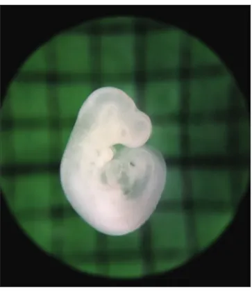
Fig 1. Normally developing embryo cultured in WRS
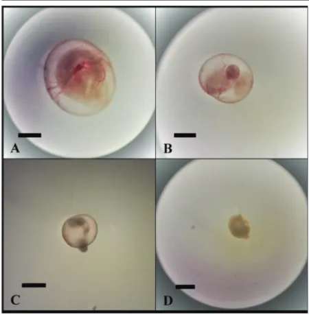
Fig 2. Embryos after 48 h culture. A: control group, B: 100 ng/mL
triclosan group, C: 200 ng/mL triclosan group, D: 300 ng/mL triclosan group; Bar: 500 µm
the control and experimental group (P<0.001). In addition to the total morphological scores, yolk sac diameter, somite numbers, and crown-rump length were significantly lower in the experimental groups than those in the control group (P<0.001 for each). While the mean yolk sac diameter was
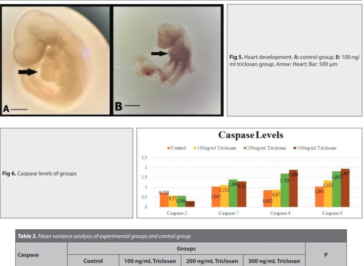
3.59±0.21 mm in the control group, it was 2.68±0.30 mm, 1.61±0.38 mm, and 1.15±0.14 mm in the experimental groups exposed to 100 ng/mL, 200 ng/mL and 300 ng/mL of TCS, respectively (Fig. 3). The mean crown-rump length was 2.95±0.2 mm in the control group; however, in the experimental groups, it gradually diminished as the dose of TCS increased (1.97±0.40 mm, 0.86±0.24 mm, and not measured for 100 ng/mL, 200 ng/ mL and 300 ng/mL, respectively). The mean somite number was 26.8±1.75 in the control group and it was also gradually diminished in the experimental groups as the dose of TCS increased (15.9±4.06, 7.1±2.02 and, not measured for 100 ng/mL, 200 ng/mL and 300 ng/mL, respectively (Fig. 4). The heart development values of embryos was also lower in the experimental groups (3±0.47, 1.7±0.82, and 0.3±0.48 for 100 ng/mL, 200 ng/mL and 300 ng/mL, respectively) compared with that in the control group (4.9±0.31) (P<0.001) (Fig. 5).
The molecular mechanisms underlying the toxic eff ects of TCS on early development of rat (whole embryo culture) was assessed via quantitative real-time-PCR and gene expression. TCS significantly altered four caspases genes (mRNA expression levels) associated with apoptosis. When caspase levels of the experimental groups were compared with the control group, TCS caused upregulation of expression of caspase-7, caspase-8, and caspase-9 and downregulation of expression of caspase-2
(Table 3). There were significant diff erences
between the experimental and control groups regarding the gene levels of caspase-2 and caspase-9 (P<0.001) (Fig. 6). However, no significant diff erence was found between the groups in terms of the gene levels of caspase-7 (P=0.566) and caspase-8 (P=0.396). According to the correlation analysis of the levels of caspase-2, caspase-7, caspase-8, and caspase-9 with all study parameters, significant positive correlations were obtained between caspase-2 and morphological score, somite number, and yolk sac diameter. Additionally, there were significant negative correlations between caspase-9 and all morphological parameters (P<0.01).
DISCUSSION
Triclosan has been widely used in personal care products or medical devices, such as sutures, owing to its antibacterial
Fig 3. Yolk sac vessel development. A: Control group, B: 100 ng/mL triclosan group, C: 200 ng/mL triclosan group, D: 300 ng/mL triclosan group; Bar: 1 mm
Fig 4. Eff ect of triclosan on total morphological score, yolk sac diameter mean,
crown-rump length and somit number mean
properties. It is known that TCS has potential risks to reproduction and development. Although the adverse eff ects of TCS on diff erent organs have been studied for decades, the mechanism of its toxicity is still not well understood, especially during the stages of embryo development [4,13]. Whole embryo culture method is a valuable model for assessing the eff ects of toxic agents on early embryogenesis. In this method, rat embryos are cultured in vitro after the gestational day 9.5-11.5, which is a critical period for organogenesis in rats and corresponds to 3-6 weeks after fertilization in human embryos. However, to the best of our knowledge, there are no studies in the literature verifying the toxic responses to TCS during embryo development in rat embryo culture. The present study highlighted the eff ects of TCS on in vitro embryonic development using cultured rat embryos and demonstrated the negative eff ect of TCS on cell viability leading to induction of apoptosis.
The in vitro embryotoxic eff ect of TCS using embryo/larvae
of zebrafish (Danio rerio) was previously reported by Oliveira et al.[4]. Embryos were exposed to diff erent doses of TCS (500 ng/mL, 700 ng/mL, and 900 ng/mL) for 6 days and embryotoxic eff ects (including delay in hatching, abnormal eye and body pigmentation, spinal malformations, cardiac edema, and body size smaller than normal) were observed [4]. In another study, Chen et al.[13] found no significant increase in the mortality after TCS treatment; however, 300 μg/L of TCS caused developmental retardation in zebrafish embryos at 24 h post fertilization. Ho et al.[25] examined the morphology of zebrafish larvae after TCS exposure and they observed that the zebrafish embryos exposed to 250 ng/mL of TCS developed normally, similar to the control groups. They concluded that low-dose TCS exposure did not aff ect general embryonic development [14]. In the study by Guo et al.[26], TCS was administrated in ovo to evaluate its toxic eff ects in the chicken embryos. Tarsus length (mm) was significantly shorter in the embryos exposed to 10 μg/g TCS compared with that in the embryos administered corn oil [26]. In another study, it was reported that exposure
Table 3. Mean variance analysis of experimental groups and control group
Caspase Groups P
Control 100 ng/mL Triclosan 200 ng/mL Triclosan 300 ng/mL Triclosan
Caspase 2 0.733±0.194a 0.570±0.167a 0.564±0.166a 0.303±0.14b <0.001
Caspase 7 1.047±0.444 1.112±0.319 1.389±0.512 1.31±0.977 >0.05
Caspase 8 0.852±0.209 0.870±0.166 1.701±0.802 1.888±3.36 >0.05
Caspase 9 1.041±0.285a 1.333±0.179a,b 1.805±0.643b 1.937±0.740b <0.001
The data are as average and standard deviation. Diff erent letters in the same row indicate diff erences between groups
Fig 5. Heart development. A: control group, B: 100 ng/
ml triclosan group, Arrow: Heart; Bar: 500 µm
to 8-10 µM TCS led to a high rate of developmental delay at 24 h post fertilization in zebrafish embryos [27]. In the present study, for the first time, effects of TCS on cultured rat embryos were examined. The results showed that the total embryonic growth was normal in the embryos cultured in the WRS only as opposed to the embryos cultured in the WRS containing TCS; this figure decreased depending on the dose of TCS. Embryonic retardations were observed in total embryonic growth, especially in the yolk sac diameter and vascularization, crown-rump length, somite number, body flexion, and neural tube development. These parameters were significantly lower in the experimental groups compared with the control group.
Researches have reported that TCS exposure can induce apoptosis in various biological systems [28-30]. In the present study, cultured embryos were also evaluated regarding apoptosis to investigate the potential role of apoptosis on the embryotoxic effects of TCS via caspases using the gene expression method. Since caspase-2, caspase-7, caspase-8, and caspase-9, which were measured to assess apoptosis, were present in all tissues, we performed RNA isolation on the whole embryo. Several studies have showed that caspase-2 is activated by DNA damage in the nucleolus [31,32]. Dubey et al.[29] reported that TCS induced apoptosis in human skin keratinocytes and responsible for DNA damage. In contrast, our data showed that TCS reduced the activity of caspase-2; if TCS had caused DNA damage, an increase in the level of caspase-2 would have occurred. Therefore, we could say that TCS did not cause DNA damage within the cell and that the decrease in caspase-2 level was associated with growth retardation. On the other hand, there is no data in the literature concerning the caspase-7 levels of TCS. Lamkanfi and Kanneganti [33] claimed that caspase-7 was related to inflammation; the activation of inflammatory caspase-1 and incorporation with caspase-7 could cause inflammation in the cell by the formation of a structure called inflammasome. Brentnall et al.[34] reported that caspase-9 and caspase-7 had different roles in intrinsic apoptosis. While intrinsic apoptosis resulted in the activation of caspase-9, caspase-7 did not have a role in intrinsic apoptosis but enabled apoptotic cell detachment [34] In the present study, as the dose of TCS increased, caspase-7 levels increased; however, no significant difference was observed between the groups. This finding suggested that TCS added to the embryo culture medium did not cause inflammation within the cell. In the in vitro study investigating the apoptotic effect of TCS on mouse neo-cortical neurons, Szychowski et al.[28] evaluated the levels of caspase-8 and caspase-9 and they found that the level of caspase-8 significantly increased as the exposure duration to TCS and the concentration of TCS increased. However, they observed the increase in caspase-9 level in the groups at high concentration of TCS. Accordingly, the researchers claimed that non-cytotoxic concentrations of TCS caused apoptosis through the extrinsic pathway; however, long-term exposure of TCS activated intrinsic
pathway via caspase-9 by releasing cytochrome-c from the mitochondria [28]. In the present study, caspase-7 and caspase-8 levels increased as the dose of TCS increased; however, the difference was not statistically significant between the groups. Caspase-9 has a key role in the early stage of mitochondrial apoptosis pathway by activating downstream caspases and initiating apoptosis [35]. Li et al.[36] reported that methyl-TCS was a dominant transformation product of TCS, exposure to which induced the increased expression of caspase-9 on cell culture. In the present study, caspase-9 levels were found to be increased among the groups in proportion to TCS. It is known that TCS causes cell apoptosis via caspase-9.
In the present study, TCS significantly decreased all growth and developmental parameters in a dose-dependent manner in the embryos of the experimental groups compared with the control embryos. This finding suggests that TCS can activate the intrinsic pathway in the cell by passing directly through the cell membrane and that the intrinsic pathway is functional in the embryonic period. In conclusion, the results of the present study revealed that TCS was a toxic agent and led to growth retardation by causing apoptosis through the intrinsic pathway without resulting in inflammation and direct DNA damage in the cell. Moreover, the current results highlighted the potential role of TCS as a strong embryotoxic in embryonic rats. These results would be an important source for the effects of TCS on embryonic development. Future studies isolating embryonic tissues and measuring the caspase levels by gene expression would also make contribution to investigate the effects of TCS on embryonic development.
C
onfliCtofi
nterestThe authors declare that there are no conflicts of interest.
A
uthorC
ontributionsD. Çayan and E. Unur conceived the ideas of the study and writing manuscript; D. Çayan, M. Nisari, D. Patat and E. Dağlı performed data collection and analysis; H. Akalın performed gene expression stages.
REFERENCES
1. Glaser A: The Ubiquitous TCS. Pest You J, 24 (3): 12-17, 2004.
2. Szychowski KA, Wnuk A, Rzemieniec J, Kajta M, Leszczyńska T, Wójtowicz AK: Triclosan-evoked neurotoxicity involves NMDAR subunits
with the specific role of glun2a in caspase-3-dependent apoptosis. Mol
Neurobiol, 56, 1‐12, 2019. DOI: 10.1007/s12035-018-1083-z
3. Cheng W, Yang S, Liang F, Wang W, Zhou R, Li Y, Feng Y, Wang Y:
Low-dose exposure to TCS disrupted osteogenic differentiation of mouse embryonic stem cells via BMP/ERK/Smad/Runx-2 signalling pathway.
Food Chem Toxicol, 127, 1‐10, 2019. DOI: 10.1016/j.fct.2019.02.038
4. Oliveira R, Domingues I, Koppe Grisolia C, Soares AMVM: Effects of
triclosan on zebrafish early-life stages and adults. Environ Sci Pollut Res, 16, 679‐688, 2009. DOI: 10.1007/s11356-009-0119-3
human health effects. J Toxicol Environ Health B Crit Rev, 20 (8): 447‐469, 2017. DOI: 10.1080/10937404.2017.1399306
6. Dhillon GS, Kaur S, Pulicharla R, Brar SK, Cledon M, Verma M, Surampalli RY: Triclosan: Current status, occurrence, environmental
risks and bioaccumulation potential. Int J Environ Res Public Health, 12 (5): 5657‐5684, 2015. DOI: 10.3390/ijerph120505657
7. Sandborgh-Englund G, Adolfsson-Erici M, Odham G, Ekstrand J: Pharmacokinetics of Triclosan following oral ingestion in humans.
J Toxicol Environ Health A, 69 (20): 1861-1873, 2006. DOI: 10.1080/
15287390600631706
8. Allmyr M, Adolfsson-Erici M, McLachlan MS, Sandborgh-Englund G: Triclosan in plasma and milk from Swedish nursing mothers and their
exposure via personal care products. Sci Total Environ, 372, 87-93, 2006. DOI: 10.1016/j.scitotenv.2006.08.007
9. Fang JL, Stingley RL, Beland FA, Harrouk W, Lumpkins DL, Howard P: Occurrence, efficacy, metabolism, and toxicity of Triclosan. J Environ
Sci Health C Environ Carcinog Ecotoxicol Rev, 28, 147-171, 2010. DOI:
10.1080/10590501.2010.504978
10. Wu Y, Chitranshi P, Loukotková L, Gamboa da Costa G, Beland FA, Zhang J, Fang JL: Cytochrome P450-mediated metabolism of Triclosan
attenuates its cytotoxicity in hepatic cells. Arch Toxicol, 91 (6): 2405‐2423, 2017. DOI: 10.1007/s00204-016-1893-6
11. Chaudhari U, Nemade H, Sureshkumar P, Vinken M, Ates G, Rogiers V, Hescheler J, Hengstler JG, Sachinidis A: Functional cardiotoxicity
assessment of cosmetic compounds using human-induced pluripotent stem cell-derived cardiomyocytes. Arch Toxicol, 92 (1): 371‐381, 2018. DOI: 10.1007/s00204-017-2065-z
12. Park BK, Gonzales ELT, Yang SM, Bang M, Choi CS, Shin CY: Effects
of triclosan on neural stem cell viability and survival. Biomol Ther (Seoul), 24 (1): 99‐107, 2016. DOI: 10.4062/biomolther.2015.164
13. Chen X, Xu B, Han X, Mao Z, Chen M, Du G, Talbot P, Wang X, Xia Y: The effects of Triclosan on pluripotency factors and development of
mouse embryonic stem cells and zebrafish. Arch Toxicol, 89, 635-646, 2015. DOI: 10.1007/s00204-014-1270-2
14. Van Maele-Fabry G, Delhaise F, Picard JJ: Morphogenesis
and quantification of the development of post-implantation mouse embryos. Toxicol In Vitro, 4 (2): 149‐156, 1990. DOI: 10.1016/0887-2333(90)90037-t
15. Augustine-Rauch K, Zhang CX, Panzica-Kelly JM: In vitro
developmental toxicology assays: A review of the state of the science of rodent and zebrafish whole embryo culture and embryonic stem cell assays. Birth Defects Res C Embryo Today, 90 (2): 87-98, 2010. DOI: 10.1002/ bdrc.20175
16. Unur E, Ülger H, Ekinci N, Hacıalioğulları M, Ertekin T, Kılıç E: Effect
of anti-basic fibroblast growth factor (anti-bFGF) on in vitro embryonic development in rat. Anat Histol Embryol, 38, 241-245, 2009. DOI: 10.1111/j.1439-0264.2009.00927.x
17. Tekinarslan İİ, Unur E, Ülger H, Ekinci N, Ertekin T, Hacıalioğulları M, Arslan S: The effects of FGF-9 on in vitro embryonic development.
Balkan Med J, 28, 18-22, 2011. DOI: 10.5174/tutfd.2009.02019.2
18. Ertekin T, Ülger H, Nisari M, Karaca Ö, Unur E, Şahin U, Elmalı F:
Effects of angiostatin on in vitro embryonic rat development. Kafkas Univ
Vet Fak Derg, 17 (5): 843-847, 2011. DOI: 10.9775/kvfd.2011.4637
19. Nisari M, Ulger H, Unur E, Karaca O, Ertekin T: Effect of interleukin
12 (IL-12) on embryonic development and yolk sac vascularisation. Bratisl
Lek Listy, 115 (9): 532-537, 2014. DOI: 10.4149/bll_2014_103
20. Toder V, Carp H, Fein A, Torchinsky A: The role of pro- and
anti-apoptotic molecular interactions in embryonic maldevelopment. Am J
Reprod Immunol, 48, 235-244, 2002. DOI: 10.1034/j.1600-0897.2002.01130.x
21. McArthur K, Kile BT: Apoptotic caspases: Multiple or mistaken
identities? Trends Cell Biol, 28 (6): 475‐493, 2018. DOI: 10.1016/j. tcb.2018.02.003
22. Aşan E, Dağdeviren A: Hücre. In, Aşan E, Dağdeviren A (Eds):
Moleküler Histoloji. 145-168, Atlas Kitapçılık, Ankara, 2012.
23. New DAT: Whole-embryo culture and the study of mammalian
embryos during organogenesis. Biol Rev Camb Philos Soc, 53, 81-122, 1978.
24. Horie Y, Yamagishi T, Takahashi H, Iguchi T, Tatarazako N:
Effects of Triclosan on Japanese medaka (Oryzias latipes) during embryo development, early life stage and reproduction. J Appl Toxicol, 38 (4): 544‐551, 2018. DOI: 10.1002/jat.3561
25. Ho JCH, Hsiao CD, Kawakami K, Tse WKF: Triclosan (TCS) exposure
impairs lipid metabolism in zebrafish embryos. Aquat Toxicol, 173, 29‐35, 2016. DOI: 10.1016/j.aquatox.2016.01.001
26. Guo J, Ito S, Nguyen HT, Yamamoto K, Tanoue R, Kunisue T, Iwata H: Effects of prenatal exposure to triclosan on the liver transcriptome in
chicken embryos. Toxicol Appl Pharmacol, 347, 23‐32, 2018. DOI: 10.1016/j. taap.2018.03.026
27. Haggard DE, Noyes PD, Waters KM, Tanguay RL: Phenotypically
anchored transcriptome profiling of developmental exposure to the antimicrobial agent, triclosan, reveals hepatotoxicity in embryonic zebrafish. Toxicol Appl Pharmacol, 308, 32‐45, 2016. DOI: 10.1016/j. taap.2016.08.013
28. Szychowski KA, Sitarz AM, Wojtowicz AK: Triclosan induces Fas
receptor-dependent apoptosis in mouse neocortical neurons in vitro.
Neuroscience, 284, 192-201, 2015. DOI: 10.1016/j.neuroscience.2014.10.001
29. Dubey D, Srivastav AK, Singh J, Chopra D, Qureshi S, Kushwaha HN, Singh N, Ray RS: Photoexcited Triclosan induced DNA damage and
oxidative stress via p38 MAP kinase signaling involving type I radicals under sunlight/UVB exposure. Ecotoxicol Environ Saf, 174, 270‐282, 2019. DOI: 10.1016/j.ecoenv.2019.02.065
30. Lee GA, Choi KC, Hwang KA: Treatment with phytoestrogens
reversed triclosan and bisphenol A-induced anti-apoptosis in breast cancer cells. Biomol Ther (Seoul), 26 (5): 503‐511, 2018. DOI: 10.4062/ biomolther.2017.160
31. O’Byrne KJ, Richard DJ: Nucleolar caspase-2: Protecting us from DNA
damage. J Cell Biol, 216 (6): 1521‐1523, 2017. DOI: 10.1083/jcb.201704114
32. Ando K, Parsons MJ, Shah RB, Charendoff CI, Paris SL, Liu PH, Fassio SR, Rohrman BA, Thompson R, Oberst A, Sidi S, Bouchier-Hayes L: NPM1 directs PIDDosome-dependent caspase-2 activation
in the nucleolus. J Cell Biol, 216 (6): 1795‐1810, 2017. DOI: 10.1083/ jcb.201608095
33. Lamkanfi M, Kanneganti TD: Caspase-7: A protease involved in
apoptosis and inflammation. Int J Biochem Cell Biol, 42, 21-24, 2010. DOI: 10.1016/j.biocel.2009.09.013
34. Brentnall M, Rodriguez-Menocal L, De Guevara RL, Cepero E, Boise LH: Caspase-9, caspase-3 and caspase-7 have distinct roles
during intrinsic apoptosis. BMC Cell Biol, 14:32, 2013. DOI: 10.1186/1471-2121-14-32
35. Galluzzi L, López-Soto A, Kumar S, Kroemer G: Caspases connect
cell-death signaling to organismal homeostasis. Immunity, 44 (2): 221-231, 2016. DOI: 10.1016/j.immuni.2016.01.020
36. Li X, An J, Li H, Qiu X, Wei Y, Shang Y: The methyl-triclosan induced
caspase-dependent mitochondrial apoptosis in HepG2 cells mediated through oxidative stress. Ecotoxicol Environ Saf, 182:109391, 2019. DOI: 10.1016/j.ecoenv.2019.109391