The diagnostic value of serum urokinase-type plasminogen
activator receptor in acute appendicitis
Ali Aygün, M.D.,1 Mücahit Günaydın, M.D.,2 Ömer Vefik Özozan, M.D.,3 Murat Cihan, M.D.,4 Murat Karakahya, M.D.5
1Department of Emergency Medicine, Ordu University Faculty of Medicine, Ordu-Turkey 2Department of Emergency Medicine, Giresun University Faculty of Medicine, Giresun-Turkey 3Department of General Surgery, Istinye University Hospital, İstanbul-Turkey
4Department of Biochemistry, Ordu University Training and Research Hospital, Ordu-Turkey 5Department of General Surgery, Ordu University Faculty of Medicine, Ordu-Turkey
ABSTRACT
BACKGROUND: To measure serum uPAR levels in patients operated with a preliminary diagnosis of acute appendicitis (AA) and to investigate whether these parameters can be used as a biochemical marker in the diagnosis of AA.
METHODS: Patients aged 18 or over, presenting to the emergency department between May and December 2018 and operated with a diagnosis of AA were enrolled. This study included 84 patients with surgical pathology results compatible with AA (Group A), 26 patients with surgical pathology results were not compatible with AA (Group B) and 55 healthy control groups. Serum uPAR levels were measured from venous blood samples taken at admission.
RESULTS: Mean uPAR levels were 4.53±3.47 ng/mL in the Group A, 1.13±1.63 ng/mL in the Group B and 0.80±1.21 ng/mL in the control group. Serum uPAR levels differed statistically significantly from Group A in Group B and the control group, (p<0.05).
CONCLUSION: uPAR was found to be significantly higher in the AA patients compared to the control group and patients with surgically determined non-AA pathologies. uPAR can be used as an aid in the diagnosis of acute appendicitis.
Keywords: Abdominal pain; acute appendicitis; adult; inflammation; urokinase-type plasminogen activator receptor.
right lower quadrant, negative surgical pathology results are encountered at a rate of 10–30% in patients operated with a preliminary diagnosis of AA.[2,4–6] Clinicians, therefore, re-quire new research to reduce increasing malpractice suits and negative appendectomy rates. Studies have, therefore, shown a relation between AA and biochemical parameters showing acute inflammation, such as white blood cell count (WBC), C-reactive protein (CRP), and procalcitonin.[2,4]
The pathophysiology of AA is associated with mucosal im-pairment caused by invasive infection and inflammation.[7] In-filtration of the intestinal wall by activating neutrophils occurs following invasion by intraluminal bacteria of the appendix wall
INTRODUCTION
Acute appendicitis (AA) is the principal cause of acute ab-domen in patients presenting to the emergency department due to abdominal pain.[1,2] Morbidity and mortality increase if AA is diagnosed late. Perforated appendix and associated peritonitis, intra-abdominal abscess, sepsis, and ileus can de-velop in the event of late diagnosis.[3] History, physical exam-ination, an increase in blood inflammatory parameters, and clinical experience occupy an important place in diagnosis. Although radiological imaging methods, such as ultrasound (USG) and computerized tomography (CT), are used in the differential diagnosis of other pathologies causing pain in the
Cite this article as: Aygün A, Günaydın M, Özozan ÖV, Cihan M, Karakahya M. The diagnostic value of serum urokinase-type plasminogen activator receptor in acute appendicitis. Ulus Travma Acil Cerrahi Derg 2019;25:467-473.
Address for correspondence: Ali Aygün, M.D.
Ordu Üniversitesi Tıp Fakültesi, Acil Tıp Anabilim Dalı, Ordu, Turkey Tel: +90 452 - 225 01 85 E-mail: [email protected]
Ulus Travma Acil Cerrahi Derg 2019;25(5):467-473 DOI: 10.14744/tjtes.2019.55623 Submitted: 01.06.2019 Accepted: 25.06.2019 Online: 21.08.2019 Copyright 2019 Turkish Association of Trauma and Emergency Surgery
with an impaired mucosal barrier.[8] Degradation of the extra-cellular matrix is important for neutrophil invasion of tissue in the inflammatory response. One study showed immunoreac-tive urokinase-type plasminogen activator (uPA), involved in the conversion of plasminogen to plasma, in inflamed appendix tissue.[9] Plasmin leads to leukocytes passing the tissue barrier by reducing pericellular matrix proteins. Urokinase-type plas-minogen activator receptor (uPAR, CD 87) is a glycosylphos-phatidylinositol-anchored protein with high uPA receptor affinity. In addition to serving as a binding point on the cell surface, uPAR also facilitates leukocyte adhesion and migra-tion.[10] Rijneveld et al.[11] determined that uPA and uPAR ex-hibit immune functions more by activating other defense cells than through fibrinolytic effects. Following an inflammatory stimulus, uPAR and proteases, such as chymotrypsin and phos-pholipase, facilitate leukocyte adhesion and migration, and ac-tivated neutrophils release the chemotactically active soluble form of uPAR from the cell surface into the circulation. With its direct chemotactic effect, uPAR facilitates the production of additional anti-inflammatory cells (generally neutrophils and macrophages) and the mobilization of hematopoietic stem cells in order to overcome bacterial invasion.[12]
This study was planned to measure serum uPAR levels in pa-tients operated with a preliminary diagnosis of AA and to investigate whether these parameters can be used as a bio-chemical marker in the diagnosis of AA.
MATERIALS AND METHODS
This study was conducted after Ordu University Medical Fac-ulty Clinical Research Ethical Committee approval (decision No. 2018/61). Patients aged 18 or over, presenting to the emergency department of a tertiary hospital between May and December 2018, and operated with a diagnosis of AA were enrolled. We planned to exclude patients with a non-AA focus of infection, acute coronary syndrome, hemorrhagic stroke, cerebrovascular disease, liver failure, acute pulmonary edema, cardiopulmonary arrest, acute mesenteric ischemia, or pulmonary thromboembolism, pregnant patients, subjects with acute trauma, or for whom consent to participate was not granted by the patient or relatives. We also intended to exclude patients for whom data deficiencies were deter-mined at the end of the study period. Healthy volunteers aged over 18 with no disease and presenting to hospital for a check-up and agreeing to participate were included as the control group in this study.
The clinical and demographic characteristics, symptoms, physical examination findings, Alvarado scores, WRP and CRP values, all abdominal USG and CT imaging results, and postoperative pathology results of the patients included in this study were recorded onto study forms. We planned to measure the serum uPAR values of the patient group and the control group. Patients with surgical pathology results com-patible with AA were assigned into Group A, and patients
with surgical pathology results not compatible with AA were assigned into Group B.
Analysis of Biochemical Parameters
Venous blood specimens were collected at the time of pre-sentation. Blood specimens were drawn into a serum sepa-rator tube until the vacuum was filled. Tubes with sepasepa-rator gel were used for serum collection, and tubes containing potassium-EDTA were used for a blood count. Plasmas were separated by centrifugation for 10 min at 3000 rpm and were stored -80 ºC. Serum CRP levels were studied spectrophoto-metrically on a closed system with a Cobas 600 series c501 modular analyzer in our laboratory. Blood WBC levels were collected with results obtained with an XN-1000 device in our laboratory. uPAR levels in human blood serum were de-termined using a Cloud Clone (USCNK) (Wuhan, China) en-zyme-linked immunosorbent assay (ELISA) kit in line with the manufacturer’s instructions. uPAR levels in specimens were calculated as ng/mL.
Statistical Analysis
Statistical software was used for data analysis. Descriptive ex-press was exex-pressed as number and percentage for categorical variables and mean, standard deviation (SD), minimum (min), and maximum (max) for numerical variables. Compatibility with normal distribution was assessed using the Kolmogorov-Smirnov test. The t-test was used for two-way comparisons of normally distributed parameters, and the Mann-Whitney U test for non-normally distributed parameters. One-way analysis of variance (ANOVA) was used to compare nor-mally distributed variables between three groups, and the Kruskal-Wallis test for non-normally distributed parameters. When significance was determined with the ANOVA test, two-group comparisons were performed using Turkey’s test if the group were homogeneous, or with Tamhane’s test if they were not homogeneous. The Mann-Whitney U test with Bonferroni correction was used for two-way comparisons when significance was determined using the Kruskal-Wal-lis test. Correlation coefficients and statistical significances were determined using Pearson’s test for normally distributed variables and Spearman’s test for non-normally distributed variables. The decision-determining characteristics of serum uPAR values in predicting the diagnosis of appendicitis were examined using Receiver Operating Characteristics (ROC) curve analysis. The sensitivity, specificity, positive predictive and negative predictive values were calculated for significant threshold values. At area under the curve (AUC) analysis, type 1 error levels less than 5% were interpreted as the sta-tistically significant diagnostic value of the test. Statistical sig-nificance was set at p<0.05.
RESULTS
One hundred ten patients aged 18 or over, presenting to the emergency department with abdominal pain and operated
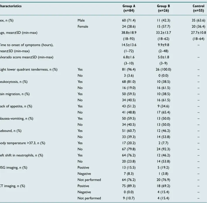
with a diagnosis of AA were included in this study. The con-trol group consisted of 55 healthy volunteers. Twenty-five patients could not be included in this study due to insufficient data. Following examination of the postoperative surgical pathology results of the patients operated with a diagnosis of AA, although pathology compatible with AA was determined in 84 cases (Group A), non-AA histopathological results were obtained in 26 (Group B) patients. Histopathological exam-ination of the Group B patients revealed normal appendix vermiformis tissue in 15 cases, mesenteric lymphadenitis in six, ovarian cyst hemorrhage in three, and diverticulitis in two. Distributions of the patient groups’ age, sex, Alvarado score and clinical characteristics are shown in Table 1. CT
was the most commonly employed diagnostic imaging modal-ity in the emergency department among the cases enrolled in the study. CT was performed on 75 Group A patients but not on the other nine. CT imaging was reported to be compatible with AA in 18 of the patients in Group B, and four patients were operated although CT imaging was not reported to be compatible with AA (Table 1).
The patient groups’ mean WBC, CRP and uPAR values were compared. Serum uPAR levels differed statistically signifi-cantly from Group A in Group B and the control group. No significant difference was observed between the serum uPAR levels of Group B and the control group (Table 2).
Table 1. The groups’ demographic characteristics
Characteristics Group A Group B Control
(n=84) (n=26) (n=55)
Sex, n (%) Male 60 (71.4) 11 (42.3) 35 (63.6)
Female 24 (28.6) 15 (57.7) 20 (36.4)
Age, mean±SD (min-max) 38.8±18.9 33.2±13.7 27.7±10.8
(18–93) (18–62) (18–64)
Time to onset of symptoms (hours), 14.5±13.6 9.9±9.8 –
mean±SD (min-max) (1–72) (2–48)
Alvarado score mean±SD (min-max) 6.8±1.6 5.0±1.8 –
(3–10) (3–9)
Right lower quadrant tenderness, n (%) Yes 81 (96.4) 26 (100.0) –
No 3 (3.6) 0 (0.0) –
Leukocytosis, n (%) Yes 68 (81.0) 10 (38.5) –
No 16 (19.0) 16 (61.5) –
Pain migration, n (%) Yes 50 (59.5) 10 (38.5) –
No 34 (40.5) 16 (61.5) –
Lack of appetite, n (%) Yes 43 (51.2) 9 (34.6) –
No 41 (48.8) 17 (65.4) –
Nausea-vomiting, n (%) Yes 50 (59.5) 13 (50.0) –
No 34 (40.5) 13 (50.0) –
Rebound, n (%) Yes 51 (60.7) 12 (46.2) –
No 33 (39.3) 14 (53.8) –
Body temperature >37.3, n (%) Yes 17 (20.2) 2 (7.7) –
No 67 (79.8) 24 (92.3) –
Left shift in neutrophils, n (%) Yes 64 (76.2) 12 (46.2) –
No 20 (23.8) 14 (53.8) –
USG imaging, n (%) Positive 13 (15.5) 5 (19.2) –
Negative 7 (8.3) 1 (3.8) –
Not performed 64 (76.2) 20 (76.9) –
CT imaging, n (%) Positive 75 (89.3) 18 (69.2) –
Negative 0 (0.0) 4 (15.4) –
Not performed 9 (10.7) 4 (15.4) –
The mean uPAR value of the AA patients with Alvarado scores less than 5 (n=17) was 1.92±4.06, compared to 4.06±3.24 in the AA patients (n=93) with Alvarado scores of 5 or more serum. The difference was statistically significant (p=0.018). The area under the curve (AUC) at ROC analysis performed to measure the diagnostic value of serum uPAR in patients was 0.88 (p<0.001, 95% confidence interval [CI] 0.80–0.97) (Fig. 1). AUC at ROC analysis performed for WBC was 0.83 (p<0.001, 95% CI 0.74–0.92), and 0.70 at ROC analysis for CRP (p<0.001, 95% CI 0.59–0.81). Analysis of correlation
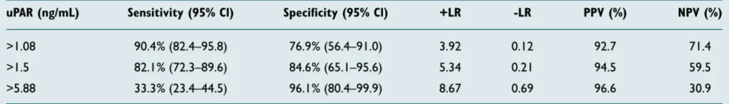
levels between uPAR values and WBC and CRP values re-vealed significant relations (r values 0.63 and 0.59, respec-tively) (p<0.001 for both). The sensitivity and specificity of uPAR in the diagnosis of appendicitis were calculated and are shown in Table 3. The specificity of uPAR in the diagnosis of appendicitis increased as uPAR values in patients’ plasma increased (Table 3).
DISCUSSION
Activation of the uPA/uPAR system plays a key role in chemo-taxis and inflammatory cell infiltration at the start of the in-flammatory reaction.[13] In addition, the uPA system is also a key factor in cell migration, tissue remodeling, wound healing, inflammation, angiogenesis, tumor invasion, and metastasis. [14,15] uPAR (CD87) is a cellular receptor for uPA and is re-leased from several cells, including leukocytes, and endothe-lial and malignant cells.[15,16] uPAR contributes to the conver-sion to plasmin of plasminogen, which leads to the proteolysis of matrix proteins, and the migration of leukocytes to the relevant region in the event of infection of inflammation.[15,17] Serum uPAR levels have been shown to increase in many in-flammatory pathologies, such as sepsis, non-septic systemic inflammatory response (SIRS), pneumonia, pancreatitis, and intestinal inflammation.[12,18–23] Additionally, an increase in serum concentrations of the soluble form of uPAR has been shown to reflect activation of the immunological system and the severity of inflammatory diseases.[24,25] Kolber et al.[25] ob-served a significant increase in serum uPAR concentrations Table 3. The sensitivity and specificity percent of uPAR in diagnosing acute appendicitis
uPAR (ng/mL) Sensitivity (95% Cl) Specificity (95% Cl) +LR -LR PPV (%) NPV (%)
>1.08 90.4% (82.4–95.8) 76.9% (56.4–91.0) 3.92 0.12 92.7 71.4
>1.5 82.1% (72.3–89.6) 84.6% (65.1–95.6) 5.34 0.21 94.5 59.5
>5.88 33.3% (23.4–44.5) 96.1% (80.4–99.9) 8.67 0.69 96.6 30.9
uPAR: Urokinase-type plasminogen activator receptor; +LR: Positive likelihood ratio; -LR: Negative likelihood ratio; PPV: Positive predictive value; NPV: Negative pre-dictive value; Cl: Confidence interval.
Table 2. Comparison of patient group blood white blood cell, C-reactive protein and uPAR levels
Characteristic Group Aa Group Bb Controlc p (n=84) (n=26) (n=55)
Mean±SD Mean±SD Mean±SD
White blood cell (cells/mm3) 14.096±3898 9953±2261 7391±1621 <0.001α
a,b<0.001γ, a,c<0.001γ, b,c<0.001γ
C-reactive protein (mg/L) 3.61±4.22 1.30±1.93 0.16±0.17 <0.001β
a,b=0.001δ, a,c<0.001δ, b,c<0.001δ
uPAR (ng/mL) 4.53±3.47 1.13±1.63 0.80±1.21 <0.001β
a,b<0.001δ, a,c<0.001δ, b,c=0.439δ
αAccording to ANOVA test; βAccording to Kruskal Wallis test; γAccording to Post Hoc Tamhane’s testi; δAccording to Bonferroni-corrected Mann-Whitney U test. uPAR:
Urokinase-type plasminogen activator receptor; SD: Standard deviation.
Figure 1. ROC analysis chart performed to measure the diagnostic
value of WBC, CRP and uPAR in patients with acute appendicitis.
Sensitivity 1 - Specificity ROC Curve 1.0 0.8 0.6 0.4 0.2 0.0 0.0 0.2 0.4 0.6 0.8 1.0
Source of the Curve uPAR WBC CRP Reference Line
from the first day of onset of symptoms of acute pancreatitis. That study also determined a significant correlation between uPAR and the neutrophil to lymphocyte ratio and reported that uPAR was associated with the severity of acute pancre-atitis.
AA is an inflammation that frequently occurs as a result of the invasion of the appendix by micro-organisms and obstruction of the lumen. An increase in inflammatory cells as a cytokine response to inflammation and an increase in biochemical in-flammation values are observed.[26] Chan et al.[27] analyzed serum soluble uPAR in the extracted intestinal segments of patients with necrotizing enterocolitis, and also in blood stud-ied simultaneously. This revealed a close relation between im-pairment of the intestinal mucosa barrier and increased uPAR levels in blood. Grøndahl-Hansen et al.[9] reported negative uPA immunostaining in normal appendix tissue and positive immunostaining in acute inflamed appendix tissue. Solberg et al.[28] reported increased uPA staining in perforated and non-perforated appendix biopsies compared to normal ap-pendix tissue biopsies. Oztan et al.[15] determined a significant increase in serum uPAR levels in perforated and non-perfo-rated cases among pediatric appendicitis patients compared to a healthy control group. They also reported sensitivity for AA in children of 85.7%, specificity of 84.3%, and an AUC of 0.90 at a uPAR cut-off value greater than 2.2 ng/mL. We also determined a significant difference in uPAR levels between adult AA patients and healthy controls (p<0.05), with an in-crease in serum uPSR levels in patients identified as AA-pos-itive in terms of postoperative histopathology, in agreement with the previous literature. At the same time, we deter-mined that the uPAR levels of patients with histopathology negative in terms of AA increased less than the levels of pos-itive patients. We, therefore, think that uPAR levels increase in appendix inflammation in a manner compatible with its role in the inflammatory response. WBC and CRP are the inflammatory markers most commonly used in the diagnosis of AA. Several studies have investigated the diagnosis of AA with these parameters. Based on the results of those studies, WBC has been reported to exhibit a sensitivity of 67–97.8% and specificity of 31.9–90.8% in the diagnosis of AA, with NPV between 77.9% and 82% and PPV between 42% and 91.8%.[2,15,29] Yu et al.[30] performed a systemic review and meta-analysis to determine the diagnostic accuracy of CRP, WBC and procalcitonin in AA patients and reported a wide range of sensitivity (39–73%) and specificity (58–97%) values for CRP. They reported large differences between CRP cut-off values in studies, and that CRP had the largest area under the ROC curve, followed by WBC and procalcitonin. Cor-relation analysis in our study revealed that uPAR exhibited a good level of correlation with WBC and CRP. This suggests that the diagnostic value of uPAR in AA will increase when used together with WBC and CRP.
Although AA is one of the most common surgical patholo-gies, difficulties continue to be experienced in diagnosis
un-der emergency conditions. Clinical scoring systems are em-ployed in addition to clinical findings in diagnosis. The most commonly employed scoring system in the diagnosis of AA is the Alvarado score.[26] The Alvarado scoring system relies on systemic symptoms, physical examination findings, and labo-ratory values. Alvarado scores of 4 or lower indicate a low probability of appendicitis, while surgery is recommended in all cases with scores ≥7.[31,32] However, prospective studies have reported that the Alvarado score alone cannot be used as a diagnostic test.[33,34] The Alvarado scores of the patients included in our study were recorded. We determined the significant difference between serum uPAR levels in patients with Alvarado scores above and below 5, suggesting a higher probability of AA in patients with high Alvarado scores and serum uPAR levels. Although history, physical examination, and Alvarado score occupy an important place in the diagno-sis of AA, general surgeons currently employ imaging tech-niques in diagnosis due to increasing malpractice suits. Ab-dominal CT is a radiological methodology with high evidential value at differential diagnosis of AA and pathologies, causing right lower quadrant pain. Although the question of which radiological imaging technique should be employed is still the subject of debate, several studies have reported that CT is more reliable in the diagnosis of AA.[29] CT was also the most commonly used method in the diagnosis of AA in the present study. However, negative laparotomy results were encoun-tered in 18 of the 93 patients with results compatible with AA at CT examination. Since atypical presentations are pos-sible in patients presenting due to the right lower quadrant pain, we think that rather than using blood infective parame-ters of imaging techniques alone in the diagnosis of AA, use should be made of all findings obtained by correlating them with one another.
In conclusion, the sensitivity and specificity levels of uPAR in the diagnosis of adult AA are compatible with previous studies investigating inflammatory parameters in AA patients. uPAR was significantly higher in the AA patients compared to the control group and patients with surgically determined non-AA pathologies. We think that serum uPAR values can be used as an assistant test in the diagnosis of adult AA pa-tients.
Limitations
The first limitation of this study is the relatively low number of patients. In addition, time to presentation to the emer-gency department after the onset of symptoms in AA pa-tients is a variable and given that this leads to differences in patients’ serum uPAR levels is another limitation.
Acknowledgements
This study was supported by the Ordu University Scientific Research Foundation (Project Number: HD-1803), Ordu, Turkey. We thank Dr. Volkan Karabacak for their help in this study.
Compliance with Ethical Standards: None of the au-thors had any financial or personal relationships with other individuals or organizations that might inappropriately influ-ence their work during the submission process.
Informed consent: Informed consent was obtained from all individual participants included in this study.
Conflict of interest: None declared.
REFERENCES
1. Sevim Y, Namdaroglu OB, Akpınar MY, Ertem AG. The diagnostic value of Neutrophil Lymphocyte ratio in acute appendicitis. Sakarymj 2014;4: 78–81. [CrossRef ]
2. Aygun A, Katipoglu B, İmamoglu M, Demir S, Yadigaroglu M, Tatli O, et al. Diagnostic value of plasma pentraxin-3 in acute appendicitis. J Invest Surg 2019;32:143–8. [CrossRef ]
3. Saraç M, Bakal Ü, TartarT, Kazez A. The role of the doctors in perforated appendicitis. Firat Med J 2014;19:126–9.
4. Mengücük ME, Ayten R, Bülbüller N, Gödekmerdan A, Başbuğ M, Mungan İ. Role of C-reactive protein, procalsitonin and neopterin in the diagnosis of acute appendicitis. Firat Med J 2010;15:40–3.
5. Gökçe AH, Aren A, Gökçe FS, Dursun N, Barut AY. Reliability of ultra-sonography for diagnosing acute appendicitis. [Article in Turkish]. Ulus Travma Acil Cerrahi Derg 2011;17:19–22. [CrossRef ]
6. Binnebösel M, Otto J, Stumpf M, Mahnken AH, Gassler N, Schumpelick V, et al. Acute appendicitis. Modern diagnostics-surgical ultrasound. [Ar-ticle in German]. Chirurg 2009;80:579–87. [CrossRef ]
7. Carr NJ. The pathology of acute appendicitis. Ann Diagn Pathol 2000;4:46–58. [CrossRef ]
8. Tsuji M, Puri P, Reen DJ. Characterisation of the local inflammatory response in appendicitis. J Pediatr Gastroenterol Nutr 1993;16:43–8. [CrossRef ] 9. Grøndahl-Hansen J, Kirkeby LT, Ralfkiaer E, Kristensen P, Lund LR,
Danø K. Urokinase-type plasminogen activator in endothelial cells dur-ing acute inflammation of the appendix. Am J Pathol 1989;135:631–6. 10. Del Rosso M, Margheri F, Serratì S, Chillà A, Laurenzana A, Fibbi
G. The urokinase receptor system, a key regulator at the intersection between inflammation, immunity, and coagulation. Curr Pharm Des 2011;17:1924–43. [CrossRef ]
11. Rijneveld AW, Florquin S, Bresser P, Levi M, De Waard V, Lijnen R, et al. Plasminogen activator inhibitor type-1 deficiency does not influence the outcome of murine pneumococcal pneumonia. Blood 2003;102:934–9. 12. Backes Y, van der Sluijs KF, Mackie DP, Tacke F, Koch A, Tenhunen JJ,
et al. Usefulness of suPAR as a biological marker in patients with sys-temic inflammation or infection: a systematic review. Intensive Care Med 2012;38:1418–28. [CrossRef ]
13. Zeng M, Chang M, Zheng H, Li B, Chen Y, He W, et al. Clinical value of soluble urokinase-type plasminogen activator receptor in the diag-nosis, progdiag-nosis, and therapeutic guidance of sepsis. Am J Emerg Med 2016;34:375–80. [CrossRef ]
14. Montuori N, Visconte V, Rossi G, Ragno P. Soluble and cleaved forms of the urokinase-receptor: degradation products or active molecules? Thromb Haemost 2005;93:192–8. [CrossRef ]
15. Oztan MO, Aksoy Gokmen A, Arslan FD, Cakir E, Sayan A, Abay E, et al. Diagnostic value of serum urokinase-type plasminogen activator receptor in children with acute appendicitis. Pediatr Emerg Care 2018. [Epub ahead of print] [CrossRef ]
16. Renckens R, Roelofs JJ, Florquin S, van der Poll T. Urokinase-type plas-minogen activator receptor plays a role in neutrophil migration during lipopolysaccharide-induced peritoneal inflammation but not during Escherichia coli-induced peritonitis. J Infect Dis 2006;193:522–30. 17. Sitrin RG, Pan PM, Harper HA, Todd RF 3rd, Harsh DM, Blackwood
RA. Clustering of urokinase receptors (uPAR; CD87) induces proin-flammatory signaling in human polymorphonuclear neutrophils. J Im-munol 2000;165:3341–9. [CrossRef ]
18. Wu XL, Long D, Yu L, Yang JH, Zhang YC, Geng F. Urokinase-type plasminogen activator receptor as a predictor of poor outcome in patients with systemic inflammatory response syndrome. World J Emerg Med 2013;4:190–5. [CrossRef ]
19. Koch A, Voigt S, Kruschinski C, Sanson E, Dückers H, Horn A, et al. Circulating soluble urokinase plasminogen activator receptor is stably ele-vated during the first week of treatment in the intensive care unit and pre-dicts mortality in critically ill patients. Crit Care 2011;15:R63. [CrossRef ] 20. Donadello K, Scolletta S, Covajes C, Vincent JL. suPAR as a prognostic
biomarker in sepsis. BMC Med 2012;10:2. [CrossRef ]
21. Kolber W, Kuśnierz-Cabala B, Dumnicka P, Maraj M, Mazur-Laskowska M, Pędziwiatr M, et al. Serum Urokinase-Type Plasminogen Activator Re-ceptor Does Not Outperform C-Reactive Protein and Procalcitonin as an Early Marker of Severity of Acute Pancreatitis. J Clin Med 2018;7pii: E305. 22. Genua M, D’Alessio S, Cibella J, Gandelli A, Sala E, Correale C, et al. The
urokinase plasminogen activator receptor (uPAR) controls macrophage phagocytosis in intestinal inflammation. Gut 2015;64:589–600. [CrossRef ] 23. Wrotek A, Jackowska T. The role of the soluble urokinase plasminogen
activator (suPAR) in children with pneumonia. Respir Physiol Neurobiol 2015;209:120–3. [CrossRef ]
24. Lipinski M, Rydzewska-Rosolowska A, Rydzewski A, Cicha M, Rydze-wska G. Soluble urokinase-type plasminogen activator receptor (suPAR) in patients with acute pancreatitis (AP) - Progress in prediction of AP severity. Pancreatology 2017;17:24–9. [CrossRef ]
25. Kolber W, Kuśnierz-Cabala B, Maraj M, Kielar M, Mazur P, Maziarz B, et al. Neutrophil to lymphocyte ratio at the early phase of acute pan-creatitis correlates with serum urokinase-type plasminogen activator re-ceptor and interleukin 6 and predicts organ failure. Folia Med Cracov. 2018;58:57–74. [CrossRef ]
26. Cesur Ö, Benli AR, Koyuncu M. Analyses of laboratory tests in cases with appendicitis in childhood. Konuralp Tıp Dergisi 2016;8:5–8. [CrossRef ] 27. Chan KY, Leung FW, Lam HS, Tam YH, To KF, Cheung HM, et al.
Immunoregulatory protein profiles of necrotizing enterocolitis ver-sus spontaneous intestinal perforation in preterm infants. PLoS One 2012;7:e36977. [CrossRef ]
28. Solberg A, Holmdahl L, Falk P, Willén R, Palmgren I, Ivarsson ML. Tissue proteolysis in appendicitis with perforation. J Surg Res 2011;169:194–201. [CrossRef ]
29. Yildirim O, Solak C, Koçer B, Unal B, Karabeyoğlu M, Bozkurt B, et al. The role of serum inflammatory markers in acute appendicitis and their success in preventing negative laparotomy. J Invest Surg 2006;19:345–52. 30. Yu CW, Juan LI, Wu MH, Shen CJ, Wu JY, Lee CC. Systematic review
and meta-analysis of the diagnostic accuracy of procalcitonin, C-reactive protein and white blood cell count for suspected acute appendicitis. Br J Surg 2013;100:322–9. [CrossRef ]
31. Konan A, Hayran M, Kılıç YA, Karakoç D, Kaynaroğlu V. Scoring sys-tems in the diagnosis of acute appendicitis in the elderly. Ulus Travma Acil Cerrahi Derg 2011;17:396–400. [CrossRef ]
32. Alvarado A. A practical score for the early diagnosis of acute appendicitis. Ann Emerg Med 1986;15:557–64. [CrossRef ]
33. Ohmann C, Yang Q, Franke C. Diagnostic scores for acute appendicitis. Abdominal Pain Study Group. Eur J Surg 1995;161:273–81.
34. Macklin CP, Radcliffe GS, Merei JM, Stringer MD. A prospective evalu-ation of the modified Alvarado score for acute appendicitis in children. Ann R Coll Surg Engl 1997;79:203–5.
OLGU SUNUMU
Akut apandisitte serum ürokinaz-tipi plazminojen aktivatör reseptörünün tanısal değeri
Dr. Ali Aygün,1 Dr. Mücahit Günaydın,2 Dr. Ömer Vefik Özozan,3 Dr. Murat Cihan,4 Dr. Murat Karakahya5 1Ordu Üniversitesi Tıp Fakültesi, Acil Tıp Anabilim Dalı, Ordu
2Giresun Üniversitesi Tıp Fakültesi, Acil Tıp Anabilim Dalı, Giresun 3İstinye Üniversitesi Hastanesi, Genel Cerrahi Anabilim Dalı, İstanbul 4Ordu Üniversitesi Eğitim ve Araştırma Hastanesi, Biyokimya Laboratuvarı, Ordu 5Ordu Üniversitesi Tıp Fakültesi, Genel Cerrahi Anabilim Dalı, Ordu
AMAÇ: Acil servise sağ alt kadran ağrısı ile başvuran erişkin hastalarda serum ürokinaz-tipi plazminojen aktivatör reseptörü (uPAR) düzeylerini ölçmek ve bu parametrenin akut apandisit (AA) tanısında bir biyokimyasal belirteç olup olamayacağını araştırmayı planladık.
GEREÇ VE YÖNTEM: Çalışmaya Mayıs 2018–Aralık 2018 tarihleri arasında acil servise başvuran ve AA tanısı konularak ameliyat edilen 18 yaş ve üzeri hastalar dahil edildi. Çalışmaya AA (Grup A) ile uyumlu cerrahi patoloji sonuçları olan 84 hasta, AA (Grup B) ile uyumlu olmayan cerrahi patoloji sonuçları olan 26 hasta ve 55 sağlıklı kontrol grubu dahil edildi. Hastalardan başvuru anında alınan venöz kan örneklerinden serum uPAR seviyeleri ölçüldü.
BULGULAR: Grup A’da ortalama uPAR düzeyleri 4.53±3.47 ng/mL, Grup B’de 1.13±1.63 ng/mL ve kontrol grubunda 0.80±1.21 ng/mL idi. Grup A hastaların serum uPAR düzeyinin Grup B ve kontrol grupların serum uPAR düzeyleri ile karşılaştırılmasında istatiksel olarak anlamlı fark bulundu (p<0.05).
TARTIŞMA: uPAR, AA hastalarında kontrol grubu ve cerrahi olarak AA dışı patoloji saptanan hastalara göre anlamlı olarak yüksek bulundu. Serum uPAR değerleri erişkin hastalarda AA tanısında yardımcı tetkik olarak kullanılabilir.
Anahtar sözcükler: Akut apandisit; erişkin; inflamasyon; karın ağrısı; ürokinaz-tipi plazminojen aktivatör reseptörü.
Ulus Travma Acil Cerrahi Derg 2019;25(5):467-473 doi: 10.14744/tjtes.2019.55623