See discussions, stats, and author profiles for this publication at: https://www.researchgate.net/publication/336959943
The impact of imiquimod on radiation-induced lung injury: Results of an
experimental study
Article · November 2019 DOI: 10.4328/ACAM.6118 CITATIONS 0 READS 42 1 author:Some of the authors of this publication are also working on these related projects:
The effect of midkine on growth factors and oxidative status in an experimental wound model in diabetic and healthy rats View project Mustafa Calik
Konya Training and Research Hospital 61PUBLICATIONS 63CITATIONS
SEE PROFILE
All content following this page was uploaded by Mustafa Calik on 09 December 2019.
707 Mustafa Calik1, Güler Yavaş2, Zeliha Esin Çelık3, Çağdaş Yavaş2, Saniye Goknil Calık4, Mustafa Fevzi Sargon5, Hidir Esme1
1Department of Thoracic Surgery, Health Sciences University, Konya Training and Research Hospital, Konya, 2Department of Radiation Oncology, Selcuk University Faculty of Medicine, Konya, 3Department of Pathology, Selcuk University Faculty of Medicine, Konya, 4Department of Emergency and First Aid, KTO Karatay University Vocational School of Health, Konya, 5Department of Anatomy, Hacettepe University Faculty of Medicine, Ankara, Turkey
Imiquimod attenuates radiation lung injury
The impact of imiquimod on radiation-induced lung injury:
Results of an experimental study
DOI: 10.4328/ACAM.6118 Received: 10.12.2018 Accepted: 02.01.2019 Published Online: 05.01.2019 Printed: 01.11.2019 Ann Clin Anal Med 2019;10(6): 707-13 Corresponding Author: Mustafa Calik, Department of Thoracic Surgery, Health Sciences University, Konya Training and Research Hospital, 42090, Meram Konya, Turkey. T.: +90 3323236709 F.: +90 3323236723 E-Mail: [email protected]
ORCID ID: https://orcid.org/ 0000-0001-9963-5724 Abstract
Aim: Radiation-induced lung injury (RILI) is a major dose-limiting factor during thoracic irradiation. Imiquimod (Imq) is a heterocyclic amine that has been shown to be effective in diseases related to inflammation and fibrosis. In this study, we aimed to evaluate the impact of Imiquimod on RILI. Material and Method: The study included 60 adult female Wistar-Albino rats (250-300 g). Rats were divided into 6 groups: Group (G) 1: control, G2: radiotherapy (RT) only, G3 and G4: 5 and 10 mg/kg Imq; G5 and G6 RT: plus 5 and 10 mg/kg Imq groups respectively. A single dose of 15 Gray (Gy) RT was given to the lungs. Imq was applied intraperitoneal. Results: The inflammation, fibrosis, and transforming growth factor (TGF)-β scores of the study groups were significantly different at 6th and 16th week of RT (p< 0.001 for all). At the 6th week of RT, inflammation, fibrosis, and TGF- β scores did not differ in both RT and non-RT groups. By the 16th week of RT inflammation, fibrosis, and TGF- β scores were significantly different between G2 and G5, and G2 and G6. Electron microscopy findings supported the results of the light microscopy. Discussion: Although Imq did not improve pneumonitis phase, Imq attenuated radiation-induced lung fibrosis. These findings should be clarified with further preclinical and clinical studies.
Keywords
Imiquimod; Fibrosis; Lung; Pneumonia; Radiotherapy
The Annals of Clinical and Analytical Medicine Original Research
| The Annals of Clinical and Analytical Medicine Imiquimod attenuates radiation lung injury
708
Introduction
Radiation-induced lung injury (RILI) is a major dose- limiting toxicity for patients receiving radiation to the thorax [1]. The mechanism of RILI is not completely understood; however, there are numerous cells, mediators and signaling pathways that are involved in the initiation and progression of RILI, sug-gesting multiple potential complex mechanisms for the preven-tion and treatment of this disease. Radiotherapy (RT) causes an oxidative stress and free radical production that leads to an inflammatory response and subsequent DNA damage. The resulting injury or apoptosis of alveolar epithelial cells and vas-cular endothelial cells then induce a series of inflammatory re-actions and chemotaxis of monocytes, lymphocytes, and granu-locytes, which gather at the site of tissue injury. Subsequently, high levels of circulating platelet-derived and basic fibroblast growth factors can be predominant and lead to fibroblast pro-liferation and migration, transforming growth factor-β (TGF-β) upregulation and collagen deposition, which eventually causes to ventilation-perfusion mismatches and late declines in pul-monary function [2-4]. It is thought that various cytokines and related factors have been implicated in the pathogenesis of ra-diation fibrosis, including interleukin-1 (IL-1), IL-4, IL-6, IL-13, Interferon-gamma (IFNγ), and tumor necrosis factor-α /TNF-α). TGF-β1 plays an integral role in fibrosis formation by promot-ing the chemo-attraction of fibroblasts and their conversion to myofibroblasts [2,4,5].
Clinically RILI is divided into two phases: (1) pneumonitis and (2) fibrosis. The radiation pneumonitis is rarely encountered during the treatment since it usually develops 1-6 months following RT (2). Radiation pneumonitis is usually manifested with low-grade fever, dyspnea and, non-productive cough. Progressive radiation pneumonitis may cause chronic pulmonary damage or acute respiratory distress that can be life-threatening. The differential diagnosis of radiation pneumonitis and, radiation fibrosis is often not possible, since these two entities may con-stitute a continuous spectrum of RILI. Moreover, the develop-ment of radiation pneumonitis may increase the risk for the development of radiation fibrosis [2, 3, 6].
Imiquimod (Imq) is a non-nucleoside, low molecular weight, heterocyclic amine (imidazoquinolone) with the chemical struc-ture 1-(2 methylpropyl)-IH-imidazo [4,5-c] quinolin-4-amine [7]. Imq was first shown to be a potent inducer of cytokines using the guinea pig model of herpes simplex virus (HSV) infection [8]. The exact mechanism of action in human beings is still un-known; however, in general application, Imq stimulates multiple proinflammatory cytokines including IFN-α, IFN- γ and (tumor necrosing factor) TNF-α, which increases collagen breakdown and reduces fibroblast-mediated collagen production via reduc-ing TGF-β [9]. The stimulation of pro-inflammatory cytokines by Imq induces a profound tumor-directed cellular immune re-sponse [7, 10]. The molecular basis of this pro-inflammatory activity of imiquimod is probably related to a toll-like receptor (TLR)-directed agonistic activity [10, 11]. It has been demon-strated that Imq has a beneficial effect in the prevention of postsurgical keloid recurrence, [12, 13, 14] solar keratosis, cu-taneous tumors including mycosis fungicides, superficial basal cell carcinoma, [15] human papillomavirus (HPV)-induced geni-tal warts, [16] non-alcoholic fatty liver disease-related
inflam-mation, and fibrosis by TLR-directed pathway, [17] induces ex-pression of various inflammatory genes in the hypothalamus [18]. The present study was designed to determine whether imiquimod treatment would attenuate RILI.
Material and Method
Study Design
The study included 60 adult female Wistar-Albino rats (250-300 g). All institutional and national guidelines for the care and use of laboratory animals were followed. In addition, all ani-mals received humane care and were used in compliance with standards established by the European Convention for Animal Care and Use of Laboratory Animals. The rats were fed with a standard pelleted diet and were allowed to access tap water ad libitum. The animals were housed in 4 per cage cages on a 12 -hour h light/dark cycle at room temperature in a humidity-controlled environment. The 60 female Wistar rats included in the study were randomly grouped into six groups containing 10 rats each. Group (G) 1 was defined as a control group in which the rats were sham irradiated. G2 was the RT only group re-ceiving a single dose of RT (15 Gy), but no medication. G3 and G4 were 5 mg/kg and 10 mg/kg Imq groups and rats in these groups were given Imq (R837; InvivoGen, San Diego, CA, USA) in a single dose of 5 mg/kg and 10 mg/kg daily intraperitoneal injection for 16 weeks respectively. G5 and G6 were defined as 5 mg/kg Imq + RT, and 10 mg/kg Imq + RT groups and rats in these groups were given Imq in a single dose of 5 mg/kg and 10 mg/kg daily intraperitoneal injection for 16 weeks, starting 48 hours before the administration of 15 Gy single dose RT (Table 1). The rats from G1 and G2 were given NaCl intraperi-toneally in the same timing procedure with Imq groups (G3, G4, G5, and G6). The local Animal Ethical Committee of this study has approved all study-related procedures. This study was ap-proved and funded by The School of Medicine Animal Care and Investigational Committee at our institution which we grate-fully acknowledge. The funders had no role in study design, data collection, and analysis, decision to publish, or preparation of the manuscript.
Irradiation protocol
RT was applied under general anesthesia with intraperitoneally administered 90 mg/kg ketamine hydrochloride (Ketanest, Pfiz-er Pharma GmbH, Karlsruhe, GPfiz-ermany) and 10 mg/kg xyalazine (Alfazyne 2%; Alfasan International. BV, Woerden, Netherlands). A single dose of 15 Gy with 6 MV photon beams was applied via a single anterior field to 2 cm depth with SAD (source-axis distance) technique. Elasto-gel bolus (1 cm) was used to build up the radiation dose on the lungs and to provide contour regu-larity. The field size was 4x4 cm and included both lungs.
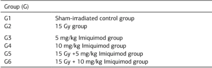
Table 1. The abbreviations used for the study groups Group (G)
G1
G2 Sham-irradiated control group15 Gy group G3 G4 G5 G6 5 mg/kg Imiquimod group 10 mg/kg Imiquimod group 15 Gy +5 mg/kg Imiquimod group 15 Gy + 10 mg/kg Imiquimod group
Imiquimod protocol
Lyophilized Imiquimod (R837; InvivoGen, San Diego, CA, USA) was reconstituted in sterile endotoxin-free water at a concen-tration of 1 mg/ml according to the manufacturer’s instructions. Doses of 5 mg/kg or 10 mg/kg were used for intraperitoneal injections [18]. Imq was started 48 hours before the irradiation and continued daily doses until the animals were sacrificed.
Morphologic Studies
At the 6th and 16th weeks of the RT 5 animals from each group were anesthetized and sacrificed by cervical dislocation, and both lungs were removed. The lung tissues were examined with both light and electron microscopies.
Histopathologic evaluation
The bilateral whole lungs of each rat were excised and fixed in 10% neutral buffered formalin. Two lobes of each lung for each rat were processed and embedded in paraffin for light mi-croscopic examination. The 4 µ thick sections obtained with microtome were stained with haematoxylin and eosin (H&E) to evaluate the inflammation, and with histochemical Masson Trichrome staining to identify the fibrosis in the lung. Extent of the chronic inflammatory cells including lymphocytes on al-veolar walls was graded on a scale from 0 (normal) to 3 (se-vere). Fibrosis was defined as the thickened alveolar walls with superimposed collagen. As a quantitative endpoint, extent of the radiation-induced fibrosis was graded on a scale from 0 (normal lung or minimal fibrous thickening) to 4 (total fibrous obliteration of the field) as described in Table 2 [19]. The pa-thologist was not aware of the treatment groups at the time of the histological examination of the specimens. After examining the whole sections for each rat, the average value of fibrosis and chronic inflammation per rat was taken as the fibrosis and inflammation scores and mean values of each group were cal-culated.
Immunohistochemistry staining and scoring procedure
Paraffin-embedded tissues of chosen slides were collected and 4 µm thick sections were prepared for immunohistochemistry. The sections were deparaffinized at 37 ºC oven overnight. Im-munohistochemical staining was performed using an automatic staining machine (Ventana, Benchmark XT). The sections were boiled at sodium citrate buffer at 95 ºC for 60 min and then incubated with primary antibody Anti TGF-β rabbit polyclonal
antibody (ABCAM, ab92486, Cambridge, UK) at a dilution of 1:100 for 52 minutes. The sections were incubated with the secondary antibody for 20 min at room temperature, incubated with Ultra I-view detection kit and counterstained with hema-toxylin for 8 min.
The immunohistochemical TGF-β stained slides were evaluated and scored by a single pathologist blinded to patients’ data. On the light microscopic evaluation of each 4 µm thick section, 10 different fields magnified 100X were reviewed. Immuno-reac-tivity scoring system (IRS) which was previously described by Wang et al. [20] was used to determine TGF-β expression lev-els. This system depends on multiplication of staining intensity and TGF-βpositive alveolar cell percentage. The percentage of positive cells was scored as follows: 0, negative; 1, 1-25%; 2, 26-50%; 3, 51-75%; 4, 76-100% and the staining intensity as 0 (-), 1 (+), 2 (++), 3 (+++).
Ultrastructural evaluation with electron microscopy
The tissue samples which were taken from the animals 6 and 16 weeks after RT were put into 2.5% glutaraldehyde for 24 hours for primary fixation. The same application was done to the rats of the Imq and control groups after the administration of the drug or NaCl in the same timing procedure. Then, these samples were washed with Sorenson’s Phosphate Buffer solu-tion (pH: 7.4) and they were post-fixed in 1% osmium tetroxide. After post-fixation, they were washed with the same buffer and dehydrated in increasing concentrations of alcohol series. Af-ter dehydration, the tissues were washed with propylene oxide and embedded in epoxy resin embedding media. The semi-thin and ultrathin sections of the obtained tissue blocks were cut with an ultramicrotome (LKB Nova, Sweden). These semi-thin sections which were 2 micrometers in thickness were stained with methylene blue and examined under a light microscope (Nikon, Japan). Following this procedure, trimming was done to the tissue blocks and their ultrathin sections which were about 60 nanometers in thickness were taken by the same ultrami-crotome. These ultra-thin sections were stained with uranyl acetate and lead citrate and they were examined under Jeol JEM 1200 EX (Japan) transmission electron microscope. The electron micrographs of the specimens were taken by the same microscope.
Statistical Method
The Statistical Package for Social Sciences (SPSS) 18.0 por-table for Windows (SPSS Inc, Chicago, Illinois, USA) was used for statistical analyses. As the pathological scores were ordinal in nature, the differences in pathological findings between the study groups were analyzed using the Kruskal-Wallis test. When an overall statistically significant difference was observed, pair-wise comparisons were performed using the Mann-Whitney U test. Bonferroni correction was used for multiple comparisons. A 5% type-I error level was used for the statistical significance cut-off for overall comparisons. P<0.05 was considered for sta-tistical significance level.
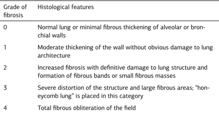
Table 2. The scoring system for lung fibrosis Grade of
fibrosis
Histological features
0 Normal lung or minimal fibrous thickening of alveolar or bron-chial walls
1 Moderate thickening of the wall without obvious damage to lung architecture
2 Increased fibrosis with definitive damage to lung structure and formation of fibrous bands or small fibrous masses
3 Severe distortion of the structure and large fibrous areas; “hon-eycomb lung” is placed in this category
4 Total fibrous obliteration of the field
Ashcroft T, Simpson JM, Timbrelli V. Simple method of estimating severity of pulmonary fibrosis on a numerical scale. J Clin Pathol. 1998;41:467-470 (20).
| The Annals of Clinical and Analytical Medicine Imiquimod attenuates radiation lung injury
710 Results
Histopathological and immunohistochemical findings at the 6th week of RT
The inflammation, fibrosis, and TGF-β scores between study groups were significantly different at the 6th week of RT (p< 0.001 for all) (Table 3). The tissue samples taken from the RT groups had higher scores of all parameters when compared to non-RT groups. Therefore, we performed pair-wise compari-sons separately for RT groups and non-RT groups. Among RT groups, the mean inflammation, fibrosis, and TGF-β scores were not significantly different (Table 4). In all RT groups there were moderate inflammation and fibrosis.
In the non-RT groups, there weren’t any pathological findings. The histopathological examination of both low-dose Imq group and high-dose Imq group was similar with the control group and there was no statistically significant difference between the non-RT groups (Table 4). The histological sections of the rats’ lung exhibited alveolar structures surrounded by the dense capillary nets. Alveoli were separated from each other by thin alveolar septums and covered by the layer of epithelial cells.
Histopathological and immunohistochemical findings at the 16th week of RT
At the 16th week of RT, histopathological and immunohisto-chemical evaluation revealed that there were significant differ-ences between the study groups in terms of the inflammation, fibrosis, and TGF-β scores (p< 0.001 for all) (Table 3). Similar to the 6th-week findings, the RT groups had higher scores of all parameters when compared to non-RT groups. The inflamma-tion, fibrosis and, TGF- β scores of RT only group were signifi-cantly higher than low-dose Imq + RT group and, high-dose Imq +RT groups (p-values were, G2-G5: 0.033, 0.041, and <0.001; and G2-G6: 0.043, 0.002 and, <0.001 for inflammation, fibrosis and, TGF-β scores respectively) (Figure 1). There wasn’t any dif-ference with respect to all parameters between low-dose Imq +RT and, high-dose Imq + RT groups (Table 4) (Figure 2). There
weren’t any pathological findings in non-RT groups at the 16th weeks of RT.
Ultrastructural findings at the 6th week of RT
Ultrastructural examination with electron microscopy revealed that there weren’t any pathologic changes in non-RT groups at the 6th week of RT. In the non-RT groups, the alveolocapil-lary membrane, bronchi, and bronchioles were ultrastructurally normal. There was no vacuole in the alveolar epithelial cells.
Table 3. The semiquantitative scoring of chronic inflammation, fibrosis and TGF-β staining at 6th and 16th weeks of RT
Group *Inflammation (mean±SD) *P **Fibrosis (mean±SD) **P ***TGF-β staining (mean±SD) ***P 6th week of RT Control Group 0.1±0.33 <0.001 0.2±0.44 <0.001 1.4±0.52 <0.001 RT only group 2±0.82 2.1±0.87 5.2±1.98 Low-dose I 0.2±0,42 0.2±0.42 1.7±0.48 High-dose-I 0.2±0,42 0.2±0,42 2.1±0.57 RT +Low-dose I 2.1±0.57 2.1±0.32 6.2±0.63 RT + High-dose I 2.2±0,52 2.2±0.42 6.1±0.74 16th week of RT Control Group 0.2±0.42 <0.001 0.3±0.48 <0.001 1.5±0.53 <0.001 RT only group 2.8±0.42 3.6±0.52 10.9±0.99 Control Group 0.4±0.52 0.3±0.48 1.5±1.052 RT only group 0.3±0.48 0.3±0.48 2.4±0.84 Low-dose I 2.1±0.51 2.8±0.63 7.3±0.82 High-dose-I 2.0±0.42 2.6±0.69 6.3±0.94
*: Scores for chronic inflammation are: 0=none, 1=mild, 2=moderate, 3=severe; **: Scores for fibrosis are: 0=none, 1=mild, 2=moderate, 3=severe, 4=total lung fibrosis; ***Scores of TGF-β are: 0 (-), 1 (+), 2 (++), 3 (+++).
Table 4. Pair-wise comparisons of the RT groups regarding to inflammation, fibrosis, and TGF-β scores at 6th and 16th weeks of RT
Pair-wise Comparison Groups 6th week p*
Group 1 Group 2 Inflammation Fibrosis TGF-β
Non-RT groups G1 G3 0.916 0.994 0.983 G1 G4 0.916 0.994 0.323 G3 G4 0.996 0.999 0.742 RT groups G2 G5 0.996 0.890 0.906 G2 G6 0.919 0.994 0.983 G5 G6 0.674 0.994 0.984
Pair-wise Comparison Groups 16th
week
p*
Group 1 Group 2 Inflammation Fibrosis TGF-β
Non-RT groups G1 G3 0.928 0.999 0.999 G1 G4 0.997 0.999 0.112 G3 G4 0.997 0.999 0.112 RT groups G2 G5 0.033 0.041 <0.001 G2 G6 0.043 0.002 <0.001 G5 G6 0.928 0.819 0.119
G=group, RT=radiotherapy, TGF-β=Transforming growth factor-beta *: Bonferroni Correction was used to evaluate the significance levels of Type-I error for pair- wise comparisons
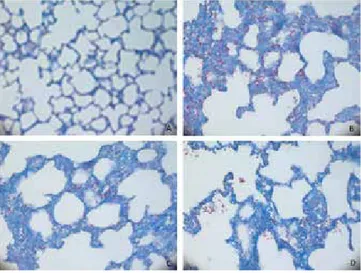
Figure 1. Histochemical Masson Trichrome staining (200X): Section from the lungs of the rats from a) Control group; b) RT only group; c) RT + 5 mg/kg imiqui-mod; d) RT + 10 mg/kg imiquimod at the 16th week of RT.
a) Control group: Normal alveolar structures with thin alveolar walls.
b) RT only group: There were thickening in the alveolar walls with severe fibrosis and partial lung obliteration.
c) RT + 5 mg/kg imiquimod: There were thickening in the alveolar walls with mod-erate fibrosis.
d) RT + 10 mg/kg imiquimod: There were thickening in the alveolar walls with moderate fibrosis.
There was no intercellular edema in the bronchioles and lower respiratory systems.
In RT only group, there were many large vacuoles in the alveo-lar epithelial cells. Additionally, small vacuoles were seen in the cytoplasm of capillary endothelial cells. There was moderate intercellular edema in-between the alveolar epithelial cells. In low-dose Imq +RT and, high-dose Imq +RT groups we observed some vacuoles in the alveolar epithelial cells in addition to cap-illary endothelial cells. Pericellular edema was present around
the alveolar epithelial cells. However, the vacuoles and intercel-lular edema were smaller than RT only group. There was not any difference between low-dose Imq +RT and, high-dose Imq +RT groups after 6 weeks of RT.
Ultrastructural findings at the 16th week of RT
The sixteenth-week examination of the rats from the non-RT groups demonstrated that there were no ultrastructural path-ological findings. The ultrastructural examination of RT only group revealed many vacuoles in the alveolar epithelial cells and capillary endothelial cells. There was severe intercellular edema in between the alveolar epithelial cells. The vacuoles were larger and intercellular edema was more severe than the 6th week findings of the RT only group. At the 16th week of RT, the low-dose Imq +RT group had the same ultrastructural find-ings with low-dose Imq +RT group at the 6th-week of RT. Simi-larly, both the 6th and the 16th- week ultrastructural findings of high-dose Imq +RT group were same (Figure 3).
Discussion
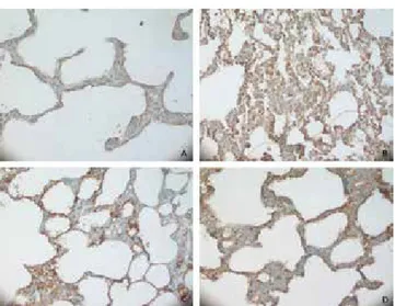
Radiation therapy is an essential treatment modality for breast cancer, lung cancer, esophageal cancer, and other thoracic can-cers. However, the efficacy of RT can be severely compromised by the side effects of RILI including pneumonitis and lung fi-brosis, which eventually deteriorate the quality of life of the patients. Modern treatment techniques, including intensity modulated RT (IMRT), image-guided RT (IGRT) and stereotactic body RT (SBRT) are still limited by the toxicity of nearby normal tissues, thus impending further gains with these technologies. Therefore, RILI is of critical importance and major dose- limit-ing factor for a radiation oncologist in attemptlimit-ing to deliver curative doses to the tumor. Imq, the first of a new class of compounds called “immune system modifiers” stimulates the innate and adaptive immune pathways and induces cytokine production. It is used for many conditions including genital and perianal warts, postsurgical keloid recurrence, solar keratosis, and cutaneous, non-alcoholic fatty liver disease. Imq induces the cytokines IFN-α and IFN- γ, TNF-α, IL-1, 5, 6, 8, 10 and 12. However, it is the induction of IFN-α and - γ, that makes Imq particularly suited for the anti-fibrotic purpose [21]. It was demonstrated Imq inhibits human fibroblast collagen produc-tion by IFN-α and – γ in a dose-dependent manner [22]. IFN-α and IFN-γ reduce both collagen and fibrosis. Moreover, IFN- γ may down-regulate the profibrotic Th2 cytokine IL-4, in addi-tion to down-regulating the expression of TGF-β, which is the master chief of the radiation-induced fibrosis [2, 21, 23]. By the light of these findings, we hypothesized that Imq may have a protective effect on RILI. To the best of our knowledge, the cur-rent study is the first one investigating the role of Imq in RILI. We demonstrated that Imq attenuated “radiation-induced lung fibrosis” without ameliorating the early pneumonitic phase. Ei-ther lower-dose or the higher-dose had the same effect. RILI, which is also named as “radiation pneumonopathy” is a continuous process and regarded as the result of an abnormal healing response. Subclinical early damage in type I pneumo-cytes progress to an acute interstitial inflammation at 6-12 weeks after the onset of RT, which is called as “pneumonitic phase”. Radiation pneumonitis may be progressive and result Figure 2. Immunohistochemical TGF-β staining (200X): Section from the lungs of
the rats from a) Control group; b) RT only group; c) RT + 5 mg/kg imiquimod; d) RT + 10 mg/kg imiquimod group at 16th week of RT.
a) Control group: There were a few TGF-β positive alveolar epithelial cells or fibro-blasts with staining of low intensity.
b) RT only group: There were intense and diffuse TGF-β positivity in alveolar epi-thelial cells or fibroblasts.
c) RT + 5 mg/kg imiquimod group: TGF-β positivity was lower than group b. d) RT + 10 mg/kg imiquimod: TGF-β positivity was lower than group b and c.
Figure 3. Ultrastructural findings with electron microscopy at the 16th week of RT (a) Control group (b) RT only group (c) RT+5 mg/kg imiquimod group (d) RT+ 10 mg/kg imiquimod group:
(a) Control group: Electron micrograph showing alveolar epithelial cells (*), nucleus of an alveolar epithelial cell (n) and an erythrocyte (e) inside the alveolar capillary in the lung of a control group rat.
(b) RT only group: Many vacuoles were observed in the alveolar epithelial cells and capillary endothelial cells. Intercellular edema was present in between the alveolar epithelial cells.
(c) RT + 5 mg/kg imiquimod group: Many vacuoles were observed in the alveo-lar epithelial cells and capilalveo-lary endothelial cells. Intercellualveo-lar edema was present in between the alveolar epithelial cells. However, the vacuoles and intercellular edematous areas were smaller than RT only group.
(d) RT + 10 mg/kg imiquimod group: Many vacuoles were observed in the alveo-lar epithelial cells and capilalveo-lary endothelial cells. Intercellualveo-lar edema was present in between the alveolar epithelial cells. However, the vacuoles and intercellular edematous areas were smaller than RT only group.
| The Annals of Clinical and Analytical Medicine Imiquimod attenuates radiation lung injury
712
in chronic pulmonary damage or acute respiratory distress syndrome which eventually may be fatal. The radiation-in-duced acute lung parenchymal changes can progress to lung fibrosis after many months and years. Fibrosis is a part of the wound-healing process. Therefore, radiation fibrosis is a form of chronic lung damage that usually evolves over 4-24 months after irradiation [5,24]. Radiation-induced lung fibrosis is char-acterized by the accumulation of fibroblasts, myofibroblasts, inflammatory cells and extracellular matrix proteins, such as collagen, with the subsequent formation of a scar, which even-tually results in impaired lung function [4, 25]. Because there are numerous contributing factors and various diagnostic set-tings for radiation-induced lung fibrosis, the reported incidence in patients who have received radiotherapy varies significantly, from 1% to 43% [26-28].
Imq, applied topically, has been used to treat morphea, a skin disease characterized by localized fibrosis, and its use has been advocated for the treatment of Dupuytren’s contracture, anoth-er fibrosing disease [10, 21, 29-31]. Dytoc et al. reported their case series on the use of Imq for morphoea [21]. Morphoea or localized scleroderma is a fibrotic disease characterized by fi-brosis of the skin with the accumulation of extracellular matrix (ECM) components, particularly collagen [21]. Research on fi-brosis in patients with localized scleroderma focuses mainly on TGF-β, which is the master-chief of radiation-induced fibrosis. The results of the case series Dytoc et al. showed that Imq suc-cessfully treated the localized scleroderma. In the current study, Imq ameliorated the “fibrotic phase” of the RILI, without affect-ing the “pneumonitic phase”. We found that TGF-β score of RT only group was significantly higher than low-dose Imq+RT; and high-dose Imq+RT groups scores.
Namazi postulated that Imq is a potential drug for Dupuytren’s contracture, which is a condition of the hand characterized by the development of new fibrotic tissue in the form of nodules and cords [29]. In the pathophysiology of Dupuytren’s contrac-ture, TGF-β is a key fibrogenic cytokine that has been shown to stimulate fibroblast proliferation and extracellular matrix deposition [32]. By the same mechanism of radiation-induced lung fibrosis, Namazi hypothesized that Imq induces the cyto-kine IFN-α, IFN- γ, TNF-α, IL-1, IL-6, IL-8, IL-10, IL-12 and it stimulate cell- mediated immunity (TH1 Pathway). IFN- γ is a TH-l cytokine that downregulates expression of TGF-β, which eventually ameliorates fibrosis process.
As mentioned before, “fibrosis” is part of the wound-healing pro-cess after an irritant stimulus in lung tissue. It was shown that adenosine receptors (AR) are required for appropriate granula-tion tissue formagranula-tion and in adequate wound healing. A2A and A2B AR stimulate both of the critical functions in granulation tissue formation including new matrix production and angio-genesis; and the A1 AR may also contribute to new vessel for-mation [33]. Angiogenesis is a feature of chronic lung diseases such as asthma and pulmonary fibrosis. Studies in adenosine deaminase (ADA)-deficient mice, characterized by elevated lung tissue levels of adenosine, strongly suggest a causal associa-tion between adenosine and an inflammatory phenotype [33-35]. Schön et al. demonstrated that Imq, at pharmacologically relevant concentrations, is an A2AAR antagonist and that this may account for its immunological effects [31]. The end result
of this additional activity of Imq would be an amelioration of fibrosis which may contribute to the anti-fibrotic effect of TGF-β-related pathway.
Our study has also some limitations that deserve to be men-tioned as well. Firstly, we studied only the TGF-β related path-way without investigating the AR related pathpath-way. We believe that, although the TGF-β related pathway seems to be the ma-jor one, AR-related pathway, which causes anti-fibrotic effect indirectly, worth to be investigated in a new study. Secondly, we did not demonstrate any difference with respect to pneu-monitic phase between the RT groups. In the pathophysiology of the RILI, it was emphasized that the fibrosis is an abnormal wound-healing process, and usually seen as a progression of pneumonic phase. However, Imq itself causes pro-inflammatory activity which may be augmented by suppression of a negative feedback mechanism mediated by adenosine receptor signal-ing. Therefore, in order to explain better both limitations, there is a need for a new study investigating the radiation fibrosis and Imq induced anti-fibrotic activity at a molecular level.
Conclusion
The current study is the first one observing the effect of Imq on RILI. We demonstrated that Imq attenuated the radiation-induced lung fibrosis without ameliorating the radiation pneu-monitis. Therefore, during the pathophysiology of RILI Imq have a limited anti-inflammatory effect and a substantial anti-fibrot-ic effect. We did not observe any difference between low-dose and high dose Imq; therefore, a lower dose may be adequate. The anti-fibrotic effects of Imq might have potentially been at-tributed to its inhibitory effects on adenosine receptors. These findings should be clarified with further preclinical and clinical studies.
Scientific Responsibility Statement
The authors declare that they are responsible for the article’s scientific content including study design, data collection, analy-sis and interpretation, writing, some of the main line, or all of the preparation and scientific review of the contents and ap-proval of the final version of the article.
Animal and human rights statement
All procedures performed in this study were in accordance with the ethical standards of the institutional and/or national re-search committee and with the 1964 Helsinki declaration and its later amendments or comparable ethical standards. No ani-mal or human studies were carried out by the authors for this article.
Funding: None Conflict of interest
None of the authors received any type of financial support that could be considered potential conflict of interest regarding the manuscript or its submission.
References
1. Kocak Z, Evans ES, Zhou SM. Challenges in defining radiation pneumoni-tis in patients with lung cancer. Int J Radiat Oncol Biol Phys. 2005; 62: 635-8. DOI:10.1016/j.ijrobp.2004.12.023
| The Annals of Clinical and Analytical Medicine 713
2. Simone CB 2nd. Thoracic Radiation Normal Tissue Injury. Semin Radiat Oncol. 2017; 27(4): 370-7. DOI: 10.1016/j.semradonc.2017.04.009
3. Tsoutsou P.G, Koukourakis M.I. Radiation pneumonitis and fibrosis: Mechanisms underlying its pathogenesis and implications for future research. Int J Radiat On-col Biol Phys. 2006; 66:1281-93. DOI: 10.1016/j.ijrobp.2007.02.045
4. Ding N, Li JJ, Sun L. Molecular mechanisms and treatment of radiation induced lung fibrosis. Curr Drug Targets. 2013; 14:1247-1356.
5. Graves PR, Siddiqui F, Anscher MS, Movsas B. Radiation pulmonary toxicity: from mechanisms to management. Semin Radiat Oncol. 2010; 20: 201-7. DOI: 10.1016/j.semradonc.2010.01.010.
6. Mazeron R, Etienne-Mastroianni B, Pérol D, Arpin D, Vincent M, Falchero L, et. Al Predictive factors of late radiation fibrosis: A prospective study in non-small cell lung cancer. Int J Radiat Oncol Biol Phys.2010; 77: 38-43. DOI: 10.1016/j. ijrobp.2009.04.019.
7. Skinner RB Jr. Imiquimod. Dermatol Clin. 2003; 21(2): 291-300.
8. Harrison CJ, Jenski L, Voychehovski T, Bernstein DI. Modification of immunologi-cal responses and cliniimmunologi-cal disease during topiimmunologi-cal R-837 treatment of genital HSV-2infection. Antiviral Res. 1988; 10: 209–23 DOI: 10.1016/0166-3542(88)90032-0. 9. Robles DT, Moore E, Draznin M, Berg D. Keloids: pathophysiology and manage-ment. Dermatol Online J. 2007; 13(3): 9.
10. Schön MP, Schön M. Imiquimod: mode of action. Br J Dermatol. 2007; 157 (2): 8-13. DOI: 10.1111/j.1365-2133.2007.08265.x
11. Hemmi H, Kaisho T, Takeuchi O, Sato S, Sanjo H, Hoshino K, et. Al. Small anti-viral compounds activate immune cells via the TLR7 MyD88-dependent signaling pathway. Nat Immunol. 2002; 3: 196–200. DOI: 10.1038/ni758
12. Berman B, Harrison-Balestra C, Perez OA, Viera M, Villa A, Zell D, et Al.. Treat-ment of keloid scars post-shave excision with imiquimod 5% cream: a prospective, double-blind, placebocontrolled pilot study. J Drugs Dermatol. 2009; 8(5): 455–58. 13. Mrowietz U, Seifert O. Keloid scarring: new treatments ahead. Actas Dermosi-filiogr. 2009; 100(2): 75–83.
14. Martin-Garcia RF, Busquets AC. Postsurgical use of imiquimod 5% cream in the prevention of earlobe keloid recurrences: results of an open-label, pilot study. Dermatol Surg. 2005; 31: 1394-98.
15. Lewis DJ, Byekova YA, Emge DA, Duvic M. Complete resolution of mycosis fungoides tumors with imiquimod 5% cream: a case series. J Dermatolog Treat. 2017; 28(6): 567-9. DOI: 10.1080/09546634.2017.1294728
16. Stanley MA. Imiquimod and the imidazoquinolones: mechanism of action and therapeutic potential. Clin Exp Dermatol. 2002; 27(7): 571-7.
17. Kim S, Park S, Kim B, Kwon J. Toll-like receptor 7 affects the pathogenesis of non-alcoholic fatty liver disease. Sci Rep. 2016; 9; 6: 27849. DOI: 10.1038/ srep27849.
18. Damm J, Wiegand F, Harden LM, Gerstberger R, Rummel C, Roth J. Fever, sickness behavior, and expression of inflammatory genes in the hypothalamus after systemic and localized subcutaneous stimulation of rats with the Toll-like receptor 7 agonist imiquimod. Neuroscience. 2012; 201: 166-83. DOI: 10.1016/j. neuroscience.2011.11.013
19. Ashcroft T, Simpson JM, Timbrelli V. Simple method of estimating severity of pulmonary fibrosis on a numerical scale. J Clin Pathol. 1998; 41: 467-70. 20. Wang J.Y, Darbinyan A, White M.K, Darbinian N, Reiss K, Amini S. Involvement of IRS‐1 Interaction With ADAM10 in the Regulation of Neurite Extension. Journal of cellular physiology. 2014; 229(8): 1039-46. DOI: 10.1002/jcp.24528. 21. Dytoc M, Ting PT, Man J, Sawyer D, Fiorillo L. First case series on the use of imiquimod for morphoea. Br J Dermatol. 2005; 153: 815–20. DOI: 10.1111/j.1365-2133.2005.06776.x
22. Jimenez SA, Freundlich B, Rosenbloom J. Selective inhibition of human diploid fibroblast collagen synthesis by interferons. J Clin Invest. 1984; 74: 1112–16. DOI: 10.1172/JCI111480
23. Jablonska S, Blaszczyk M. Scleroderma and sclerotic skin conditions: unap-proved treatments. Clin Dermatol. 2002; 20: 634–7.
24. Trott KR, Herrmann T, Kasper M. Target cells in radiation pneumopathy. Int J Radiat Oncol Biol Phys. 2004; 58: 463-9.
25. Sime PJ, O’Reilly KM. Fibrosis of the lung and other tissues: new concepts in pathogenesis and treatment. Clin Immunol. 2001; 99: 308–19. DOI: 10.1006/ clim.2001.5008
26. Kong FM, Ten Haken R, Eisbruch A, Lawrence TS. Non-small cell lung cancer therapy-related pulmonary toxicity: an update on radiation pneumonitis and fibro-sis. Semin Oncol. 2005; 32(Suppl. 3): S42–54.
27. Movsas B, Raffin TA, Epstein AH, Link CJ. Pulmonary radiation injury. Chest.1997; 111: 1061–76.
28. Arpin D, Mahe MA, Servois V, Claude L. Predictive factors for acute radia-tion pneumonitis. Rev Pneumol Clin. 2009; 65: 177–86. DOI: 10.1016/j.pneu-mo.2009.03.011.
29. Namazi H. Imiquimod: a potential weapon against Dupuytren contracture. Med Hypotheses. 2006; 66(5): 991-2. DOI: 10.1016/j.mehy.2005.09.052
30. Sambo P, Baroni SS, Luchetti M, Paroncini P, Dusi S, Orlandini G, et al. Oxida-tive stress in scleroderma: maintenance of scleroderma fibroblast phenotype by the constitutive up-regulation of reactive oxygen species generation through the NADPH oxidase complex pathway. Arthritis Rheum. 2001; 44: 2653–64. 31. Schön MP, Schön M, Klotz KN. The small antitumoral immune response modi-fier imiquimod interacts with adenosine receptor signaling in a TLR7- and TLR8-independent fashion. J Invest Dermatol. 2006; 126: 1338–47. DOI: 10.1038/ sj.jid.5700286
32. Bayat A, Watson JS, Stanley JK, Ferguson MW, Ollier WE. Genetic susceptibility
to Dupuytren disease: association of ZF-9 Transcription factor gene. Plast Re-constr Surg. 2009; 11(7): 2133–9. DOI: 10.1097/01.PRS.0000060531.98180.32 33. Feoktistov I, Biaggioni I, Cronstein BN. Adenosine receptors in wound heal-ing, fibrosis and angiogenesis. Handb Exp Pharmacol. 2009; (193): 383-97. DOI: 10.1007/978-3-540-89615-9_13.
34. Blackburn MR, Volmer JB, Thrasher JL, Zhong H, Crosby JR, Lee JJ, et al. Meta-bolic consequences of adenosine deaminase deficiency in mice are associated with defects in alveogenesis, pulmonary inflammation, and airway obstruction. J Exp Med. 2000; 192:159–70.
35. Blackburn MR. Too much of a good thing: adenosine overload in adenosine-deaminase-deficient mice. Trends Pharmacol Sci. 2003; 24: 66–70. DOI: 10.1016/ S0165-6147(02)00045-7.
How to cite this article:
Calik M, Yavaş G, Çelık ZE, Yavaş Ç, Calık SG, Sargon MF, Esme H. The impact of imiquimod on radiation-induced lung injury: Results of an experimental study. Ann Clin Anal Med 2019;10(6): 707-13.