Phylogenetic Analysis of Bovine Respiratory Syncytial Virus from
Calves with Respiratory Disorders
İlke KARAYEL HACIOĞLU
1,a
Nüvit COŞKUN
2Selda DURAN YELKEN
3Seçil SEVİNÇ
4Feray ALKAN
1,b 1 Ankara University, Faculty of Veterinary Medicine, Department of Virology, TR-06110 Ankara - TURKEY2 Kafkas University, Faculty of Veterinary Medicine, Department of Virology, TR-36100 Kars - TURKEY 3 Siirt University, Faculty of Veterinary Medicine, Department of Virology, TR-56100 Siirt - TURKEY 4 Selcuk University, Faculty of Veterinary Medicine, Department of Virology, TR-42003 Konya - TURKEY
a ORCID: 0000-0003-1566-630X; b ORCID: 0000-0003-3854-6503
Article Code: KVFD-2018-20819 Received: 12.05.2018 Accepted: 21.12.2018 Published Online: 22.12.2018 How to Cite This Article
Karayel Hacioğlu İ, Coşkun N, Duran Yelken S, Sevinç S, Alkan F: Phylogenetic analysis of Bovine Respiratory Syncytial Virus from calves with
respiratory disorders. Kafkas Univ Vet Fak Derg, 25 (2): 251-256, 2019. DOI: 10.9775/kvfd.2018.20819
Abstract
Bovine respiratory disease (BRD) causes economic losses related to a reduction in weight gain of affected animals, veterinary treatment costs, death, etc. One of the important respiratory tract disease viruses is the bovine respiratory syncytial virus (BRSV). In this study, it is aimed to report the molecular characterization of detected BRSVs. Therefore, nasal samples from three calves in a herd seen severe respiratory disorders were examined for BRSV and other possible viral etiological agents by PCRs and the amplicons were sequenced. In the phylogenetic tree, BRSV circulating in this herd is clustered with the genetic subgroup III BRSVs deposited in GenBank from some other countries. This study on the molecular characterization of BRSV circulating in calves would contribute for future studies on the epidemiology of this infection and the development and/or choice the effective vaccines in Turkey.
Keywords: BRSV, Calves, Genotype, Subgroup III
Solunum Sistemi Hastalığı Olan Buzağılarda Saptanan Bovine Respiratory
Syncytial Virusun Filogenetik Analizi
Öz
Sığırlarda solunum sistemi hastalıkları, etkilenen hayvanların kilo alımında azalma, veteriner tedavi masrafları, ölüm gibi sebeplerle ekonomik kayıplara neden olmaktadır. Bovine Respiratory Syncytial Virus (BRSV), solunum sistemini etkileyen viral etkenlerin başında gelmektedir. Bu çalışmada BRSV’nin saptanması ve tespit edilen etkenlerin moleküler karakterizasyonlarının ortaya konulması amaçlanmıştır. Bu amaçla, buzağılarda ağır solunum yolu sistemi bulguları gözlenen bir işletmedeki üç buzağıdan alınan burun akıntısı örnekleri BRSV ve diğer viral etkenler yönünden PCR ile test edilmiştir. RT-PCR sonucunda pozitif bulunan örneklerin dizin bilgileri elde edilmiştir. Yapılan filogenetik analiz sonucunda bu sürüde tespit edilen BRSV’nin genetik olarak subgrup III içerisinde yer aldığı ortaya konulmuştur. Buzağılarda saptanan BRSV’nin moleküler karakterizasyonu üzerine yapılan bu çalışma, bu enfeksiyonun epidemiyolojisi ve etkili aşıların geliştirilmesi ve/veya seçimiyle ilgili gelecekteki çalışmalara katkıda bulunacaktır.
Anahtar sözcükler: BRSV, Buzağı, Genotip, Subgrup III
INTRODUCTION
Bovine respiratory syncytial virus (BRSV), bovine para-influenza virus type 3 (BPIV3), bovine herpesvirus type 1 (BHV-1), bovine adenovirus (BAV), bovine coronavirus (BCoV) and bovine viral diarrhea virus (BVDV) are important patho- gens associated with the bovine respiratory disease complex (BRDC) [1]. It is known that each of these agents can cause infection either alone or in combination with the bacterial
agents and the other viruses [2]. Economic effects of BRSV infection together with the other BRDC agents to the cattle industry should not be underestimated due to the mortality, treatment expenses, and slower growth of affected animals [1,3]. BRSV infection could mostly cause mild disease in adult cattle, the infection could be quite a severe infection in calves and even leads to outbreaks and deaths [4]. Bovine respiratory syncytial virus belongs to the genus
İletişim (Correspondence)
+90 312 3170315/4363Orthopneumovirus in the family Pneumoviridae and contains 10 genes encoding 11 proteins. Three of them are trans-membrane glycoproteins and located on the surface of the viral envelope. These are the large attachment glycoprotein (G), which mediates viral attachment to the host cell; the fusion protein (F), which enables fusion of virus with the host cell, and the small hydrophobic protein (SH). Other proteins are nucleocapsid associated nucleoprotein (N), phosphoprotein (P), the viral RNA-dependent polymerase protein (L), M2-1, and M2-2 and the matrix protein M. Finally, there are 2 non-structural (NS) proteins that accumulate in infected cells, NS1 and NS2 [5].
Based on the reaction patterns with monoclonal antibodies, BRSV has been divided into four antigenic subgroups, designated as A, B, AB and untyped although there is only one serotype of BRSV [6,7]. Further studies on the nucleotide sequencing of the G protein gene and the reaction with neutralizing antibodies, the antigenic subgroups were confirmed, although there is genetic variability in these subgroups [1]. Moreover, the genetic and antigenic hetero-geneity of G protein along with the analysis of the genes-encoding N and F protein has been used to characterize BRSV strains [8,9]. Currently, seven genetic subgroups of BRSV strains have been determined by the nucleotide sequencing of attachment G protein [8].
Despite the serological and virological data on the presence of BRSV and contribution on respiratory infections [2,10,11], the circulating genotypes were not previously reported in Turkey. The aims of the study were i) to investigate the etiological agent(s) causing severe respiratory disorders in a herd and ii) to describe the molecular characterization of BRSVs detected in Turkey.
MATERIAL and METHODS
History of Infection and Study Design
At the beginning of September of 2016, a severe outbreak characterized by respiratory distress, fever, cough, sneeze, etc. in calves, aged 7-85 days, was seen in a herd including approximately 3000 Brown Swiss cattle in Eskişehir. This herd was closed and had been restocking only from internal animal source. Animals over 1 year of age are kept under the same roof, although newborns are grouped under a different roof for every 3 months of age. However, they do mingle freely during the grazing. Animals analyzed in this study were vaccinated with a commercial vaccine (Elite 9-HS; Boehringer Ingelheim Vetmedica, Inc., Germany). The morbidity and the mortality (rate up to 50% and 20%, respectively) became higher during the outbreak, notably between November and February, despite the treatment using antibiotics and other drugs to prevent symptoms and also the infection.
Extraction and Screening of Samples By PCR
Nasal secretions were collected from three calves with
clinical signs using sterile swabs and immediately after collection viral transport medium (1 mL) was added into swabs. The samples were sent to the laboratory on ice and stored at -80°C, until analyses. Following the submission of nasal swabs to our laboratory for diagnostic purposes, all samples were examined for BRSV and other possible etiological agents such as BCoV, BPIV3, BVDV, and BHV-1 by PCRs. Viral RNA/DNA was purified using Trizol LS reagent (Thermo Scientific, USA) according to manufacturer’s instructions. Briefly, synthesis of cDNA was achieved following denaturation of RNA at 70°C for 5 min. The cDNA was synthesized using Moloney Murine Leukemia Virus (MMLV) reverse transcriptase (RT) (Thermo Scientific, USA) and random hexamers (Thermo Scientific, USA) by incubating at 25°C for 10 min, and thereafter at 37°C for 1 h. MMLV-RT was therefore inactivated at 70°C for 10 min. PCR/RT-PCRs for the detection of mentioned viruses were performed by using the specific primers according to the protocols reported elsewhere [12-16]. BRSVs in nasal samples were detected by nested RT-PCR using the primer sets B1/B2A: 5’-AATCAACATGCGTGCAGTTAG-3’/5’-TTTGGTCATTCGTTATAGGCAT-3’ (711 bp) and B3/B4A: 5’-GTGCAGTTAGTAGAGGTTATCTTAGT-3’/5’-TAGTTCTTTAG ATCAAGTACTTTGCT-3’ (481 bp) targeting F gene region [15]. The PCR mixture was incubated for 5 min at 96°C. Then, 40 amplification cycles (1 min at 94°C, 1 min at 55°C and 1 min at 72°C) were performed followed by a final extension step at 72°C for 10 min. The nested PCR was performed under the same reaction conditions with inner primers. To molecularly characterize of BRSVs, primers (G2.5/F2.7: 5’-AGACATTAAAGAGGGCTTGGA-3’/5’-CTGCA CTGCATGTTGATTGA-3’) targeting G gene region were used with an expected amplicon size of 1030 bp [9]. The mixture for PCR was preheated at 98°C for 30 sec, subjected to 35 cycles of 30 sec at 98°C, 30 sec at 60°C, 30 sec at 72°C and a final 7 min incubation at 72°C. An isolate previously identified in our department was used as the positive control virus. The PCR amplicons of the samples and the positive control virus were analyzed by 1% agarose gel electrophoresis and visualized under UV light.
Sequence and Phylogenetic Analysis
Purification of the amplicons and sequencing were performed from by a commercial company. Following the sequencing of these amplicons, multiple sequence alignments were prepared by the MUSCLE algorithm as implemented in AliView Software [17,18]. Cognate sequences of reference BRSVs representing different subgroups for the G protein gene were retrieved from Gen Bank through the BLAST engine. Phylogenetic analysis of nucleotide sequences of the G protein gene was performed using MEGA 6.06 software [19]. The phylogenetic tree was constructed using the maximum likelihood method and Tamura-Nei nucleotide substitution model and the statistical significance was estimated by bootstrap analysis (1000 replicates). The nucleotide identity table was
computed using online tools (SIAS, http://imed.med.ucm. es/Tools/sias.html).
RESULTS
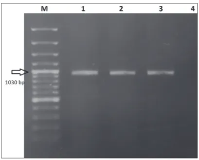
All nasal swab samples (n=3) were positive for BRSV by RT-PCR targeting F gene region while they were negative other mentioned viruses tested (Fig. 1). Additionally, herd manager reported that they sent all samples to another routine diagnostic laboratory and the samples were negative for bacterial agents. RT-PCR targeting G protein gene region for the detection of a genetic subgroup of BRSV produced correct size amplicons (1030 bp) only in two nasal samples (Fig. 2). Genome sequences of G protein
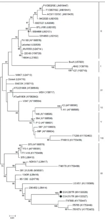
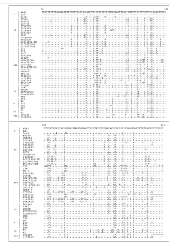
gene of BRSVs were deposited in GenBank under the following accession numbers: MH133326 and MH133327. In the phylogenetic tree (Fig. 3) both BRSVs were classified in the same cluster and they determined as belonging to subgroup III. Additionally, deduced amino acid sequences of G protein of two Turkish BRSVs were compared to the other deduced amino acid sequences of G protein of BRSVs belonging to different countries and vaccines (Fig. 4). The partial nucleotide and amino acid sequences of the G protein gene of two BRSVs were compared with each other and with the sequences of BRSVs from other countries acquired from GenBank. The identities of the nucleotide sequences and also predicted amino acid sequences of two Turkish BRSVs were 100%. They shared the closest genetic relationship with the USA strain 236-652 (94.51%). The nucleotide sequence and deduced amino acid sequence similarities of the G protein regions between our field virus and other BRSVs identified as subgroup III, ranged from 85.4 to 100% and from 79.61-93.33%, respectively. The comparison of the deduced amino acid sequences of the G protein of two Turkish BRSVs with other BRSVs belonging to different countries and vaccines showed that the all four cysteine (Cys) residues (Cys 173-Cys 186 and Cys 176-Cys 182) in the immunodominant region were conserved in both Turkish BRSVs without any substitutions. However, Asn 179 and of Ala205 replaced by Ser and Iso in both strains, respectively, differing from the other BRSVs in all subgroups (Fig. 4).
DISCUSSION
This study investigated viruses in bovine respiratory disorders in calves, including BRSV, BPIV3, BHV-1, BCoV, and BVDV. Results of PCRs and sequencing data showed that all of the nasal swab samples were positive for F protein gene of BRSV while they were negative for the other tested viral agents. Also, bacteriological investigations of the samples using routine diagnostic procedures in other lab did not provide evidence for any bacterial pathogens. BRSV was determined as the causative agent in severe acute respiratory system disease seen in calves, along with or without other possible agents which were not investigated in this study.
BRSV infection in calves is widespread in America [20,21], many European countries [1,8,22-24] and Turkey according to serologic [10,11] and virologic [2] evidence. There is only one study on the investigation with virological methods of BRSV and also other some viruses causing the respiratory disease in calves. Alkan et al.[2], reported that nasal swab samples from 95 cattle with signs of respiratory disease housed in eleven different herds were tested for BHV-1, BPIV 3, BRSV
Fig 2. The results of the amplification of G gene region. M: 100 bp DNA ladder,
1: Positive control; 2,3,: Positive samples. 4: Negative control
Fig 1. The results of the amplification of F gene region. M: 100 bp DNA ladder,
and BVDV using direct immunofluorescence technique and BRSV detected as one of the causative agents especially along with BHV-1 and BPIV-3. However, there is no data about the molecular characterization of BRSV(s) in Turkey to date.
In this study, partial G protein gene region of BRSVs was successfully able to be amplified for only two nasal samples and they were sequenced. Based on the sequence analysis, both of our field viruses clustered in the genetic subgroup III, including the USA and Italian BRSVs [8,9]. Although the
small number of samples was examined in this study, the obtained data suggest that subgroup III is circulating in our country.
It is suggested that there is an interaction between the genetic subgroups of BRSVs and geographic regions [23]. Thus, in the studies on the BRSV in Europe, subgroups II, V, and VI in France; subgroups II, IV and V in Belgium; subgroups III and VII in Italy; subgroup I in Switzerland; subgroup II in Poland, Norway, Sweden, and Denmark were detected [8,9,22,25,26]. Whereas Turkey is geographically close to Europe, the sequencing data of the G protein-encoding gene of BRSVs showed that they are genetically different from the most European strains but similar with the USA and Italian BRSVs [6,8,9]. The current data of Turkish BRSVs belong to the genetic subgroup III along with the USA, Italian and also Japanese strains [8,9,23] does not fully support the theory of geographical and temporal clustering of BRSV. However, due to the fact that a lot of animals were imported from the USA and some European countries to Turkey, this result needs to be investigated. It is thought that further investigation of the BRSV infection will contribute to a more precise assessment of the diversity of Turkish BRSVs and determine the possible source of viruses circulating.
The G protein is significantly important because of its immunodominant region (cysteine-rich and between 174 and 188 aa position) in the central conserved domain which is externally accessible for the neutralizing anti-bodies [5]. In this study, deduced amino acid sequences of G protein of two Turkish strains were compared to the other deduced amino acid sequences of G protein of BRSVs belonging to different countries and vaccines. Based on the sequencing data of the immunodominant region, all 4 cysteines (Cys) residues (Cys 173-Cys 186 and Cys 176-Cys 182) were conserved in our BRSV strains with no substitutions, however, one of the quite important amino acid for the antibody binding, Asn 179, was replaced by Ser in both strains differing from the other strains in all subgroups (Fig. 2). A previous study [27] reported that the role of Asn is to stabilize helix and type I turn and also the role of Asn could also be performed by Ser which is the only residue exists as frequently as Asn at N-cap position of the helix, based on the statistical analysis. The point mutations at 180 and 205 aa determines BRSV subgroups [27]. At the central conserved region, the mutation of Ala205 to Thr determines the subgroup and distinguishes the subgroup I and II from those in the other subgroups [9]. It is demonstrated that only the mutation of Ala205 to Thr allowed escaping from antibody binding [27]. Amino acid residue 205 of the Turkish strains is an Iso, while the other isolates in subgroup III Thr are present. It is not known, this situation (Thr205 to Iso) could change the protein structure and alter the biological function of the G protein. The biological importance of antigenic subgroups is not clear. It is reported that the mutations in the
immuno-Fig 3. Phylogenetic tree of the G protein gene of BRSVs identified in this study
with those of other BRSVs selected in GenBank. Our BRSVs are indicated by black dots. The statistical significance was estimated by bootstrap method (1000 pseudo-replicates) and values of <50% are omitted
dominant region of the G protein may contribute to the lack of cross-protection between vaccine and field isolates [5]. The sequence and structure of the G protein should be considered when designing vaccination strategies and choosing viral strains for the construction of the vaccine. Furze et al.[28] showed that polyclonal sera obtained from calves vaccinated with the BRSV G protein from subgroup A virus recognized a different subgroup A BRSV, less
recognized subgroup AB, but not subgroup B or an untyped isolate. We did not investigate the antigenic features of the field strains by using monoclonal or polyclonal antibodies. However, based on our phylogenetic analysis and the results of other studies on BRSV subgroup III we could indicate that our strains belong to subgroup A. Unfortunately, it was not possible to know the BRSV strain included in a commercially inactivated vaccine had been
Fig 4. Sequences of amino acids 53 to 215 of the G protein of BRSVs. Designations on the left indicate the codes
used in this herd although we made an effort to contact the company. Although BRSV could also reinfect seropositive calves [29], it is noted that the level of the possible maternal immunity to BRSV in sampled calves is not tested in this study. Thus, it was not possible to make an interpretation of the antigenic similarity between the vaccine and field virus, and also to speculate on the efficacy of vaccination. This study provides an information on the molecular characterization of BRSV in Turkey which may have important implications for the planning the future studies on the epidemiology of this infection and the development and/ or choice the effective vaccines. The genetic analysis and evolution of BRSVs should be monitored regularly in calves and adult animal populations for the choice of vaccine to control the infection.
C
onfliCtofi
nterestThe authors declare that they have no conflict of interest. REFERENCES
1. Socha W, Larska M, Rola J: Molecular characterisation of the first polish
isolates of bovine respiratory syncytial virus. Bull Vet Inst Pulawy, 53, 569-574, 2009.
2. Alkan F, Ozkul A, Bilge Dagalp S, Yesilbag K, Oguzoglu TC, Akça Y, Burgu I: Virological and serological studies on the role of PI-3 virus, BRSV,
BVDV and BHV-1 on respiratory infections of cattle. I. The detection of etiological agents by direct immunofluorescence technique. Dtsch Tierarztl
Wochenschr, 107 (5): 193-195, 2000.
3. Klem TB, Kjæstad HP, Kummen E, Holen H, Stokstad M: Bovine
respiratory syncytial virus outbreak reduced bulls’ weight gain and feed conversion for eight months in a Norwegian beef herd. Acta Vet Scand, 58: 8, 2016. DOI: 10.1186/s13028-016-0190-y
4. Larsen LE, Tjørnehøj K, Viuff B: Extensive sequence divergence among
bovine respiratory syncytial viruses isolated during recurrent outbreaks in closed herds. J Clin Microbiol, 38 (11): 4222-4227, 2000.
5. Valarcher JF, Taylor G: Bovine respiratory syncytial virus infection. Vet
Res, 38 (2): 153-180, 2007. DOI: 10.1051/vetres:2006053
6. Prozzi D, Walravens K, Langedijk JPM, Daus F, Kramps JA, Letesson JJ: Antigenic and molecular analyses of the variability of bovine respiratory
syncytial virus G glycoprotein. J Gen Virol, 78 (2): 359-366, 1997. DOI: 10.1099/0022-1317-78-2-359
7. Schrijver RS, Daus F, Kramps JA, Langedijk JPM, Buijs R, Middel WGJ, Taylor G, Furze J, Huyben MWC, van Oirschot JT: Subgrouping of bovine
respiratory syncytial virus strains detected in lung tissue. Vet Microbiol, 53 (3-4): 253-260, 1996. DOI: 10.1016/S0378-1135(96)01223-0
8. Bertolotti L, Giammarioli M, Rosati S: Genetic characterization of
bovine respiratory syncytial virus strains isolated in Italy: Evidence for the circulation of new divergent clades. J Vet Diagn Invest, 30 (2): 300-304, 2018. DOI: 10.1177/1040638717746202
9. Valarcher JF, Schelcher F, Bourhy H: Evolution of bovine respiratory
syncytial virus. J Virol, 74 (22): 10714-10728, 2000. DOI: 10.1128/JVI.74.22. 10714-10728.2000
10. Yeşilbağ K, Güngör B: Seroprevalence of bovine respiratory viruses
in North-Western Turkey. Trop Anim Health Prod, 40 (1): 55-60, 2008. DOI: 10.1007/s11250-007-9053-x
11. Burgu I, Toker A, Akca Y, Alkan F: A seroepidemiologic study of bovine
respiratory syncytial virus (BRSV) in Turkey. Dtsch Tierarztl Wochenschr, 97 (2): 88-89, 1990.
12. Cho KO, Hasoksuz M, Nielsen PR, Chang KO, Lathrop S, Saif LJ:
Cross-protection studies between respiratory and calf diarrhea and winter
dysentery coronavirus strains in calves and RT-PCR and nested PCR for their detection. Arch Virol, 146 (12): 2401-2419, 2001. DOI: 10.1007/ s007050170011
13. Esteves PA, Dellagostin OA, Pinto LS, Silva AD, Spilki FR, Ciacci-Zanella JR, Hübner SO, Puentes R, Maisonnave J, Franco AC, Rijsewijk FAM, Batista HBCR, Teixeira TF, Dezen D, Oliveira AP, David C, Arns CW, Roehe PM: Phylogenetic comparison of the carboxy-terminal region
of glycoprotein C (gC) of bovine herpesviruses (BoHV) 1.1, 1.2 and 5 from South America (SA). Virus Res, 131 (1): 16-22, 2008. DOI: 10.1016/j. virusres.2007.08.004
14. Vilček S, Herring AJ, Herring JA, Nettleton PF, Lowings JP, Paton DJ: Pestiviruses isolated from pigs, cattle and sheep can be allocated into
at least three genogroups using polymerase chain reaction and restriction endonuclease analysis. Arch Virol, 136 (3-4): 309-323, 1994. DOI: 10.1007/ BF01321060
15. Vilček S, Elvander M, Ballagi-Pordany A, Belak S: Development of
nested PCR assays for detection of bovine respiratory syncytial virus in clinical samples. J Clin Microbiol, 32 (9): 2225-2231, 1994.
16. Maidana SS, Lomonaco PM, Combessies G, Craig MI, Diodati J, Rodriguez D, Parreno V, Zabal O, Konrad JL, Crudelli G, Mauroy A, Thiry E, Romera SA: Isolation and characterization of bovine parainfluenza virus
type 3 from water buffaloes (Bubalus bubalis) in Argentina. BMC Vet Res, 8 (1): 83, 2012. DOI: 10.1186/1746-6148-8-83
17. Edgar RC: MUSCLE: Multiple sequence alignment with high accuracy
and high throughput. Nucleic Acids Res, 32 (5): 1792-1797, 2004. DOI: 10.1093/nar/gkh340
18. Larsson A: AliView: A fast and lightweight alignment viewer and editor
for large datasets. Bioinformatics, 30 (22): 3276-3278, 2014. DOI: 10.1093/ bioinformatics/btu531
19. Tamura K, Stecher G, Peterson D, Filipski A, Kumar S: MEGA6:
Molecular evolutionary genetics analysis version 6.0. Mol Biol Evol, 30 (12): 2725-2729, 2013. DOI: 10.1093/molbev/mst197
20. Contreras-Luna MJ, Ramírez-Martínez LA, Sarmiento Silva RE, Cruz Lazo C, Pérez Torres A, Sánchez-Betancourt JI: Evidence of respiratory
syncytial virus and parainfluenza-3 virus in Mexican sheep. Virusdisease, 28 (1): 102-110, 2017. DOI: 10.1007/s13337-016-0354-4
21. Affonso IB, De Souza A, Martini MC, Dos Santos MMAB, Spilki FR, Arns CW, Samara SI: Detection of an untyped strain of bovine respiratory
syncytial virus in a dairy herd. Semin Agrar, 35 (5): 2539-2550, 2014. DOI: 10.5433/1679-0359.2014v35n5p2539
22. Bidokhti MRM, Tråvén M, Ohlson A, Zarnegar B, Baule C, Belák S, Alenius S, Liu L: Phylogenetic analysis of bovine respiratory syncytial
viruses from recent outbreaks in feedlot and dairy cattle herds. Arch Virol, 157 (4): 601-607, 2012. DOI: 10.1007/s00705-011-1209-3
23. Yaegashi G, Seimiya YM, Seki Y, Tsunemitsu H: Genetic and antigenic
analyses of bovine respiratory syncytial virus detected in Japan. J Vet Med
Sci, 67 (2): 145-150, 2005. DOI: 10.1292/jvms.67.145
24. Nettleton PF, Gilray JA, Caldow G, Gidlow JR, Durkovic B, Vilcek S:
Recent isolates of Bovine respiratory syncytial virus from Britain are more closely related to isolates from USA than to earlier British and current mainland European isolates. J Vet Med B, 50 (4): 196-199, 2003. DOI: 10.1046/ j.1439-0450.2003.00647.x
25. Klem TB, Rimstad E, Stokstad M: Occurrence and phylogenetic
analysis of bovine respiratory syncytial virus in outbreaks of respiratory disease in Norway. BMC Vet Res, 10 (1): 15, 2014. DOI: 10.1186/1746-6148-10-15
26. Larsen LE, Tegtmeier C, Pedersen E: Bovine respiratory syncytial virus
(BRSV) pneumonia in beef calf herds despite vaccination. Acta Vet Scand, 42 (1): 113-121, 2001. DOI: 10.1186/1751-0147-42-113
27. Langedijk JP, Meloen RH, Taylor G, Furze JM, van Oirschot JT:
Antigenic structure of the central conserved region of protein G of bovine respiratory syncytial virus. J Virol, 71 (5): 4055-4061, 1997.
28. Furze JM, Roberts SR, Wertz GW, Taylor G: Antigenically distinct G
glycoproteins of BRSV strains share a high degree of genetic homogeneity.
Virology, 231 (1): 48-58, 1997. DOI: 10.1006/viro.1997.8490
29. van der Poel WH, Kramps JA, Middel WG, van Oirschot JT, Brand, A: Dynamics of bovine respiratory syncytial virus infections: A longitudinal