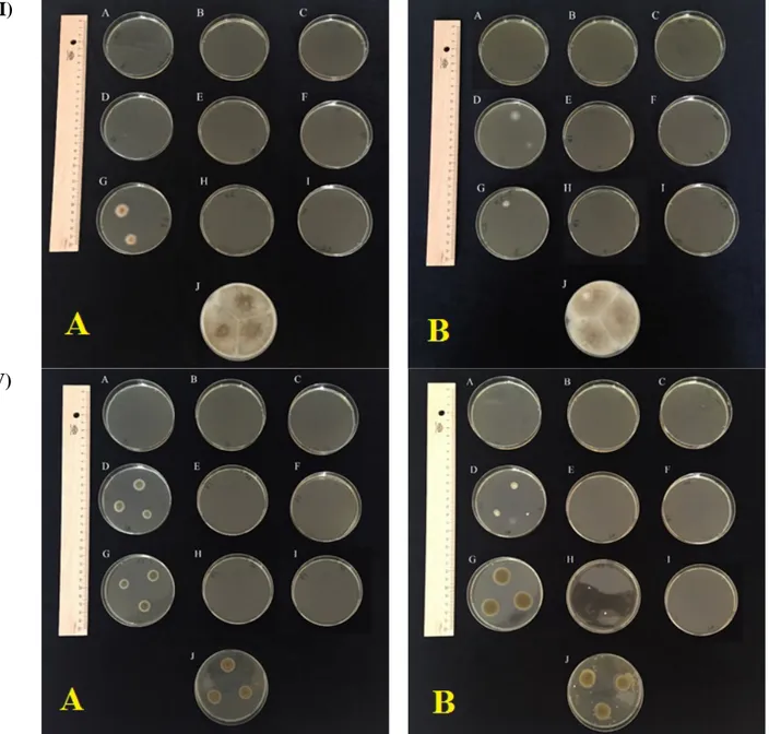
Effect of combinations of salt and temperature on morphological characteristics of microfungi
Tam metin
Şekil
Benzer Belgeler
It was retrospectively evaluated whether there was a difference in the severity and course of stroke in acute ischemic stroke patients diagnosed with type-2 DM and taking
While thinking that compassion will bring negative consequences to him, perceiving compassion as weakness and obedi- ence to others, not wanting to remember past memories of
Whenever some conflicts occurred between the nationalists elements and the government, in which education was also involved directly, two things happened:.. One, there
The adsorbent in the glass tube is called the stationary phase, while the solution containing mixture of the compounds poured into the column for separation is called
Adamović constructs a relatively long chain (as many as five links) of unattested variants in order to connect çağa to çocuk, without allowing for çacuk and –
Keywords: search and retrieval in medical databases, Kendall’s rank correlation coefficient, brain MR image analysis, brain iron deposition, hypointense features, principal
In this study, both state and trait anxiety levels of college administrators were assessed and the administrators were compared based on their anxiety
In fact, even though I exaggerated, I mention “the Anatolian Journal of Cardiology owes its level to devoted efforts of serious referees and frequent trainings of authors provided
