Contents lists available atScienceDirect
Am J Otolaryngol
journal homepage:www.elsevier.com/locate/amjoto
Therapeutic e
ffects of metformin for noise induced hearing loss
Özge Gedik
a, Remzi Do
ğan
b,⁎, Mehmet Ali Babademez
c, Ersin Karata
ş
d, Mehmet
Şerif Aydın
e,
Abdurrahim Koçyi
ğit
f, Mukaddes E
şrefoğlu
g, Orhan Özturan
baBezmialem Vakif University, Faculty of Health Sciences, Audiology Department, Fatih, Istanbul, Turkey bBezmialem Vakif University, Department of Otorhinolaryngology, Fatih, Istanbul, Turkey
cAnkara Yildirim Beyazit University, Department of Otorhinolaryngology, Ankara, Turkey
dGebze Technical University, Faculty of Basic Sciences, Department of Moleculer Biology and Genetics, Kocaeli, Turkey eIstanbul Medipol University, Regenerative and Restorative Medicine Research Center, Beykoz, Istanbul, Turkey fBezmialem Vakif University, Department of Basic Medical Sciences, Medical Biochemistry, Fatih, Istanbul, Turkey gBezmialem Vakif University, Department of Basic Medical Sciences, Histology and Emryology, Fatih, Istanbul, Turkey
A R T I C L E I N F O
Keywords: ABR DNA damage DPOAE
Noise-induced hearing loss Metformin
Rat
A B S T R A C T
Objective: This study aimed to investigate the healing effect of metformin on noise induced hearing loss (NIHL) by measuring audiological, biochemical and histological parameters.
Materials and methods: 32 rats were divided into four groups (Group 1: Noise, Group 2: Noise + Metformin, Grup 3: Metformin, Grup 4: Control). Broadband noise was applied to Group 1 and Group 2 after basal measurements. Measuring audiological (distortion product otoacoustic emission (DPOAE) and Auditory Brainstem Response (ABR)), biochemical (total antioxidant status (TAS), total oxidant status (TOS), oxidative status index (OSI), DNA damage, IL-1 beta, IL-6, TNF alfa, HSF-1 and COX-2) and histological parameters.
Results: Group 2 had significant decreases in ABR thresholds on day 7 and day 14 compared to day 1. DPOAE values of Group 2 on the 7th and 14th days were significantly higher than the post-noise levels. DNA damage, TOS and OSI values of Group 1 were significantly higher than the other groups. The Cox-2 value of Group 1 was higher than all other groups. The HSF-1 value of Group 2 was significantly higher than that of Group 1. In terms of IL-1 Beta, IL-6 and TNF-alpha values, there was no significant difference between groups 2, 3 and 4 and these values were significantly lower than group 1. In histopathological results of our study, no significant difference was found between the groups being exposed to noise and the control group.
Conclusion: This study showed that early period of Metformin treatment has therapeutic effect on NIHL.
1. Introduction
Noise-induced hearing loss (NIHL) is the most common cause of sensorineural hearing loss (SNHL). The World Health Organization re-ported that 360 million people worldwide experienced the problem of hearing loss and that 91% of them were predicted to be adults and 9% predicted to be children [1].
The noise-induced cochlear injury is considered to be a reflection of mechanical and metabolic injury. Mechanical injury is the direct de-struction to the cellular and intracellular structures of the organ of corti. Whereas metabolic injury is caused by the injury in the metabolic process required to maintain homeostasis in the inner ear [2].
Commonly accepted mediator of hair cell injury is ROS and these free radicals cause injury by reacting chemically with cells, DNA, pro-teins, cytosolic molecules, cell surface receptors and numerous
compounds in membrane lipids, and thereby, causing damage and thus affecting many intracellular processes [3].
The prophylactic use of certain antioxidant substances was proven to reduce NIHL. It was also indicated that, in the NIHL model, the co-chlea is mechanically damaged by severe noise, that metabolic stress caused the death of hair cells whereas antioxidant substances protected the cochlea from hair cell loss after exposure to severe noise by redu-cing ROS [4].
Metformin is an oral antihyperglycemic agent used in the treatment of type 2 diabetes. It produces metabolic activity in the liver through the regulation of glucose and fat metabolism. Metformin [5], one of the hypoglycemic drugs used in the world, is also taken into consideration for preventing and treating cancer [6].
Metformin also has beneficial effects on endothelium. Metformin therapy suppresses the production of proinflammatory cytokines. It
https://doi.org/10.1016/j.amjoto.2019.102328
Received 4 October 2019
⁎Corresponding author.
E-mail address:[email protected](R. Doğan).
0196-0709/ © 2019 Elsevier Inc. All rights reserved.
increases the synthesis of endothelial nitric oxide and production of nitric oxide in the endothelial cells. This helps to reduce the rate of atherosclerotic injuries. It regulates mitochondrial homeostasis and thereby, reduces apoptosis by preventing oxidative stress and cell death. It supports the antioxidant system and mitochondrial function complementing each other [5].
The diabetic patients receiving and not receiving metformin were compared and according to this result that the oxidative status of the patients receiving metformin improved, it was suggested that met-formin might have had antioxidant properties or regulated ROS pro-duction in stress situations [7].
Metformin was proven to reduce cell death in cortical neurons and prevent oxidative stress. The in vitro studies indicated that metformin increased survival in the cochlear auditory hair cells rows following the administration of gentamicin by reducing the production of ROS [7]. 2. Material and method
2.1. Animals
This study was initiated after receiving approval from the Local Ethics Board for Animal Experiments. 32 Spraque Dawley female rats were used in the study. Rats were PND 90 and their average weight ranged from 200 to 240 g. All rats' ears were examined by an ENT doctor. If there was ear wax or otitis in the rat, the animal was excluded from the study. All of the rats' outer ear canals and tympanic mem-branes were normal. Rats were divided into 4 groups.
2.2. Groups
Group 1 (n = 8): exposed to only noise
Group 2 (n = 8): exposed to noise and administered metformin Group 3 (n = 8): only administered metformin
Group 4 (n = 8) (control group): not exposed to noise and not ad-ministered metformin
2.3. Noise exposure
After the basal audiological measurements were completed, all of the rats in Group 1 and Group 2 were exposed to wide band noise for 8 h via two loudspeakers in a sound proof room.
2.4. Metformin application
Group 2 and Group 3 rats were given Metformin via the oral gavage, at a dosage of 70 mg/kg/day during thefirst 14 days of noise exposure. 2.5. Audiological evaluation
At the beginning of the study, DPOAE and ABR measurements were performed on all of the rats. Before the DPOAE and ABR measurements the rats were anesthetized using intraperitoneal ketamin hydrochloride (40 mg/kg) and xylazine (5 mg/kg). After the noise exposure session, on the 1st, 7th and the 14th day of DPOAE and ABR measurements were performed on all of the rats. Group 3 and Group 4 rats were not exposed noise exposure because of this 1st day of DPOAE and ABR measure-ments were not performed none of them in Group 3 and Group 4 rats. Intelligent Hearing System (IHS) device was used in DPOAE and ABR measurements. The measurements were done in a sound proof room.
2.6. DPOAE
During the measurements a ratio of 1.2 for f2/f1 was chosen and the intensity level was set at L2 = L1− 10, where L2 = 55 dB and L1 = 65 dB. During the measurements, SNR ratio of 2f1-f2 for the
frequency and DPOAE amplitudes of 6 dB and above were accepted as positive. DPOAE measurements were done at 2834, 4002, 5636, 7988, 11.288, 15.991, 22.608 Hz. SNR ratio was taken in consideration to evaluate DPOAE results.
2.7. ABR
In ABR measurements subcutaneous needle electrodes were used. Passive electrode was put in vertex, active electrodes were put in under both mastoid skin and ground electrode was put in forehead. Neonate probe was used during measurements. All measurements were done in a sound proof room. Click stimulus, 8, 16, 20 and 32 kHz tone burst sti-mulus were used. Alternate polarity, for averaging 400 sweep, for fil-tering 30–3000 Hz were used. 37.1/s was chosen for rate. During the measurements, ipsilateral recording was used. ER 3 insert earphones were used for click and 8 kHz tone burst stimulus and high frequency transducers were used for 16, 20 and 32 kHz tone burst stimulus. Stimulus started at 80 dB SPL level and decreased in increments of 20 dB until the threshold of II. waves was reached.
3. Biochemical evaluation 3.1. Preparation of tissue
Rat cochlea tissues were taken from rat inner ears and homogenized by steel beads in %0,9 NaCl. Than centrifuged (Beckman Coulter, Krefeld, Germany) at 14.000 RPM for 10 min at 4 °C, and thefinal su-pernatant was used as total protein. Protein quantities measured with Bradford method (1976).
3.2. Comet assay
Blood samples were processed within 2 h. Lymphocyte isolation for the comet assay was performed using the Histopaque 1077 (Sigma). 1 ml heparinized blood was carefully layered over 1 ml Histopaque and centrifugal for 35 min at 500 ×g at room temperature. The interface band containing lymphocyte were washed with phosphate buffered saline (PBS) and then collected by 15 min centrifugation at 400 ×g. The comet assay was performed according to Singh et al., with the following modifications. Thus, 10 μl of fresh blood (around 20,000 cells) were mixed with 80μl of 0.7% low-melting agarose in PBS at 37 °C. Subsequently, 80μl of mixture was layered onto a slide pre-coated with thin layers of 1% normal melting point agarose (NMA), and im-mediately covered with a coverslip. Slides were left for 5 min at 4 °C to allow the agarose to solidify. After removing the coverslips, the slides were submersed in freshly prepared cold (4 °C) lysing solution (2.5 M NaCl, 100 mM EDTA-2Na, 10 mM Tris–HCl, pH 10–10.5, 1% Triton X-100 and 10% DMSO added just before use) for at least 1 h. Slides were than immersed in freshly prepared alkaline electrophoresis buffer (0.3 mol/l NaOH, and 1 mmol/l Na2ETDA, pH > 13) at 4 °C for un-winding (40 min) and then electrophoresed (25 V/300 mA, 25 min). All the steps were carried out under minimal illumination. After electro-phoresis, the slides were stained with ethidium bromide (2μg/ml in distilled H2O; 70μl/slide), covered with a coverslip and analyzed using a fluorescence microscope (Nikon). Images of 100 randomly elected cells (50 cells from each of two replicate slides) were analyzed visually from each subject. Each image was classified according to the intensity of thefluorescence in the comet tail and was given a value of either 0, 1, 2, 3 or 4 (form un damaged class 0 to maximally damaged class 4), so that the total score of slide could be between 0 and 400 arbitrary units (AU).
3.3. ELISA
Samples were thawed and enzyme immunoassay was used for the quantitative measurement IL-6, IL-1 Beta, TNF Alpha in rat serum
samples. Biochemical analyzes were performed in a plate reader (Thermo Scientific Multiskan FC, 2011-06, USA) using Enzyme-Linked Immunosorbent Assay (ELISA) using rat serum samples for study. Each assay was performed according to the instructions on its prospectus.
3.4. TAS, TOS, OSI
Totatl antioxidant status (TAS) and Total oxidant status (TOS) levels were determined using commercially available kits (Rel Assay Diagnostics; Mega Tip, Gaziantep, Turkey), and the Oxidative stress index (OSI) was calculated. Serum TAS and TOS levels were determined colourimetrically method by using a Varioskan Flash Multimode Reader; (Thermo Fisher Scientific Inc., Waltham, Massachusetts, USA) and the measurement methods developed by Erel [8,9].
4. Western blot
Western blots were used to analyze the expression HSF1 and Cox-2 and ß-Actin proteins. Cellular proteins were prepared from cochlea cells in inner ear. The total sample volume was 25μL at a concentration of 2μg/μL. All cellular proteins were electrophoresed on 4–12% SDS-PAGE acrylamide gels, transferred onto polyvinylidenefluoride mem-branes (PVDF), and incubated for 1 h in tris-buffered saline and 5% Tween (TBS-T) containing 5% skim milk (Sigma). Membranes were then incubated overnight at 4 °C with primary antibodies against HSF1 (Santa Cruz, 1:1000) and Cox-2 (Santa Cruz 1:1000), β-Actin (Santa Cruz 1:1000) washed in TBS-T, and incubated for 1 h at room tem-perature with corresponding HRP conjugated anti-rabbit antibodies (Santa Cruz Biotechnology, 1:5000). The membranes were developed with the Western Blotting Luminol Reagent (Santa Cruz Biotechnology, Santa Cruz, CA, USA) and each membrane wasfilmed with a chemi-luminescent imaging system (Fusion Fx7; Vilber Lourmat), and the blots were quantified using Image J (Glyko, Novato, CA, USA) software package.
5. Histopathological Assesment
For light microscopic investigations, samples were placed in 10% formaldehyde, dehydrated in ascending alcohol series (70%, 90%, 96% and 100%), cleared in xylene and embedded in paraffin. Tissue sections (5μm) were stained with hematoxylin and eosin (H&E) and examined with a light microscope (Nikon Eclipse i5, Tokyo, Japan) coupled with a camera (Nikon, DS-Fi1c).
5.1. Statistical analysis
The statistical analysis was carried out using Statistical Package for the Social Sciences 13.0 version package (SPSS Inc., Chicago, Illinois, USA) in the windows program. All quantitative variables were calcu-lated using the central location (such as mean and median) and dis-tribution measures (such as standard deviation (SD)). The normality of the distribution was checked using the Kolmogorov– Smirnov tests.
For comparing the DPOAE and ABR values within the groups themselves, one-way analysis of ANOVA was used in repeated mea-surements (p < .05 was considered significant). The Tukey HSD test was used for the post-hoc analysis and Bonferroni correction was per-formed (p < .008 was considered significant).
ANOVA was used for the comparison of intergroup biochemical and histopathological data (p < .05 were considered significant). For the post-hoc analysis, the Tukey HSD test was used and Bonferroni cor-rection was performed (p < .008 was considered significant). 6. Results
6.1. Audiological results 6.1.1. DPOAE
A statistically significant difference between basal and 1th, 7th and 14th days of noise exposure was found in DPOAE results in Group 1 (p < .008). A statistically significant difference between basal and 1th day of noise exposure and between 1th and 7th days of noise exposure
were found in DPOAE results (p < .008). A statistically significant difference between 7th and 14th days of noise exposure was not found in DPOAE results in Group 2. (p > .008). A statistically significant difference between basal and 7th and 14th days of implementation of Metformin was not found in DPOAE results in Group 3 (p > .05). A statistically significant difference between basal, 7th and 14th days was not found in DPOAE results in Group 4 (p > .05) (Fig. 1).
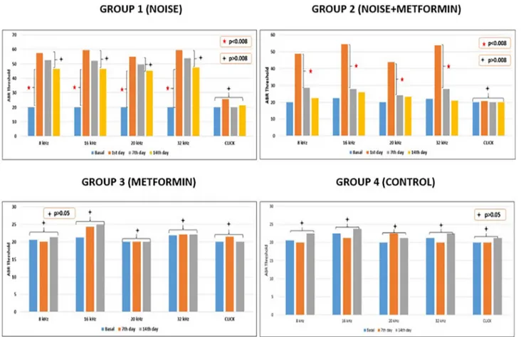
6.1.2. ABR
A statistically significant difference between basal and 1th, 7th and 14th days of noise exposure at 8, 16, 20 and 32 kHz tone burst stimulus ABR thresholds was found in Group 1 (p < .008). A statistically sig-nificant difference between basal and 1th days of noise exposure and between 1th day and 7th and 14th days of noise exposure at 8, 16, 20 and 32 kHz tone burst stimulus ABR thresholds was found in Group 2 (p < .008). A statistically significant difference between basal and 7th and 14th days of metformin implementation at 8, 16, 20 and 32 kHz tone burst stimulus ABR thresholds was not found in Group 3 (p > .05). A statistically significant difference between basal, 7th and 14th days at 8, 16, 20 and 32 kHz tone burst stimulus ABR thresholds was not found in Group 4 (p > .05). A statistically significant differ-ence between basal and 1th, 7th and 14th days of noise exposure at click stimulus ABR thresholds was not found in any groups (p > 0,008) (Fig. 2).
In the comparison of ABR thresholds between groups; There was no significant difference between basal measurements at 8, 16, 20,32 kHz (p > .05). Noise was applied only to group 1 and group 2. There was no significant difference between group 1 and group 2 between ABR thresholds after noise application (p > .05). On the 7th day ABR threshold (8,16,20,32 kHz), the ABR thresholds of Group 1 were sig-nificantly higher than the other groups (p < .008). There was no sig-nificant difference between ABR thresholds of group 2 and ABR thresholds of groups 3 and 4 (p > .05). On the 14th day, ABR
thresholds of Group 1 were significantly higher than those of the other groups (p < .008). There was no significant difference between ABR thresholds of group 2 and ABR thresholds of groups 3 and 4 (p > .05). There was no significant difference between the groups in the basal, 7th and 14th day evaluations of ABR with click stimulus (p > .05). 6.2. Biochemical results
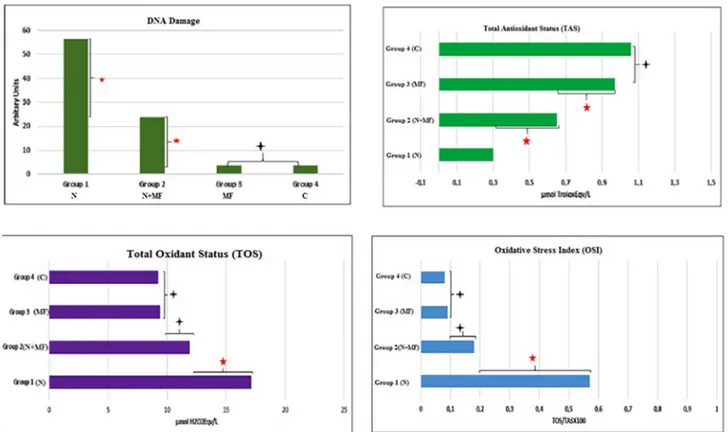
Group 1 had the biggest DNA damage of the groups. Group 2 had bigger DNA damage than Group 3 and 4 (p < .008). Group 1 had the lowest TAS level of the groups. Group 2 had higher TAS level than Group 1. Group 3 had higher TAS level than Group 2 (p < .008). A statistically significant difference at TAS levels and DNA damage be-tween Group 3 and 4 was not found (p > .008). Group 1 had the highest TOS level of the groups (p < .008). Group 1 had the highest OSI level of the groups (p < .008). A statistically significant difference between Group 2, 3 and 4 at OSI and TOS levels was not found (p > .008) (Fig. 3).
Group 1 had the highest Cox-2 level of the groups (p < .008). Group 2 had higher Cox-2 level than Group 3 and 4 (p < .008). A statistically significant difference between Group 3 and 4 at Cox-2 le-vels was not found (p > .008). Group 1 had the lowest HSF 1 level of the groups. A statistically significant difference between Group 2 and 3 was not found and this level was higher than the HSF 1 level of Group 1. Group 4 had the highest HSF 1 level (p < .008) (Fig. 4).
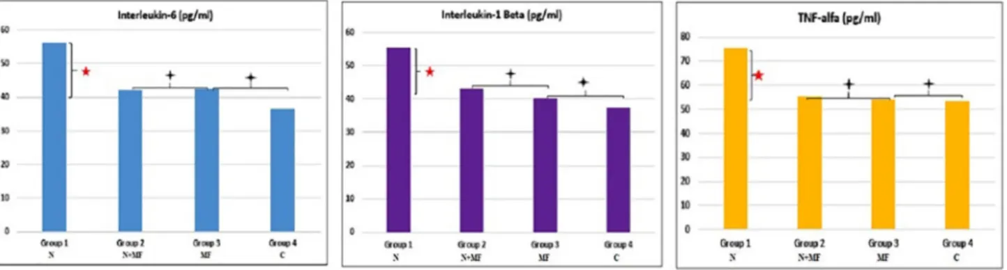
Group 1 had the highest IL-6, IL 1 Beta and TNF alpha levels of the groups (p < .008). A statistically significant difference between Group 2, 3 and 4 at IL-6, IL 1 Beta and TNF alpha levels was not found (p > .008) (Fig. 5).
6.3. Histological results
There was no difference between control group and the other groups
Fig. 2. Alterations in the ABR values of DPOAEs results in over time of all groups.
on stria vascularis, spiral ganglion and organ of corti via the light mi-croscopic examination.
There was no apoptotic cell in stria vascularis, spiral ganglion and organ of corti in none of the groups (Fig. 6).
7. Discussion
The literature reported that the hair cell damage in the organ of corti started as a small lesion, that the lesion spread during the exposure to the noise and continued to progress also after the cessation of ex-posure. Also, it was noted that the lesion started in the characteristic frequency part corresponding to the dominant frequency of the noise, the damage at onset was composed of one or several lesions and that it spread to the apical and basal part of the cochlea [10].
In the study by Lu et al. (2014), a substantial improvement was
always observed in the DPOAE levels at every time point in the animals of the antioxidant treatment group compared to the control group, this effect was striking especially on the seventh and twenty-first days. The effect of antioxidant treatment was demonstrated by a reduction in the number of loss of both outer and inner hair cells in the basal part of the cochlea after 21 days of exposure to noise [11]. In our study, a statis-tically significant decrease was observed in the DPOAE SNR values in the DPOAE measurement performed on the first day after noise ex-posure compared to the baseline measurements in the group which received metformin at the dose of 70 mg/kg/day for 14 days after 8 h of exposure to 100 dB SPL (Group 2). Whereas the measurements made on the seventh and fourteenth days after exposure to noise revealed no difference in the DPOAE SNR values compared to the baseline mea-surement values. A statistically significant decrease was noted in the DPOAE SNR values compared to the baseline measurements made on
Fig. 3. DNA damage, TAS, TOS and OSI values of all groups.
the first, seventh, fourteenth days after noise exposure in the group which was exposed to noise only (Group 1). These results indicate that metformin, an antioxidant derivative, provided an improvement in hearing thresholds.
Animal experimental studies showed that exposure to severe noise caused structural changes in the outer hair cells of the cochlea and involved the inner and outer hair cells by progressingfirstly to the first row, then to the second row and third row [12].
In our study, no significant difference was found in the DPOAE SNR values compared to the baseline values in Group 3 (metformin) and Group 4 (control group) in the 14-day follow-up period. According to this, no change was detected in the DPOAE SNR values secondary to metformin in the group receiving metformin only. This suggests that metformin has no negative effect on the outer hair cell function, which can be assessed by the DPOAE test.
Similar to our study, 8, 16 and 32 kHz tone burst stimuli were used and an increase was observed in all stimulus types in the ABR thresh-olds after noise exposure in the study by Xiong et al. [13]. Unlike this study, there was no significant difference in the threshold values in the ABR measurements made with the click stimulus on thefirst, seventh and fourteenth days compared to the baseline measurement threshold value in our study. In the literature, the area most severely affected by
noise was reported to be thefirst turn [14–16]. Whereas Strose et al. analyzed the number of damaged hair cells in each cochlear turn and found that the most damaged part of the cochlea secondary to noise was the third turn, that the second turn was moderately damaged, and that thefirst turn (basal) was damaged at the least. When the outer hair cell damage in each row was observed, they stated that the third row was mostly affected by noise trauma and that the number of damaged ste-reocilia cells in thefirst and second row were almost equa [17].
In our study, a statistically significant difference was found in the ABR threshold values of 8, 16, 20 and 32 kHz made on thefirst day of the 14-day follow-up period in the rats of Group 1 and Group 2 com-pared to the basal measurement results. Whereas in the ABR mea-surement made with the click stimulus, there was no significant dif-ference in the threshold values measured on the first, seventh and fourteenth days in both groups compared to the baseline measurements of threshold values. No significant difference was detected in the ABR threshold values obtained at 8, 16, 20 and 32 kHz on the seventh, fourteenth day in Group 2 compared to the baseline measurements of ABR threshold values. Whereas in Group 1, no significant difference was noted in the ABR threshold values obtained on the seventh and fourteenth days compared to those obtained on thefirst day. When compared with Group 1, metformin was observed to provide a
Fig. 5. IL-6, IL-1 Beta, TNF alfa values of all groups.
Fig. 6. Histological images of all groups.
significant improvement in noise-induced hearing thresholds obtained on the fourteenth day after metformin treatment.
It was reported in the literature that hair cell damage spread to both apical and basal parts of the organ of corti but the basal spread was more prominent than the apical spread. It was also stated that the spread of active cell death pattern spread from the junction point of the first and second turns to the basal part of the cochlea within the first two days following the noise exposure at 110 dB SPL, that it showed structural and metabolic differences between the apical and basal parts of the cochlea as the damage spread towards the basal part of the organ of corti, and, in this respect, the basal part showed a lesser antioxidant capacity and, as a result, this region suffered more from the oxidative stress [10].
No significant difference was found in terms of the ABR threshold values in the ABR measurements made at 8, 16, 20 and 32 kHz with tone burst and click stimuli in Group 3 (metformin) and Group 4 (control group) during the 14-day follow-up period compared to the baseline values. According to this, no change was detected in the ABR threshold values secondary to metformin in the group receiving met-formin only. These results indicate that metmet-formin has no negative impact on the auditory function.
In all tested frequencies on the rats, amongfive waves forming the ABR, wave II had the highest amplitude, followed by the formation of I, IV and V waves, respectively and these waves had similar amplitudes. Wave III was reported to have the smallest amplitude [18]. In our study, consistently with the literature, the most apparent wave was observed to be wave II in all rats and at all frequencies.
According to the literature, the number of ROS positive spiral ganglion cells, apoptotic spiral ganglion cells and outer hair cells cov-ered with stereocilia and the changes of ABR threshold were positively correlated [13]. Consistently with the literature, the DNA damage value was found to be higher in Group 1 and Group 2 DNA compared to those of Group 3 and Group 4 in our study.
It was reported in the literature that metformin rapidly affected the blood-brain barrier and accumulated in different manners and thus metformin helped to reduce vascular inflammation. Metformin therapy was indicated to provide a functional benefit without significantly al-tering the volume of the big infarct area or blood glucose level at the beginning. Metformin therapy was stated to improve phagocytosis in the dead tissue by helping to improve recovery and increasing the ratio of surviving cells and contributing to a decrease in the ratio of in-tracranial pressure [19].
Generally, the thresholds were observed to worsen gradually and morphological damage increased after noise exposure. Although these changes were present for the next 30 days after exposure to noise, it is an important period in thefirst 10-day acute acoustic trauma treatment after exposure to noise. 10 days after exposure to noise, the ROS /RNS formation reaches the maximum level and permanent threshold change and loss of outer hair cells reach the plateau. It is possible to observe the benefits of antioxidant treatment for 10 days due to the increase in free radicals in the next 7–10 days after exposure to noise [20]. Consistently with the literature, the DNA damage in Group 1 was found to be higher compared to all other groups of our study. The DNA damage value in Group 2 was found to be significantly higher than Group 3 and 4. ROS initiating the apoptosis of spiral ganglion cells was proven to exist in the cochlea exposed to severe noise. These results suggest that metformin treatment may be an antioxidant with healing feature for noise-induced hearing loss since the DNA damage was lesser in the group receiving metformin therapy for 14 days compared to the group exposed to noise only as well as the DPOAE SNR amplitudes and ABR thresholds were improved.
In response to exposure to noise, it was stated that ROS production could reduce bloodflow and that the vasodilator agents reducing va-soconstriction improved NIHL and that antioxidants converted ROS into less dangerous forms [21]. In our study, the TAS values in Group 2 were found to be significantly higher than Group 1. Oxidative stress factors
were reported to cause DNA damage leading to hearing loss, degrada-tion of lipids and proteins, and death of triggering cells and thus, hearing loss occurred [22]. In our study, the OSI value of Group 1 was found to be significantly higher than the other groups. These results indicated that metformin administered at the dose of 70 mg/kg/day via oral gavage for 14 days improved the antioxidant capacity of the tis-sues. Auditory function impairment is considered to occur with the increase in the oxidative stress parameters in the group exposed to noise only but did not receive metformin.
In our study, the Cox-2 value of Group 1 was found to be sig-nificantly higher than the other groups. The Cox-2 values of Group 2 were found to be significantly higher than Group 3 and 4. However, no significant difference was found between Group 3 and 4 in terms of the Cox-2 value.
In the literature, it was reported that heat shock transcription factor 1 (HSF 1) is an important transcription factor controlling the inducible stress response and that it protected the cells and tissues from major cellular and environmental stresses. Also, it was noted that this re-sponse was often named as heat shock rere-sponse which protected hu-mans from bacteria during development, that stress factors activated HSF 1, relocated the nuclei, and bound the heat shock elements in the genes for the heat shock proteins. Stress factors of both heat and noise were stated to activate the heat shock response in the cochlea and protect it from noise trauma [23]. Consistent with the literature, the HSF 1 value of Group 1 was found to be significantly higher than all other groups in our study. There was no significant difference between the HSF 1 values of Group 2 and 3 and these values were found to be significantly higher than Group 1. The HSF 1 value of Group 4 was found to be significantly higher than all other groups. According to these results obtained, it was concluded that the fact that HSF 1 was found to be higher in the group receiving metformin at the dose of 70 mg/kg/day for 14 days than the group exposed to noise only pro-vided the increase in HSF 1 which is known to play a protective role against cochlear stressors.
The literature indicated that stress-activated the endocrine, neu-ronal and immunological systems and caused the release of certain active biological compounds such as hormones, neurotransmitters, and cytokines. Also, stress was reported to stimulate the production of proinflammatory cytokines called as tumor necrosis factor-α (TNF-α), interleukin 1β (IL-1-β) and interleukin 6 (IL-6) [24]. In our study, the values of IL-6, IL-1β, and TNF-α in Group 1 were found to be sig-nificantly higher than all other groups. No significant difference was found in Group 2, 3 and 4 in terms of these values. The higher levels of cytokines obtained in the group not receiving metformin were con-sidered to impair auditory function in accordance with the DPOAE and ABR results obtained. Whereas the lower levels of cytokines in the group receiving metformin were considered to originate from the sup-pression of cytokine production by metformin which is considered to have antioxidant feature.
Sekiya et al. (2012) reported that after 9 weeks of exposure to noise in rats exposed to 137 dB of noise at 4 kHz for 1 min, the outer hair cells completely disappeared while the inner hair cells were proven to be partially preserved in the apical part and to be completely lost in the basal and middle part [25]. In our study, no difference was observed in the cells of stria vascularis, spiral ganglion and organ of corti at the light microscopic level morphologically in the groups exposed to Noise, Metformin and Noise+Metformin compared to the control group. The examination performed with the caspase-3 immunohistochemical staining method in an attempt to detect apoptotic cells revealed no apoptotic cell within the stria vascularis, spiral ganglion, and organ of corti. Chen et al. stated that pathological changes in the cochlea were not significant under the light microscope [26]. We suggest that mor-phological differences undetected under the light microscope in the morphological analysis can be detected by electron microscopy.
In the literature, it was reported that the apoptosis and necrosis of hair cells were important factors in the hearing loss caused by exposure
to noise, that apoptosis started on thefirst day after exposure to noise and that the increase in the ABR thresholds and decrease in the DPOAE amplitudes were the consequences of the apoptosis after exposure to noise [27].
Xiong et al. reported that they did not find immunoreactivity for Cas-3 in the spiral ganglion in the groups that were not exposed to severe noise and that they observed a strong immunoreactivity for Cas-3 in the spiral ganglions of the groups exposed to severe noise. They proved that they could not detect a visible morphologic change in the hair cells of the rats of the groups not exposed to noise in the electron microscope and that normal appearing intact hair cell rows with ste-reocilia were observed in every turn of the cochlea. However, they noted that a substantial outer hair cell with stereocilia was observed in thefirst and second turns of the cochlea in the group exposed to noise, that a great portion of the damage was observed in thefirst and second of rows of the hair cells whereas the inner hair cells preserved their morphological integrity throughout the cochlea [13].
It was reported in the literature that the therapeutic effects of an-tioxidant drugs depended on the timing and duration of treatment and that the therapeutic effect window appeared within 1–4 h after ex-posure to noise. Although the target and effect of each antioxidant agent treat and prevent the acute acoustic trauma with different injury mechanism, it was reported that the efficacy could be maximum in combination with other antioxidant agents [20].
8. Conclusion
Metformin, known to have an antioxidant feature, has a healing effect for the hearing loss occurring after exposure to noise.
Funding source,financial disclosures None.
Declaration of competing interest None.
References
[1] Taneja MK. Noise-induced hearing loss. Indian J Otol 2014;20:151–5.
[2] Suzuki M, Yamasoba T, Ishibashi T, Miller JM, Kaga K. Effect of noise exposure on blood-labyrinth barrier in guinea pigs. Hear Res 2002;164:12–8.
[3] Kurabi A, Keithley EM, Housley GD, Ryan AF, Wong AC. Cellular mechanisms of noise-induced hearing loss. Hear Res 2016;349:129–37.
[4] Tziridis K, Korn S, Ahlf S, Schulze H. Protective effects of ginkgo biloba extract EGb 761 against noise trauma-induced hearing loss and tinnitus development. Neural Plast 2014;2014:427298.
[5] Janjua A, Waheed A, Bakhtiar S. Protective effect of metformin against gentamicin
induced nephrotoxicity in rabbits. Pak J Pharm Sci 2014;27:1863–72.
[6] Trombini AB, Franco CC, Mİranda RA, de Oliveira JC, Barella LF, Prates KV, et al. Early treatment with metformin induces resistance against tumor growth in adult rats. Cancer Biol Ther 2015;16:958–64.
[7] Mujica-Mota MA. Otoprotection of metformin in radiation-induced sensorineural hearing loss. 150(5). 2014. p. 859–65.
[8] Erel O. A novel automated direct measurement method for total antioxidant capa-city using a new generation, more stable ABTS radical cation. Clin Biochem 2004;37:277–85.
[9] Erel O. A new automated colorimetric method for measuring total oxidant status. Clin Biochem 2005;38:1103–11.
[10] Hu B. Noise-induced structural damage to the cochlea. In: Le Prell CG, Henderson D, Fay RR, Popper AN, editors. Noise-induced hearing loss. London: Springer; 2012. p. 57–86.
[11] Lu J, Li W, Du X, Ewert DL, West MB, Steawart C, et al. Antioxidants reduce cellular and functional changes induced by intense noise in the inner ear and cochlear nucleus JARO. J Assoc Res Otolaryngol 2014;15:353–72.
[12] Colombari GC, Rossato M, Feres O, Hyppolito MA. Effects of hyperbaric oxygen treatment on auditory hair cells after acute noise damage. Eur Arch Oto-Rhino-Laryngology 2011;268:49–56.
[13] Xiong M, He Q, Lai H, Wang J. Oxidative stress in spiral ganglion cells of pigmented and albino guinea pigs exposed to impulse noise. Acta Otolaryngol
2011;131:914–20.
[14] White DR, Boettcher FA, Miles LR, Gratton MA. Effectiveness of intermittent and continuous acoustic stimulation in preventing noise-induced hearing and hair cell loss. J Acoust Soc Am 1998;103:1566–72.
[15] Fernandez EA, Ohlemiller KK, Gagnon PM, Clark WW. Protection against noise-induced hearing loss in young CBA/J mice by low-dose kanamycin. JARO - J Assoc Res Otolaryngol 2010;11:235–44.
[16] Clark WW, Bohne BA, Boettcher FA. Effect of periodic rest on hearing loss and cochlear damage following exposure to noise. J Acoust Soc Am 1987;82:1253–64. [17] Strose A, Colombari GC, Rossato M, Hyppolito MA, de Oliveira JA. Gentamicin
conditioning confers auditory protection against noise trauma. Eur Arch Oto-Rhino-Laryngology 2014;271:2641–8.
[18] Alvarado JC, Fuentes-Santamaria V, Jareno-Flores T, Blanco JL, Juiz JM. Normal variations in the morphology of auditory brainstem response (ABR) waweforms: a study in wistar rats. Neuroscience 2012;73:302–11.
[19] Venna VR, Li J, Hammond MD, Mancini NS, Mccullough LD. Chronic metformin treatment improves post-stroke angiogenesis and recovery after experimental stroke. Eur J Neurosci 2014;39:2129–38.
[20] Choi CH, Chen K, Du X, Floyd RA, Kopke RD. Effects of delayed and extended antioxidant treatment on acute acoustic trauma. Free Radic Res 2011;45:1162–72. [21] Sjostrand AP, Dogan R, Kocyigit A, Karatas E, Budak BB, Ozturan O. Therapeutic
efficacy of Ginkgo biloba for early-period noise-induced hearing loss: an experi-mental animal study. Am J Otolaryngol Head Neck Med Surg 2016;37:2–10. [22] Yuan BC, Su FM, Wu WT, Liu WS, Chiu KH. A predictive model of the association
between gene polymorphism and the risk of noise-induced hearing loss caused by gunfire noise. J Chinese Med Assoc 2012;75:36–9.
[23] Lomax MI, Gong TW. Genes that influence susceptibility to noise-induced hearing
loss. In: Le Prell CG, Henderson D, Fay RR, Popper AN, editors. Noise-induced hearing loss. London: Springer; 2012. p. 179–204.
[24] Mazurek B, Haupt H, Joachim R, Klapp BF, Stöver T, Szczepek AJ. Stress induces transient auditory hypersensitivity in rats. Hear Res 2010;259:55–63.
[25] Sekiya T, Viberg A, Kojima K, Sakamoto T, Nakagawa T, Ito J, et al. Trauma-specific insults to the cochlear nucleus in the rat. J Neurosci Res 2012;90:1924–31. [26] Chen W, Wang J, Chen J, Chen J, Chen Z. Relationship between changes in the
cochlear bloodflow and disorder of hearing function induced by blast injury in guinea pigs. Int J Clin Exp Pathol 2013;6:375–84.
[27] Aksoy F, Dogan R, Yenigun A, Veyseller B, Ozturan O, Ozturk B. Thymoquinone treatment for inner-ear acoustic trauma in rats. J Laryngol Otol 2015;129:38–45.