Corresponding author: Selin TURAL EMON E-mail: [email protected]
Original Investigation
Published Online: 27.07.2016is usually young (16-30 years) (18,19,21)but their survival is expected to be long, requiring a prevention system to avoid undesirable life quality decrease and high healthcare costs due to secondary injury following primary SCI. At the moment of trauma, inevitable primary injury occurs whereas secondary injury develops over time involving greater tissue damage (14). The factors influencing secondary damage are local inflammation, excitatory toxicity, and significant oxidative stress. This damage occurs after the primary injury within days
█
INTRODUCTION
S
pinal cord injury (SCI) can occur as a result of severe neural trauma that sometimes causes a chronic functional deformity. There are approximately 330,000 people living with SCI in the European Union, with 11,000 new cases each year (19,21). In the United States, there are 11,000 new SCI cases each year adding to present disabled SCI injuries of 450,000 people (19,21). The patient populationSelin TuRAl EMON
1, Serap uSlu
2, Elif IlGAZ AYDINlAR
3, Arzu IRBAN
4, umit INCE
5, Metin ORAKDOGEN
1,
Guldal GulEC SuYEN
61Haydarpasa Numune Training and Research Hospital, Department of Neurosurgery, Istanbul, Turkey 2Medeniyet University, School of Medicine, Department of Histology and Embryology, Istanbul, Turkey 3Acibadem University, School of Medicine, Department of Neurology, Istanbul, Turkey
4Medipol University, School of Medicine, Department of Anesthesiology and Reanimation, Istanbul, Turkey 5Acibadem University, School of Medicine, Department of Pathology, Istanbul, Turkey
6Acibadem University, School of Medicine, Department of Physiology, Istanbul, Turkey
Effects of Ozone on Spinal Cord Recovery via the Wnt/
β-Catenin Pathway Following Spinal Cord Injury in Rats
ABSTRACT
AIm: At the cellular level, spinal cord injury (SCI) provokes an inflammatory response that generates substantial secondary damage
within the spinal cord but may also contribute to its repair. Besides intracellular antioxydant increase after exactly estimated oxidative stress; oxygen formation and transport is also advanced by ozone. The Wnt family of proteins contributes to the development of the nervous system, influencing cell proliferation. In the present study we evaluated the effect of ozone on spinal cord injury in rats.
mATERIAl and mEThODS: Twenty-one male Sprague–Dawley rats were used. The rats were randomly allocated into three groups
(control, trauma and trauma+ozone). SCI was inflicted using Allen’s spinal cord trauma method. The study was performed to determine the effects of ozone therapy on rats with SCI in terms of locomotor strength clinically and neuronal injury, white matter cavitation, edema, number of blood vessels, and expression of β-catenin immunohistochemically.
RESUlTS: Comparison of the locomotor strength scores revealed a significant improvement on day 7 in trauma+ozone group.
The groups were compared with regard to edema, neuronal injury, and white matter cavitation. Average β-catenin levels were significantly different between the control group (68.11 ± 0.43), trauma+ozone group (37.96 ± 2.16), and trauma group (25.46 ± 1.07) (F = 1677.74, df = 2, p < 0.0005).
CONClUSION: The results of this study indicated that ozone therapy accelerates the healing process, increases vascularity, and
reduces neuronal damage in rodents, suggesting that ozone therapy may be an adjuvant treatment in patients with SCI.
and weeks (12).The reaction of inflammation in the acute injury can be considered as a host defense mechanism that acts as a barrier to remove pathogens and tissue debris. Inflammatory cells travel towards the wound site to trigger wound healing to end with recovery. However, this can result in an excess of undesirable toxic molecules produced by inflammatory cells during the acute and chronic phases of spinal cord injury causing unexpected tissue damage (4).
In the early stages of neural development, the Wnt family of proteins play an important role in influencing cell proliferation and patterning, cell polarity and motility, axonal guidance, neuronal survival and connectivity, and cell–cell adhesion (7,8,17).Three Wnt signaling pathways have been characterized: the canonical Wnt pathway, the non-canonical planar cell polarity pathway, and the non-canonical Wnt/ calcium pathway. The canonical Wnt pathway leads to regulation of gene transcription, the non-canonical planar cell polarity pathway regulates the cytoskeleton that is responsible for the shape of the cell, and the non-canonical Wnt/calcium pathway regulates calcium levels within the cell (7).These categories differ in that the canonical pathway involves the protein β-catenin, while the non-canonical pathway operates independently. Numerous supportive literature points to the role of Wnt signaling pathways in the adult central nervous system (CNS) homeostasis and diseases (eg.SCI) (8).β-catenin is a dual-function protein, which regulates the coordination of cell–cell adhesion and gene transcription; it is a subunit of the cadherin protein complex and acts as an intracellular signal transducer in the Wnt signaling pathway.
Ozone is a chemical compound consisting of three oxygen atoms. Medical ozone is obtained via an ozone generator in which oxygen (O2) molecules obtained from a 100% oxygen source are divided into two oxygen atoms after passing through an electric field and temporarily recombined in groups of three oxygen atoms. Then, ozone is obtained as a gas. Systemic application of ozone leads to delivery of super-enriched oxygen at a cellular level and optimizes cell function. Many biological effects have been attributed to ozone, such as increased glycolysis, effects on red blood cells, effects on rheology, bactericidal, fungicidal, and virustatic, immunomodulatory actions, and analgesic and anti-inflammatory effects. Application of low-dose ozone inhibits prostaglandin synthesis, release of bradykinin and algogenic substances, and proteinase secretion from macrophages and polymorphonuclear leukocytes (6).
The present study was performed to determine the effects of ozone therapy on rats with SCI in terms of locomotor strength clinically and neuronal injury, white matter cavitation, edema, number of blood vessels, and expression of β-catenin immunohistochemically.
█
mATERIAl and mEThODS
Experimental Design
The experimental study was carried out according to Animal Care and Use Committee at Marmara University School of Medicine, Istanbul, Turkey approval protocol. Twenty-one
male Sprague–Dawley rats were used, each weighing 250–300 g and housed in a separate cage at the Experimental Animal Implementation and Research Center (DEHAMER) of Marmara University. The animals were provided a standard rodent chow diet and water ad libitum and kept at a constant temperature (22°C) under a 12:12 h light:dark cycle.
In all of the surgical groups, the animals were anesthetized by intraperitoneal injection of ketamine hydrochloride (90 mg/kg, Ketalar; Pfizer, Istanbul, Turkey) and xylazine hydrochloride (10 mg/kg, Rompun 2%; Bayer United German Pharmaceutical Factories, Istanbul, Turkey) and then placed on an operating board in the prone position. During surgical preparation, the dorsal hair of each rat was closely shaved using an electric razor, and the surgical field was cleaned and disinfected with 10% povidone–iodine solution and draped with sterile towels. A posterior midline incision was made between the 5th and 10th thoracic vertebrae. Following paravertebral muscle dissection, laminectomy of the 9th thoracic vertebra was carried out. The rats were randomly separated into three groups (seven rats per group) using sealed envelopes selected by a physician. In Group C (control group; n=7): only T9 laminectomy was performed. Group T (trauma group; n=7): SCI was inflicted using Allen’s spinal cord trauma method (2). Group O (trauma + ozone group; n=7): SCI was inflicted using Allen’s spinal cord trauma method, and ozone was applied. In all groups, the wound sites were closed using non-absorbable sutures. After surgery, medical air in Groups C, and trauma or 0.7 mg/kg ozone in Group O was insufflated rectally once a day for 5 consecutive days (Medozon Compact; Herrmann Apparatebau GmbH, Kleinwallstadt, Germany). Routine bladder emptying was forced manually until spontaneous voiding was reached out. Ketorolac was given to all rats at a dose of 50 mg/kg intraperitoneally for postoperative analgesia for 5 days.
Allen’s Spinal Cord Trauma method
The instrument that had a 10-cm guide tube and inner rod (stainless steel, 5 g) was placed perpendicular to the spinal cord. The trauma produced a dorsal surface spinal cord injury by an impact effect of 50 g/cm2 (2).
locomotor Strength
The locomotor strength of the animals was rated using the Basso, Beattie, Bresnahan (BBB) scale (21 points: ranging from no movement to full range) and recorded by a neurosurgeon blinded to the groups on days 0, 3, 7, 14, 21, 28, and 35 following SCI (3).
Tissue Sampling
On day 35, all rats were perfused with intracardiac fixative solution (10% neutral buffered formalin), and only the injury site spinal cord tissue samples were then dissected and fixed in 10% formalin solution for 24 hours before tissue processing. After embedding in paraffin, spinal cord tissue sections 4 μm thick were stained with hematoxylin-eosin (H&E) to assess spinal cord morphology and the number of blood vessels. The spinal cord was evaluated for neuronal injury (3 points: mild
to severe), white matter cavitation/cyst formation (3 points: absent, few, > 5 in a single field), blood vessel ingrowth (5 points: absent, few, > 3), edema (+/–), demyelinization (3 points: mild to severe), inflammation (+/–), and hemorrhage (+/–) by a histologist blinded to the groups.
histomorphometric Analysis
Five serial sections from each sample and a minimum of five adjacent fields from each section were quantified using a ×40 magnification. The number of blood vessels was evaluated histomorphometrically using a semi-automatic image analysis system (University of Texas Health Science Center at San Antonio (UTHSCSA) image tool for Windows version 1.28) (1).
Immunohistochemical Analysis
Spinal cord sections 5 μm thick were cut from paraffin blocks. The tissue blocks were selected carefully after histological assessment of sections stained with H&E (hematoxylin acc. to Gill III, cat. no. 1.05174; eosin solution 0.5% alcoholic, cat. no. 1.02439; Merck, Darmstadt, Germany). Following deparaffinization and incubation of sections with primary anti-β-catenin antibody (1:100, C2206; Sigma-Aldrich, St. Louis, MO, USA), immunohistochemistry was performed using the streptavidin–biotin method in a fully automated system (Benchmark Ultra; Ventana, Tucson, AZ, USA). In the negative controls, the primary antibodies were omitted. All sections were mounted with mounting medium (Shandon EZ-Mount; Thermo Fisher Scientific, Waltham, MA, USA), and immunoreactive cells were evaluated using a Zeiss Axiovert A1 microscope and imaged using the AxioCam ICc5 (Thermo Fisher Scientific, Waltham, MA, USA). The presence of a brown precipitate indicated a positive reaction with the primary antibodies. To determine the immunoreactivity of the sections, a series of semi-quantitative analyses were performed. The H-score was calculated using the following equation: H-score = ∑Pi (i + 1), where i is the labeling intensity of value 1, 2, or 3, (weak, moderate, or strong, respectively) and Pi is the percentage of labeled cells for each intensity, varying from 0 to 100% (22).
Statistical Analysis
The Mann–Whitney U test was used to compare inconsistencies with the normal distribution of the data. Averages of the groups
were compared using one-way ANOVA. Scheffé’s test was used for post hoc analysis. In all analyses, p<0.05 was taken to indicate statistical significance. Bonferroni’s correction was used when necessary. SPSS 16 software was used for all analyses (SPSS, Chicago, IL, USA). All data were evaluated in a blinded fashion and expressed as means ± SD.
█
RESUlTS
Comparison of the BBB scores revealed a significant improvement on day 7 in Group O (U=5.5, z=–2.52, p=0.012). However, after the 35-day follow-up period, comparison of the BBB scores showed no significant differences among the groups (U=24.5, z=0.00, p=1.00).
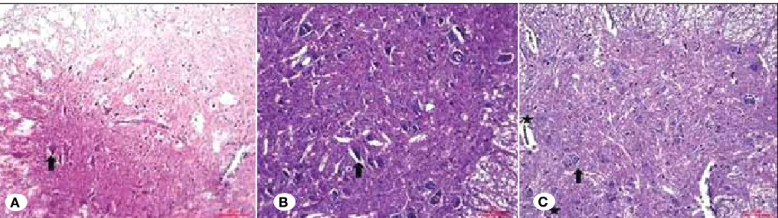
The groups were compared with regard to edema, neuronal injury, and white matter cavitation. Neuronal injury and white matter cavitation were grade 2 in Group T and grade 1 in Group O. Edema was not detected in any of the groups (Figure 1A-C, Table I).
The average numbers of blood vessels were significantly different among Group O (20.89±3.03), Group T (17.32±1.68), and Group C (8.57±0.43) (F=69.097, df=2, p<0.0005). Post hoc comparisons indicated significant differences among all three groups (Group O > Group T > Group C) (Scheffé’s test,
p<0.014) (Figure 2).
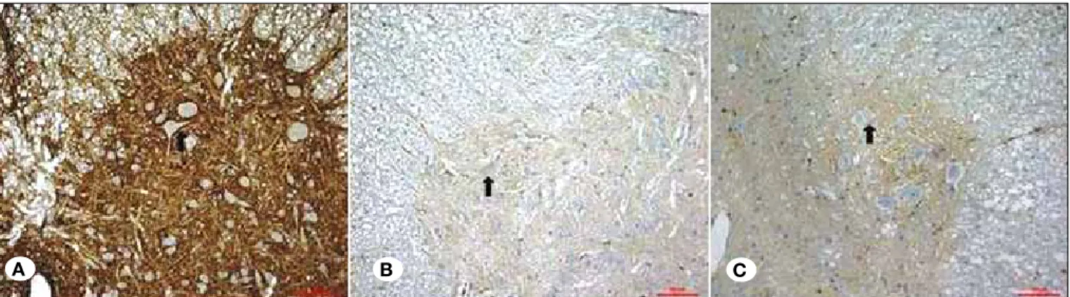
Average β-catenin levels were significantly different among Group C (68.11±0.43), Group O (37.96±2.16), and Group T (25.46±1.07) (F=1677.74, df=2, p<0.0005). Post hoc comparisons indicated significant differences among all three groups (Group C > Group O > Group T) (Scheffé’s test,
p<0.0005) (Figures 3A-C, 4).
Table I: Histological Evaluation of the Groups
Group C Group T Group O
Neuronal injury 0 2(+) 1(+)
White matter cavitation 0 2(+) 1(+)
Edema 0 0 0
Figure 1: Hematoxylin and eosin staining; A) Group C; B) Group T; C) Group O. (Black arrows indicate neurons, stars indicate blood
vessels).
in necrotic neural tissue due to inflammation and apoptosis (5,8).Our histopathological evaluation showed that the extent of neuronal injury and white matter cavitation in group O was less than that in group T on day 35. Edema was not detected in any of the groups in the present study. The lack of edema may have been due to the long post-trauma follow-up period in our study (15,19).
Disruption of blood circulation and increased free radical levels are major problems in the healing process following SCI (5,15,16).Previous studies concluded that angiogenic activity leads to improved functional outcomes in SCI animal models (13).Chen et al. (5) reported that hypoxia-inducible factor 1 improves the influx of oxygen towards injured site of the spinal cord, promoting angiogenesis thus improving the microenvironment for reformation of spinal cord functioning following SCI. Loy et al. (15) reported that vessel density increases after SCI in rats, and results of autoradiographic studies indicated that the spinal cord vasculature remains until day 28 following SCI. Angiogenic and anti-angiogenic therapies have been shown to have functional efficacy in animal models (15).Assessment of spinal cord vascularization indicated that Group O had a significantly greater number of vessels than did Group T. These observations indicated that ozone accelerates the healing process, increases the vascularity, and reduces the neuronal damage associated with SCI.
Wnt/β-catenin signaling influences the proliferation or differentiation of stem cell and progenitor populations (23). Recent studies have shown that the expression of Wnt ligands, their inhibitors, and components of their intracellular signaling pathways are prolonged in the adult spinal cord, as is activation of the canonical pathway, suggesting that the Wnt family of proteins plays a role in spinal cord function and physiology (8,10).Active β-catenin is expressed strongly in the gray matter of the non-injured spinal cord (8,10).Experimental studies suggested an important role of the Wnt/β-catenin pathway following CNS and spinal cord trauma (8,10,23). Fernández-Martos et al. (8)demonstrated that trauma induces a dramatic and time-dependent change in the physiological pattern of Wnt mRNA expression in a rat SCI model. We showed that the β-catenin level was significantly higher in group O on
█
DISCUSSION
Injury caused by SCI has many distinct factorial aspects including primary mechanical damage, secondary cell apoptosis, reactive gliosis, and axons inability to regenerate (8).The core of the SCI has the necrotic neural death characteristics, whereas secondary tissue damage is also significant in the penumbra zone. In penumbra zone processes like ischemia, inflammation, hypoxia, excitotoxicity, protease release, and free radical formation occur (8,11,20).
One of the most serious effects is functional deterioration following SCI. Chen et al. (5) reported that the first 28 days are a critical period for neural regeneration and functional recovery in rats following SCI. Our results indicated a better BBB score in group O than group T on day 7, but with no difference on day 35, suggesting that ozone rapidly improves the BBB scores during the early stages of treatment. We found that ozone accelerates the healing process.
Another serious effect of SCI is neural degeneration along with the death of neurons and oligodendrocytes, resulting
Figure 2: Numbers of blood vessels in the different groups
(*p<0.0005).
Figure 3: Immunohistochemical staining for β-catenin. A) Group C; B) Group T; C) Group O. (Black arrows indicate positive staining for
β-catenin in neurons). A B C Groups 25.00 20.00 15.00 10.00 5.00
Number of blood vessels
2. Allen AR: Remarks on the histopathological changes in the spinal cord due to impact. An experimental study. J Nerv Ment Dis 4:141-147, 1914
3. Basso DM, Beattie MS, Bresnahan JC: Graded histological and locomotor outcomes after spinal cord contusion using the NYU weight-drop device versus transection. Exp Neurol 139: 244-256, 1996
4. Bethea JR: Spinal cord injury-induced inflammation: A dual-edged sword. Prog Brain Res 128: 33–42, 2000
5. Chen MH, Ren QX, Yang WF: Influences of HIF-lα on Bax/ Bcl-2 and VEGF expressions in rats with spinal cord injury. Int J Clin Exp Pathol 15: 2312-2322, 2013
6. Curry TB, Bacon DR, Rho RH: The history of subcutaneous oxygen therapy. J Clin Anesth 18: 388-395, 2006
7. David MD, Cantí C, Herreros J: Wnt-3a and Wnt-3 differently stimulate proliferation and neurogenesis of spinal neural precursors and promote neurite outgrowth by canonical signaling. J Neurosci Res 88: 3011-3023, 2010
8. Fernández-Martos CM, González-Fernández C, González P, Maqueda A, Arenas E, Rodríguez FJ: Differential expression of Wnts after spinal cord contusion injury in adult rats. PLoS One 6(11):e27000, 2011
9. Frosini M, Contartese A, Zanardi I, Travagli V, Bocci V: Selective ozone concentrations may reduce the ischemic damage after a stroke. Free Radic Res 46: 612-618, 2012
10. González-Fernández C, Fernández-Martos CM, Shields S, Arenas E, Javier Rodríguez F: Wnts are expressed in the spinal cord of adult mice and are differentially induced after injury J Neurotrauma 15: 565-581, 2014
11. Hausmann ON: Post-traumatic inflammation following spinal cord injury. Spinal Cord 41: 369-378, 2003
12. Huang WL, King VR, Curran OE, Dyall SC, Ward RE, Lal N, Priestley JV, Michael-Titus AT: A combination of intravenous and dietary docosahexaenoic acid significantly improves outcome after spinal cord injury. Brain 130: 3004-3019, 2007 13. Kim HM, Hwang DH, Lee JE, Kim SU, Kim BG: Ex vivo VEGF
delivery by neural stem cells enhances proliferation of glial progenitors, angiogenesis, and tissue sparing after spinal cord injury. PLoS One 4(3):e4987, 2009
14. Konya D, Gercek A, Akakin A, Akakin D, Tural S, Cetinel S, Ozgen S, Pamir MN: The effects of inflammatory response associated with traumatic spinal cord injury in cutaneous wound healing and on expression of transforming growth Factor-Beta1 (TGF-Beta1) and Platelet-Derived Growth Factor (PDGF)-A at the wound site in rats. Growth Factors 26: 74-79, 2008
15. Loy DN, Crawford CH, Darnall JB, Burke DA, Onifer SM, Whittemore SR: Temporal progression of angiogenesis and basal lamina deposition after contusive spinal cord injury in the adult rat. J Comp Neurol 445: 308-324, 2002
16. Mautes AE, Weinzierl MR, Donovan F, Noble LJ: Vascular events after spinal cord injury: Contribution to secondary pathogenesis. Phys Ther 80: 673-687, 2000
17. McCrea PD, Turck CW, Gumbiner B: A homolog of the armadillo protein in Drosophila (plakoglobin) associated with E-cadherin. Science 254:1359-1361, 1991
day 35 after trauma. In contrast, White et al.(23)reported that the number of cells with β-catenin signaling increased in the cortex and subcallosal zone following traumatic brain injury but not following SCI.
Frosini et al. (9)demonstrated the neuroprotective effects of ozone in an in vitro model of brain ischemia. Oxygen induces plasma hyperoxygenation and saturates hemoglobin with oxygen, whereas ozone is figured as a pro-drug. Ozone is a rapidly dissolving molecule–more than oxygen-intensely in aqueous compartment of the plasma, reacts with solutes such as hydrosoluble anti-oxidants and polyunsaturated fatty acids bound to albumin. Erythroid 2-related factor is a released nuclear factor. Entering the nucleus and binding to the antioxidant response element, erythroid 2-related factor triggers the transcription of various antioxidants and phase II detoxifying enzymes (9).They concluded that after exactly calculated oxidative stress, ozone corrects oxygen-glucose release and delivery and coordinates intracellular antioxidant enzymes’ upregulation.
█
CONClUSION
The present study indicated a rapid recovery in the BBB score during the early period and neural recovery and increased blood vessel number and β-catenin levels in neural tissue during the late period of ozone treatment, suggesting that ozone treatment increases blood vessel density and stem cell proliferation and differentiation after SCI in rodents. Based on these observations, ozone therapy may be a useful adjuvant treatment in patients with spinal cord trauma.
█
REFERENCES
1. Aktug H, Uslu S, Terek MC, Terzi H, Turgut M, Ozsener S, Bilgin O: Effects of ovariectomy and tamoxifen on rat bone tissue: Histomorphometric and histopathologic study. Anal Quant Cytol Histol 28: 207-212, 2006
Figure 4: Distribution of β-catenin levels (H-score) among groups
(*p<0.0005). Groups 70.00 60.00 50.00 40.00 30.00 5.00 Beta-catenin levels
21. Samantaray S, Sribnick EA, Das A, Thakore NP, Matzelle D, Yu SP, Ray SK, Wei L, Banik NL: Neuroprotective efficacy of estrogen in experimental spinal cord injury in rats. Ann N Y Acad Sci 1199: 90-94, 2010
22. Uslu S, Uysal A, Bilir A, Soner BC, Oktem G: Hepatic progenitor cell inhibition during embryonic period with high dose verapamil; liable joint to the cancer therapy. Bratisl Lek Listy 114: 369-375, 2013
23. White BD, Nathe RJ, Maris DO, Nguyen NK, Goodson JM, Moon RT, Horner PJ: Beta-catenin signaling increases in proliferating NG2+ progenitors and astrocytes during post-traumatic gliogenesis in the adult brain. Stem Cells 28: 297-307, 2010
18. Michael-Titus AT: Omega-3 fatty acids and neurological injury. Prostaglandins Leukot Essent Fatty Acids 77: 295-300, 2007 19. Onose G, Anghelescu A, Muresanu DF, Padure L, Haras MA,
Chendreanu CO, Onose LV, Mirea A, Ciurea AV, El Masri WS, von Wild KR: A review of published reports on neuroprotection in spinal cord injury. Spinal Cord 47: 716-726, 2009
20. Profyris C, Cheema SS, Zang D, Azari MF, Boyle K, Petratos S: Degenerative and regenerative mechanisms governing spinal cord injury. Neurobiol Dis 15: 415-436, 2004