Turk Kardiyol Dern Ars 2019;47(3):228-231 doi: 10.5543/tkda.2018.69679
Successful transcatheter mitral valve replacement in a patient with
bioprosthetic valvular degeneration and severe regurgitation
Biyoprotez kapak dejenerasyonu ve ciddi yetersizliği olan bir hastada
başarılı transkateter mitral kapak replasmanı
Department of Cardiology, İstanbul Medipol University Faculty of Medicine, İstanbul, Turkey Beytullah Çakal, M.D., Sinem Çakal, M.D., İbrahim Oğuz Karaca, M.D.,
Mehmet Onur Omaygenç, M.D., Aydın Yıldırım, M.D.
Özet– Transkateter aort kapak, dejenere mitral biyoprotez kapak işlev bozukluğu olan ve tekrar ameliyat riski yük-sek olgularda dünyada yüzlerce hastada başarılı bir şekil-de kullanılmıştır. Transseptal yaklaşım teknik olarak daha zor olsa da transapikal yaklaşıma kıyasla daha az inva-ziv, muhtemel daha düşük mortaliteye sahip olup hastanın daha hızlı toparlanması ile ilişkilidir. Burada, dejenere bi-yoprotez kapağa sahip bir olguda transseptal yaklaşımla başarılı mitral kapak değişimini anlatan ülkemizdeki nadir uygulanmış olan olguyu bildiriyoruz. Bu yazıda, dejenere biyoprotez mitral kapak varlığında güvenli ve etkin bir şe-kilde perkütan kapak içi kapak implantasyonundan bahse-dilmiştir.
Summary– The implantation of aortic transcatheter heart valves has been successfully performed throughout the world in hundreds of patients with severe dysfunction of a degenerated mitral bioprosthesis or those at high surgi-cal risk for re-operation. The transseptal approach may be more technically challenging, but is a less invasive proce-dure and may have a lower mortality rate compared with a transapical approach, and also offers a quick patient reco-very. This report is a description of a rare case in Turkey: a successful transseptal mitral valve replacement in a case of a failed bioprosthetic valve. This case illustrates the feasibil-ity and safety of percutaneous valve-in-valve implantation to treat a degenerated bioprosthesis.
228
T
he majority of the patients with severe sympto-matic mitral regurgitation with concomitant left ventricular dysfunction and previous cardiac surgery do not undergo redo surgery, though it may be rec-ommended in the current guidelines.[1,2] Theoff-la-bel use of standard aortic transcatheter heart valves (THV) has emerged as an exciting new frontier in the treatment of a failed mitral bioprosthesis or surgical ring, as well as for patients with severe annular calci-fication.[3,4] These platforms provide anchoring for an
expandable balloon or the newer aortic THVs. Hun-dreds of patients worldwide have been treated with a transcatheter mitral valve-in-ring or valve-in-valve procedure using a bioprosthetic aortic valve.[5,6]
A Sapien valve (Edwards Lifesciences; Irvine, CA, USA) has been implanted in the majority of
cases. This report is a description of a transseptal mitral valve replacement in a case of a failed sur-gical bioprosthesis.
CASE REPORT
A 74-year-old woman was referred for evaluation and treatment of severe mitral regurgitation. Her his-tory included bioprosthetic aortic replacement with a 27-mm Carpentier-Edwards mitral valve (Edwards Lifesciences; Irvine, CA, USA) replacement 10 years earlier. Two years after the surgery, she underwent per-cutaneous mitral paravalvular leak closure. Echocar-diography revealed severe transvalvular mitral
regur-Received:October 22, 2017 Accepted: February 20, 2018
Correspondence: Dr. Beytullah Çakal. İstanbul Medipol Üniversitesi, Kardiyoloji Ana Bilim Dalı, Bağcılar, İstanbul, Turkey.
Tel: +90 212 - 460 77 77 e-mail: [email protected]
© 2019 Turkish Society of Cardiology
Abbreviations: LV Left ventricular
LVOT Left ventricular outflow tract TEE Transesophageal echocardiography THV Transcatheter heart valve
gitation, an estimated left ventricular (LV) ejection fraction of 0.45, and a pulmonary arterial systolic pressure of 60 mm Hg. Since the patient was highly symptomatic and was not a good candidate for surgery, percutaneous mitral valve replacement was scheduled.
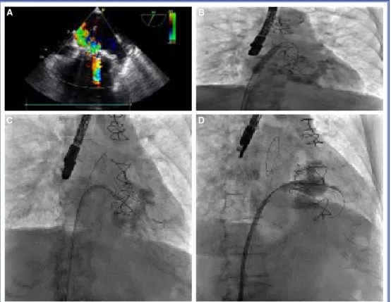
Evaluation of mitral valve regurgitation was per-formed using 2-dimensional transesophageal echocar-diography (2D-TEE) and valve sizing was determined based on the previous bioprosthetic valve size (Fig. 1a, Video 1*). The ideal THV size for the
valve-in-valve procedure was selected based on the true inter-nal diameter (which determines prosthesis anchoring), which is typically 1 to 2 mm smaller than the diam-eter of the surgical valve size reported by the man-ufacturer. The commercially available Mitral V-in-V smartphone application, designed by Vinayak Bapat,[7]
was also used to verify the ideal valve size. The pro-cedures were performed with general anesthesia, and percutaneous femoral venous access was used. After a transseptal puncture in the inferoposterior portion of the fossa ovalis with 2D-TEE and flouroscopic
guid-ance using a transfemoral approach, balloon dilatation (12-mm x 60-mm) of the septum was performed to facilitate the crossing of the septum (Fig. 1b). The transseptal sheath and dilator were advanced over a 0.035-inch J guidewire under fluoroscopy guidance into the left atrium. A soft, straight-tip, 0.032-inch wire, which was positioned using a deflectable left atrial sheath (Agilis NxT steerable introducer; St Jude Medical, St. Paul, MN, USA) was advanced through the mitral prosthesis (Fig. 1c, Video 2*). Two Amplatz
Super Stiff guidewires and a Safari wire (Boston Sci-entific Corp., Marlborough, MA, USA) were placed through a pigtail catheter in the left ventricular apex after removing the straight-tip wire. Movement during deployment was minimized with coaxial guidewires without pacing or apnea, and the second wire outside the THV delivery system was pulled back preceding deployment of the valve. The ventricular edge of a Sapien-XT stent frame was positioned at the ventricu-lar edge of the pre-existing bioprosthesis. The 26-mm Sapien-XT valve was slowly mounted in the opposite direction to the transfemoral aortic THV within the
Figure 1. (A) A transesophageal echocardiogram showing degenerative bioprosthetic mitral re-gurgitation. (B) Septum dilatation with a 12-mm balloon. (C) The catheter is flexed to point to-ward the mitral valve using fluoroscopic and transesophageal echocardiography guidance, and the mitral valve is crossed with a 0.032-inch, straight-tip guidewire. (D) Implantation of a 26-mm, balloon-expandable valve within the degenerated mitral bioprosthesis.
A
C
B
D
degenerated mitral prosthesis (Fig 1d, Video 3*). TEE
demonstrated complete resolution of mitral regurgi-tation (Video 4*) and the transvalvular mean gradient
across the mitral prosthesis was measured as 4 mm Hg. The patient was discharged from on day 2 with a New York Heart Association functional class II desig-nation and daily use of clopidogrel 75 mg and aspirin 100 was initiated.
DISCUSSION
Transcatheter mitral valve replacement has been per-formed for patients with a regurgitant mitral valve, a failing mitral valve bioprosthetic or ring, or a cal-cified mitral annulus, since these patients may have multiple comorbidities, an increased risk of operative mortality, and an extended hospital stay.[8]
The data regarding implantation of an aortic THV within a degenerated bioprosthetic mitral valve (valve-in-valve) or a mitral valve ring (valve-in-ring) is generally limited to case series using the Edwards Sapien series, the Boston Scientific Lotus valves, the Medtronic Melody products (Medtronic Inc., Min-neapolis, MN, USA), and Direct Flow Medical valves (Direct Flow Medical Inc, Santa Rosa, CA, USA) with no long-term results.[9] The anatomical challenges
associated with mitral THV implantation have been clearly defined: the valve system must be delivered to, anchored, and sealed within a large, non-circu-lar, saddle-shaped, highly dynamic, and sometimes calcific annulus to overcome the loading conditions of the LV. The complex, highly individualized, sub-valvular apparatus and the mitral annulus proximal to the left ventricular outflow tract (LVOT), the coronary sinus, and the left circumflex coronary artery pose a risk for impingement.[10] These anatomical challenges
have delayed mitral THV development. Malposition, paravalvular leak, embolization, LVOT obstruction, leaflet thrombosis, hemolysis, aortic valve injury, reduced ejection fraction, limited large transcatheter aortic valve size, and stroke are possible complica-tions of mitral THV replacement.
A venous access procedure was performed in our case while avoiding the potential complications of bleeding, apical injury, and lung injury due to thoracotomy, as well as a long hospital stay. The stan-dard surgical approach for a mitral valve replacement is via a left atriotomy; however, a transseptal surgi-cal approach via a right atriotomy has been preferred
for patients with a small left atrium, friable tissues, heavily calcified mitral valves, or when there is the need to perform combined tricuspid and mitral pro-cedures.[10] In our patient, the previous left atriotomy
made a transseptal approach easier. A transapical approach might be preferred to a transseptal alterna-tive, as it enables greater control of the delivery sys-tem, but it is more invasive. A single-center report found that among 24 patients who underwent a mitral transcatheter valve-in-valve procedure, the patients treated with a transseptal approach had a significant increase in cardiac output and improved survival when compared with patients treated with a transapi-cal approach.[11] Our patient recovered quickly and
was discharged on the second day after the procedure. Another important factor contributing to success-ful valve-in-valve implantation is comprehensive un-derstanding of the dimensions listed by the manufac-turer or the Mitral V-in-V smartphone application, the measurements obtained from the CT scan, the type of valve pathology (stenosis or regurgitation), and the risk of LVOT obstruction.[12,13] Since a stented surgical
bioprosthesis provides good surgical landmarks, and an appropriate semirigid and circular landing zone is selected easily under fluoroscopic guidance, we did not perform CT before the intervention. The size of the THV should be sufficiently greater than the inter-nal diameter of the implanted prosthesis for it to be anchored inside the previous stent. There is a high risk of embolization or atrial migration with an undersized valve, and significant oversizing may result in distor-tion of the transcatheter valve leaflets.
To our knowledge, this is a rare case of successful transcatheter valve-in-valve implantation for a failed prosthetic mitral valve, an off-label use in Turkey. This less invasive therapy may be an alternative or even a better method than surgery in selected cases. Long-term clinical trials are required to confirm the efficacy and safety.
Conclusion
Despite being primarily designed as a valve for the aortic position and the lack of clearly defined long-term valve outcomes, a transcatheter aortic valve used in reverse position for mitral valve-in-valve im-plantation is an option for patients with failed mitral prosthetic valve dysfunction who are at high risk for surgical valve replacement.
Turk Kardiyol Dern Ars
Transcatheter mitral valve replacement 231
*Supplementary video file associated with this article can be found in the online version of the journal. Peer-review: Externally peer-reviewed.
Conflict-of-interest: None.
Informed Consent: Written informed consent was
ob-tained from the patient for the publication of the case report and the accompanying images.
Authorship contributions: Concept: B.Ç.; Design:
A.Y.; Supervision: A.Y.; Materials: I.O.K.; Data collec-tion: B.Ç.; Literature search: M.O.O.; Writing: B.Ç.
REFERENCES
1. Tribouilloy C, Rusinaru D, Grigioni F, Michelena HI, Vanoverschelde JL, Avierinos JF, et al.; Mitral Regurgitation International Database (MIDA) Investigators. Long-term mortality associated with left ventricular dysfunction in mitral regurgitation due to flail leaflets: a multicenter analysis. Circ Cardiovasc Imaging 2014;7:363–70. [CrossRef]
2. Nishimura RA, Otto CM, Bonow RO, Carabello BA, Erwin JP 3rd, Guyton RA, et al.; American College of Cardiology/ American Heart Association Task Force on Practice Guide-lines. 2014 AHA/ACC guideline for the management of pa-tients with valvular heart disease: executive summary: a re-port of the American College of Cardiology/American Heart Association Task Force on Practice Guidelines. J Am Coll Cardiol 2014;63:2438–88. [CrossRef]
3. Webb JG, Wood DA, Ye J, Gurvitch R, Masson JB, Rodés-Cabau J, et al. Transcatheter valve-in-valve implantation for failed bioprosthetic heart valves. Circulation 2010;121:1848– 57. [CrossRef]
4. Maisano F, Alfieri O, Banai S, Buchbinder M, Colombo A, Falk V, et al. The future of transcatheter mitral valve inter-ventions: competitive or complementary role of repair vs. re-placement? Eur Heart J 2015;36:1651–9. [CrossRef]
5. Dvir D, Webb J, Brecker S, Bleiziffer S, Hildick-Smith D, Colombo A, et al. TCT-797 Transcatheter Mitral Valve-in-Valve / Valve-in-Valve-in-Ring Implantations For Degenerative Post
Surgical Valves: Results From The Global Valve-in-Valve Registry. J Am Coll Cardiol 2012;60:B232. [CrossRef]
6. Dvir D. Transcatheter Mitral Valve and Valve-in-Ring Implantations. Data presented at Transcatheter Cardio-vascular Therapeutics; 2016.
7. Bapat V. Valve-in-valve apps: why and how they were devel-oped and how to use them. EuroIntervention 2014;10 Suppl U:U44-51.
8. Vohra HA, Whistance RN, Roubelakis A, Burton A, Barlow CW, Tsang GM, et al. Outcome after redo-mitral valve replace-ment in adult patients: a 10-year single-centre experience. In-teract Cardiovasc Thorac Surg 2012;14:575–9. [CrossRef]
9. Wilbring M, Alexiou K, Tugtekin SM, Arzt S, Ibrahim K, Matschke K, et al. Pushing the limits-further evolutions of transcatheter valve procedures in the mitral position, includ-ing valve-in-valve, valve-in-rinclud-ing, and valve-in-native-rinclud-ing. J Thorac Cardiovasc Surg 2014;147:210–9. [CrossRef]
10. Mylotte D. Transcatheter mitral valve replacement: looking beyond the implant. EuroIntervention 2017;13:e1011–e1012. 11. Frerker C, Schmidt T, Schlüter M, Bader R, Schewel J,
Schewel D, et al. Transcatheter implantation of aortic valve prostheses into degenerated mitral valve bioprostheses and failed annuloplasty rings: outcomes according to access route and Mitral Valve Academic Research Consortium (MVARC) criteria. EuroIntervention 2016;12:1520–6. [CrossRef]
12. Santibáñez Escobar F, Serrano Gallardo G, Ramirez Marro-quin S, Lopez Soriano F, Barragán García R. The transseptal approach for mitral valve replacement revisited. Tex Heart Inst J 1997;24:209–14.
13. Guerrero M, Salinger M, Pursnani A, Pearson P, Lampert M, Levisay J, et al. Transseptal transcatheter mitral valve-in-valve: A step by step guide from preprocedural plan-ning to postprocedural care. Catheter Cardiovasc Interv 2018;92:E185–E196. [CrossRef]
Keywords: Mitral regurgitation; mitral valve-in-valve; mitral valve
re-placement; transcatheter valve replacement
Anahtar sözcükler: Mitral yetersizliği, mitral valve-in valve; mitral