Abstract
Minocycline, a semisynthetic tetracycline-derived antibiotic, has been shown to exert anti-apoptotic, anti-inflammatory, and antioxidant effects. Furthermore, there is rapidly growing evidence suggesting that minocycline may have some neuroprotective activity in various experimental models such as cerebral ischemia, traumatic brain injury, amyotrophic lateral sclerosis, Parkinson’s disease (PD), Huntington’s disease, and multiple sclerosis. In this perspective review, we summarize the
preclinical and clinical findings suggesting the neuroprotective role of minocycline in PD.
Keywords: minocycline, neurodegeneration, neuroprotection, Parkinson’s disease.
Citation
Cankaya S, Cankaya B, Kilic U, Kilic E, Yulug B. The therapeutic role of minocycline in Parkinson’s disease. Drugs in Context 2019; 8: 212553. DOI: 10.7573/dic.212553
Seyda Cankaya MD1, Baris Cankaya MD2, Ulkan Kilic PhD3, Ertugrul Kilic PhD4, Burak Yulug MD1
1Department of Neurology, Faculty of Medicine, Alaaddin Keykubat University, Alanya, Turkey; 2Department of Anesthesiology
and Reanimation, Marmara University Pendik Education and Research Hospital, Istanbul, Turkey; 3Department of Medical
Biology, Faculty of Medicine, University of Health Sciences, Istanbul, Turkey; 4Department of Medical Physiology,
Faculty of Medicine, Istanbul Medipol University, Istanbul, Turkey
The therapeutic role of minocycline in Parkinson’s disease
Introduction
Although the molecular cell death mechanisms involved in neurodegenerative diseases, such as amyotrophic lateral sclerosis (ALS), Alzheimer’s disease (AD), Parkinson’s disease (PD), and Huntington’s disease (HD), are still not fully understood, progress in the knowledge of neuronal cell death in the common pathways has led to new approaches for neuroprotective therapies. The common pathways may include several pathogenic processes, such as energy (mitochondrial) impairment, secondary glutamate excitotoxicity, oxidative stress, and inflammatory mechanisms.1 Minocycline has been shown to exert its
neuroprotective effect on common pathways in several neurodegenerative diseases, including PD.2–5 Among the
neurodegenerative disorders, PD remains the only one that responds well to symptomatic therapy. Although levodopa treatment has been accepted as the gold standard of treatment, its chronic use is associated with potentially disabling motor complications. Furthermore, although it may improve some of the most disabling symptoms of PD, patients are faced with increased substantial disability over time and a future of dependency. In addition, there is no evidence that levodopa can halt or slow the progressive degeneration of dopaminergic (DA) neurons in the substantia nigra (SN). Moreover, there are controversial findings regarding the toxic
actions of levodopa on the remaining dopaminergic neurons. Thus, there is a compelling need to develop treatments that might slow down the motor and nonmotor symptoms such as cognitive impairment in PD. Thus, one of the most important therapeutic needs in PD is the development of effective disease-modifying or neuroprotective therapies.6–8
In the light of these findings, researchers have spent an enormous effort to develop candidate neuroprotective agents for PD patients for the last 20 years. Recent findings of abnormal protein folding, coupled with oxidative stress and neuroinflammation, provide a scientific rationale for novel therapeutic strategies designed to slow disease progression in PD. In this context, recent research has been focused on the possible neuroprotective effects of anti-inflammatory drugs. So far, numerous studies, including the genetic, neurotoxic animal models, and clinical trials of minocycline in PD, have indicated that it may exert neuroprotective activity through inhibition of microglial activation, neuroinflammation, and neuronal apoptosis. Despite these promising results, no therapy has yet been proven to halt or slow disease progression of PD.
Minocycline has been chosen for this perspective review because of its capacity to decrease oxidative injury and neuroinflammation and so exert a potential neuroprotective activity, which has been proven in various in vivo and in vitro
animal models. In this respect, our rationale was to evaluate minocycline’s neuroprotective effect in PD and to compare relevant studies, including clinical results, pathologic findings, and cognitive outcomes of minocycline. We have included retrospectively recorded results from animal studies, clinical trials, and also reviews in Medline via PubMed, Scopus, and ISI Web of Science. We used ‘Minocycline and Parkinson’s disease’, ‘minocycline and neuroprotection’, ‘minocycline’, and ‘neurodegeneration’ as key search items. From 292 potentially eligible reviews and studies, we selected 92 articles for full-text analysis.
Minocycline
Minocycline is a second-generation, semisynthetic tetracycline analogue, which is a highly lipophilic molecule and can easily penetrate the blood–brain barrier.9 It shows a better
pharmacokinetic profile than the first-generation tetracyclines. However, it is rapidly and completely absorbed, even in elderly populations, with a longer half-life and excellent tissue penetration.10,11 In addition, it is a highly lipophilic molecule
that can easily pass through the blood-brain barrier12, thus
promoting its accumulation in the cerebrospinal fluid (CSF) and central nervous system (CNS)9,13,14 and enabling its use in the
treatment of several CNS diseases.15–17 In rodents, minocycline
readily crosses the blood-brain barrier with a rate of at least fivefold higher than doxycycline, another compound of the same family.18,19
Thereby, minocycline has been extensively studied in treating neurodegenerative diseases17,20,21 and has been reported to
exert neuroprotective effects on various experimental models. These studies based on minocycline’s ability to inhibit microglia activation, which is a process that has deleterious effects on neurogenesis and neuronal survival could justify its potential effectiveness in the treatment of neuroinflammatory and/or neurodegenerative disorders.
Mechanisms of action of minocycline
Parkinsonian symptoms are caused by degeneration of dopaminergic neurons in the substantia nigra pars compacta (SNpc) and the consequent loss of their projecting nerve fibers in the striatum.22 Neuroinflammation and microglial
activation, a hallmark of neuroinflammation that has been generally considered as an integral component of the progressive neurodegenerative process, has been increasingly implicated in the PD pathogenesis.23 It is
noteworthy that minocycline is a potent inhibitor of
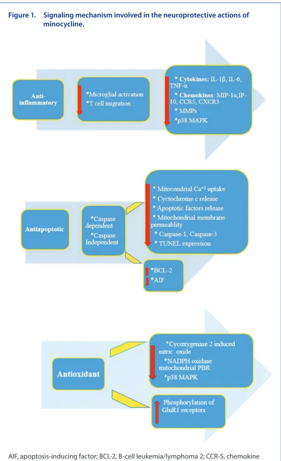
microglial activation and of apoptotic pathways. Considering all of this evidence, it is not unreasonable to assume that minocycline exerts neuroprotective activity on animal models with PD via protecting the nigrostriatal pathway. Modes of actions of minocycline are summarized in Figure 1 and Box 1.
The anti-inflammatory effect
of minocycline
Until now, studies have already indicated that neuroinflammation is mediated by microglia and astrocytes, which produce
inflammatory cytokines, reactive oxygen species, and other toxic materials in the CNS.25 Here, reactive microgliosis,
astrocytosis, an increase in proinflammatory mediators, and activation of inflammation-associated protein kinases are features common to many neurodegenerative disorders. For instance, a chronic release of proinflammatory
cytokines by activating astrocytes and microglia leads to the exacerbation of DA neurons degeneration in the SNpc. These findings together suggest that microglia are one of the major cell types that are involved in the inflammatory response in the CNS. In parallel with that, it has been proposed that activated microglia may
be beneficial to the host, at least in the early phase of the neurodegeneration process.26–28
The above-mentioned chronic neuroinflammation is also one of the hallmarks of PD pathophysiology. Beyond preclinical studies suggesting that there is a very similar chronic neuroinflammation process in PD, clinical data indicating the role of neuroinflammation are also rapidly replicating in PD.
Postmortem analyses of human PD patients indicate that the activation of glial cells and increases in
proinflammatory factor levels are common features of the PD brain. However, long-term overactivation of microglia in the PD brain significantly upregulates the expression of a large group of proinflammatory cytokines, including tumor necrosis factor-alpha (TNF-α), interleukin-6 (IL-6), IL-1 beta (β), and interferon gamma (IFN-γ), which contribute to the acceleration of nigral DA neuron degeneration.29–31
The main biological effects of minocycline, which are involved in the pathogenesis of several neurodegenerative disorders, include inhibition of microglial activation, attenuation of apoptosis, and suppression of reactive oxygen species (ROS) production. These effects appear completely different from its antimicrobial action.32
Minocycline exerts its neuroinflammatory actions by
modulating microglia, immune cell activation, and subsequent release of cytokines, chemokines, lipid mediators of
inflammation, MMPs, and NO release.33 Proinflammatory
cytokines, such as TNF-α, IL-1β, and IL-6, are produced by microglial cells, astrocytes, neutrophils, and macrophages and augment both inflammation and following immune responses.33 Thus, any potential beneficial effects of
minocycline in neurodegenerative diseases might well be exerted through its actions on the molecular pathways involving the modulation of one or more of these above-mentioned inflammatory factors.
Figure 1. Signaling mechanism involved in the neuroprotective actions of minocycline.
AIF, apoptosis-inducing factor; BCL-2, B-cell leukemia/lymphoma 2; CCR-5, chemokine receptor type 5; CXCR3, chemokine (CXC motif) receptor; GluR, glutamate receptor; IP-10, interferon-inducible protein; MAPK, mitogen-activated protein kinase; MIP-1α, macrophage inflammatory protein 1α; MMP, matrix metalloprotease; NADPH, nicotinamide-adenine dinucleotide phosphate; PBR, peripheral benzodiazepine receptor; TNF, tumor necrosis factor; TUNEL, terminal deoxynucleotidyl transferase dUTP (2´-deoxyuridine, 5´-triphosphate).
patients.39–41 Tetracycline and its derivatives, especially
minocycline, are a group of semisynthetic antibiotic agents that, besides their anti-inflammatory and antimicrobial properties, interfere with the activation of enzymes in the apoptotic pathway.42 Minocycline decreased apoptotic
neuronal cell death observed under various experimental models of neurodegenerative diseases such as ischemia, HD, PD, AD, and ALS.43,44 This apoptotic effect is mainly
being exerted on mitochondria. By reducing mitochondrial calcium overloading, minocycline stabilizes the mitochondrial membrane and inhibits release of cytochrome c and
other apoptotic factors into the cytoplasm, which finally results with decreased caspase activation and nuclear damage45,46 that play a critical role in the apoptotic cell
death cascade.
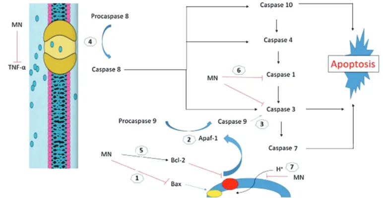
Furthermore, Caspase 8 activates a series of other caspases that finally lead to a series of lethal cleavages resulting in apoptosis (Figure 2).47 It has been revealed that B-cell leukemia/
lymphoma 2 (Bcl-2) exerts an anti-apoptotic effect through stabilizing the mitochondrial membrane. From this point of view, minocycline treatment also exerts a neuroprotective effect through shifting the Bcl-2 family proteins toward the anti-apoptotic ratio in neuron/microglia co-cultures.48 This is
consistent with the anti-apoptotic action of minocycline that is associated with its modulating effect on the expression of Bcl-2 proteins in various models.49–52
In gerbils with global brain ischemia, minocycline increased the survival of hippocampal neurons and this protection was associated with reduced caspase-1 expression.53 Minocycline
inhibits both caspase-dependent (cytochrome c and Smac/ DIABLO) and caspase-independent anti-inflammatory
Signaling pathways involved in
minocycline’s neuroprotective actions
Signaling mechanisms are also involved in the neuroprotective actions of minocycline. Minocycline shows its neuroprotective actions on p38 mitogen-activated protein kinase (MAPK)-dependent mechanism in the signaling pathway.
P38 MAPKs are serine–threonine kinases that play significant roles in signaling for a vast number of cellular functions including cell migration, proliferation, and differentiation. It is initially identified as a stress-activated phosphorylated in response to inflammatory cytokines.34 The reduction effect
of minocycline in p38 MAPK activities has inspired studies for validating minocycline’s neuroprotective effect.
It has been shown that minocycline increased neuronal survival in mixed spinal cord cultures, reduced NO-induced death of rats cerebellar granule neurons,35,36 and attenuated cell death
by 50% in hypoxic-ischemic injury-induced death of a motor neuron cell line37 by reducing microglia activation through p38
MAPK-dependent mechanisms.
The anti-apoptotic effect of minocycline
A characteristic of many neurodegenerative diseases is neuronal cell death. Cell death occurs by necrosis or apoptosis.38 Although there were controversial data on
the special cell death form of PD,39 recent studies support
that apoptotic rather than the necrotic degeneration of dopaminergic cells in the SN plays a critical role in the pathogenesis of PD. Consistently, apoptosis has been demonstrated in the SN of postmortem brains of PD
Box 1. Modes of action of minocycline.24
A. Anti-inflammatory actions
Modulation of microglia:
- To reduce the proliferation/activation of resting microglial cells, subsequently decreasing the release of cytokines, chemokines, lipid mediators of inflammation, MMPs, and nitric oxide (NO) release
Alteration of immune cell activation:
- To inhibit transmigration of T lymphocytes B. Anti-apoptotic actions
Caspase-dependent anti-apoptotic actions:
- Inhibition of cytochrome c release from mitochondria by attenuating mPT - Inhibition of caspase-1 and -3 expression
Caspase-independent anti-apoptotic actions:
- Increase in the expression of Bcl-2
- Inhibition of AIF release from mitochondria C. Involvement in the signaling pathways
Inhibition of p38 MAPK activation in microglia, thereby attenuating the production of IL-8, superoxide generation, and neutrophil chemotaxis
AIF, apoptosis-inducing factor; Bcl-2, B-cell leukemia/lymphoma 2; IL-8, interleukin-8; MAPK, mitogen-activated protein kinase; MMPs, matrix metalloprotease; mPT, mitochondrial permeability transition.
Figure 2. Schematic representation of apoptosis and proposed sites of inhibition. 1. Free oxygen radicals, lipid peroxidation, and increased cytosolic calcium cause mitochondrial depolarization and an opening of the mitochondrial permeability transition pore on the inner membrane and an outer membrane pore in comprising products of the Bcl-2 family member, Bax. This causes the release of cytochrome c into the cytosol. 2. Cytochrome c combines with Apaf-1 forming a complex called the apoptosome. Procaspase 9 is eventually cleaved on the apoptosome forming active caspase 9 (initiator caspase). 3. Caspase 9 activates the executioner caspase 3. Caspase 3 causes DNA damage, PPAR-1 activation and finally apoptosis. 4. Caspase 8 is activated by the DISC, which is attached to the death receptor (belongs to the family of TNF receptors). 5. Minocycline inhibits cytosolic calcium overload, binds with free oxygen radicals (ROS), inhibits Bax, and activates Bcl-2 (antiapoptotic gene) 6. It also inhibits caspase 1, and activation and reactivation of caspase 3. 7. It stabilizes the mitochondrial membrane. ‘Activation’ is shown by black arrows and ‘inhibition’ is shown by red arrows. H+
Apaf-1, apoptotic protease activating factor; Bax, Bcl-2-associated X-apoptosis regulator; Bcl-2, B-cell leukemia/lymphoma 2; DISC, death-inducing signaling complex; DNA, deoxyribonucleic acid; PPAR-1, peroxisome proliferator-activated receptors-1.
activity in mitochondrial death pathways16 that is in line with its
inhibitory effect on cytochrome c release, caspase-3 activation, and inflammation as shown by in vivo and in vitro models of HD and ALS.16,54
Many studies have revealed that PD, cerebral ischemia, ALS, and HD share common pathophysiological pathways (i.e., oxidative injury, neuroinflammation) during the neurodegenerative process. As specifically mentioned earlier for the neuroprotective actions of minocycline, it has become a candidate for slowing or halting the degenerative process in the CNS.
Minocycline and neuroprotection
Yrjanheikki and colleagues first described minocycline as a neuroprotective agent that inhibits the ischemia-induced
activation of microglia in an animal model of ischaemia.53
The neuroprotective role of minocycline was confirmed with further studies, which showed that minocycline inhibited neuronal death and reduced neurodegenerative disease progression including in different animal models such as PD, HD, MND, AD, and multiple sclerosis. For instance, in AD models, minocycline inhibited Aβ fibril formation,55 attenuated
amyloid-induced microglial activation,56 and reduced the
inflammatory events that were associated with the prevention of cognitive deficits.57 In line with this, minocycline also
reduced the N-methyl d-aspartic acid (NMDA) toxicity in animal models of ischemic stroke35,58 and slowed the
neurodegenerative progression in a mice model of HD.3,16,59
Unfortunately, despite promising results in experimental models of ALS,60,61 patients on minocycline declined more
Neuroprotective effects of minocycline in
animal models of Parkinson’s disease
Oxidative stress, mitocondrial dysfunction, inflammation, excitotoxicity, and abnormal processing of mutant proteins have played a critical role of the cell death of PD.42 Although
the underlying mechanisms of the neuroprotective effect of minocycline are unclear, it is reasonable to assume
that minocycline might exert its effect through an interactive signaling network including the mitochondria, oxidative stress, excitotoxicity, PARP-1, and apoptosis.42
To reveal minocycline’s protective effects in PD, animal models with 1-methyl-4-phenyl-1,2,3,6-tetrahydropyridine (MPTP) are widely used. MPTP reproduces some of the basic PD features in primates and rodents. Thereby, it is regarded as a well-established model.66 In one MPTP model,
minocycline treatment blocked dopamine depletion in the nucleus accumbens after MPTP administration.5 This
quickly than those on a placebo in clinical trials (phase III stage).62
It should also be noted that a recent study has revealed that treatment with minocycline improved spatial memory that was mediated through reduced the levels of Aβ and microglial activation in a mice model of AD.63 In addition, a clinical study
has suggested that minocycline reduced the risk of conversion of clinically isolated syndrome to multiple sclerosis (MS) in a randomized, placebo-controlled trial of early multiple sclerosis patients at 6 months.64 The results of this clinical study were
confirmed with a recent experimental study suggesting that minocycline enhanced the self-renewal capability of neural stem cells into the oligodendrocytes that were independent of its inhibitor effect on microglial activation in an autoimmune encephalomyelitis that is an animal model for multiple sclerosis.65 The neuroprotective and cognitive effects of
minocycline in animal and human studies in PD have been summarized in Table 1.
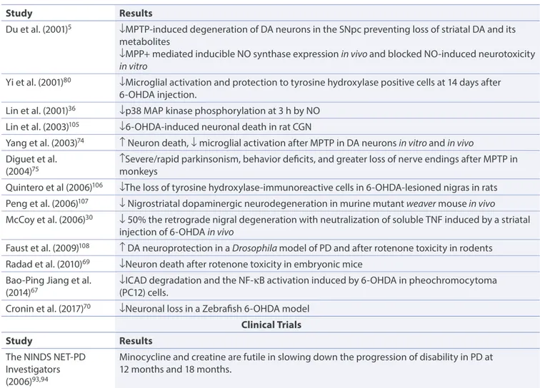
Table 1. Results of experimental studies on minocycline treatment for Parkinson’s disease.
Study Results
Du et al. (2001)5 ↓MPTP-induced degeneration of DA neurons in the SNpc preventing loss of striatal DA and its
metabolites
↓MPP+ mediated inducible NO synthase expression in vivo and blocked NO-induced neurotoxicity
in vitro
Yi et al. (2001)80 ↓Microglial activation and protection to tyrosine hydroxylase positive cells at 14 days after
6-OHDA injection.
Lin et al. (2001)36 ↓p38 MAP kinase phosphorylation at 3 h by NO
Lin et al. (2003)105 ↓6-OHDA-induced neuronal death in rat CGN
Yang et al. (2003)74 ↑ Neuron death, ↓ microglial activation after MPTP in DA neurons in vitro and in vivo
Diguet et al.
(2004)75 ↑Severe/rapid parkinsonism, behavior deficits, and greater loss of nerve endings after MPTP in monkeys
Quintero et al (2006)106 ↓The loss of tyrosine hydroxylase-immunoreactive cells in 6-OHDA-lesioned nigras in rats
Peng et al. (2006)107 ↓ Nigrostriatal dopaminergic neurodegeneration in murine mutant weaver mouse in vivo
McCoy et al. (2006)30 ↓ 50% the retrograde nigral degeneration with neutralization of soluble TNF induced by a striatal
injection of 6-OHDA in vivo
Faust et al. (2009)108 ↑ DA neuroprotection in a Drosophila model of PD and after rotenone toxicity in rodents
Radad et al. (2010)69 ↓Neuron death after rotenone toxicity in embryonic mice
Bao-Ping Jiang et al.
(2014)67 ↓ICAD degradation and the NF-κB activation induced by 6-OHDA in pheochromocytoma (PC12) cells.
Cronin et al. (2017)70 ↓Neuronal loss in a Zebrafish 6-OHDA model
Clinical Trials
Study Results
The NINDS NET-PD Investigators (2006)93,94
Minocycline and creatine are futile in slowing down the progression of disability in PD at 12 months and 18 months.
6-OHDA, 6-hydroxydopamine; CGN, cerebellar granule eurons; DA, dopaminergic neuron; ICAD, inhibitor of caspase-activated DNase; MAPK, mitogen-activated protein kinase; MPP+, 1-methyl-4-phenylpyridinium; MPTP, 1-methyl-4-phenyl-1,2,3,6-tetrahydropyridine; NET-PD, neuroprotective exploratory trials in Parkinson disease; NF-κB, nuclear factor kappa light chain enhancer of activated B cells; NINDS, National Institute of Neurological Disorders and Stroke; NO, nitric oxide; PD, Parkinson’s disease; SNpc, substantia nigra pars compacta.
For instance, although minocycline inhibited microglial
activation, minocycline significantly exacerbated MPTP-induced damage to DA neurons74 that is due to the inhibition of DA
and 1-methyl-4-phenylpyridinium (MPP+) uptake into striatal vesicles. Additionally, Diguet and colleagues have shown the deleterious effect of minocycline on a mean motor score that was associated with impaired histopathologic outcomes in an MPTP mouse model of PD.75 It is difficult to estimate the
cause of these inconsistent preclinical results; however, it can be hypothesized that different neurodegeneration and neuroinflammatory pathways could be responsible for failed positive minocycline study results in the literature. Despite these negative trials, minocycline has been proposed as a potential therapeutic agent for the treatment of neurodegenerative diseases17,76 due to its wide range of protective effects in
different animal models of brain diseases.2,3,5,22,43,53,54,77 In line
with this, the significant beneficial effects of minocycline on neuroinflammatory processes have been shown in PD (Table 2) as well as stroke animal models and in other neurodegenerative disease model.4,2,35,78–80
Procognitive effects of minocycline
in animal models of Parkinson’s
disease
To the best of our knowledge, no study in the literature has evaluated cognitive outcomes in a PD animal
model. As neuroinflammatory processes are implicated in neurodegenerative disorders characterized by prominent cognitive impairment, minocycline could be a promising candidate. Animal data would give us important clues regarding the possible procognitive effect of minocycline that is probably related to its anti-inflammatory effect through modulating the lipopolysaccharide (LPS).
It has been already revealed that LPS, a bacterial endotoxin, a commonly used inducer of neuroinflammation, can lead to cognitive impairment.87 In this context, it is interesting
to note that minocycline attenuates LPS-induced cognitive impairments in mice are associated with its action to suppress the neuroinflammation. From this point of view, it can be neuroprotective effect of minocycline was associated with
marked reductions in inducible NO synthase (iNOS), caspase 1, mature interleukin-1, and the activation of NADPH–oxidase that are microglial-derived cytotoxic mediator expression.5
The same group has also shown that mutant iNOS-deficient mice treated with minocycline are more resistant to MPTP than iNOS-deficient mice not treated with minocycline.22
They commented the microglial-related inflammatory events’ crucial role in the MPTP neurotoxic process and suggested that minocycline may be a valuable neuroprotective agent for the treatment of PD.22 The neuroprotective effect of minocycline
was also shown against the NO-induced phosphorylation of p38 mitogen-activated protein (MAP) kinase36 as well as
the 6-hydroxydopamine (6-OHDA)-induced cell toxicity.67
Accordingly, the treatment of minocycline stimulated the differentiation of newly generated neuroblasts into mature striatal oligodendrocyte by increasing the activity of adult stem cells in a mice model of PD that suggested the possible role of oligodendrogenesis responsible for the behavioral improvements in the PD symptoms due to the increased stability and efficiency of axonal function.68
Additionally, minocycline was found to produce significant neuroprotection in primary dopaminergic cultures69 and
zebrafish 6-OHDA model that was associated with the prevention and reversion of the locomotor deficits related to 6-OHDA.70
Minocycline’s protective effect on striatal dopamine loss was suggested with a different study model of SIV/macaque model of HIV-associated CNS disease showing that minocycline prevented striatal dopamine loss via elevated monoamine oxidase activity and antioxidative effect.71 In line with this,
Eunju and colleagues have recently shown that minocycline also inhibited Prothrombin Kringle-2 (pKr-2) driven microglial activation in vivo. Similar antioxidant effects of minocycline have been also defined in oxidative impairment due to cypermethrin.72 The neuroprotective effects of minocycline
were suggested following transgenic and MPTP studies including the Parkin null mutation23 and paraquat-induced
mouse models.73 Despite these promising results, there are
controversial preclinical data regarding the neuroprotective effect of minocycline.
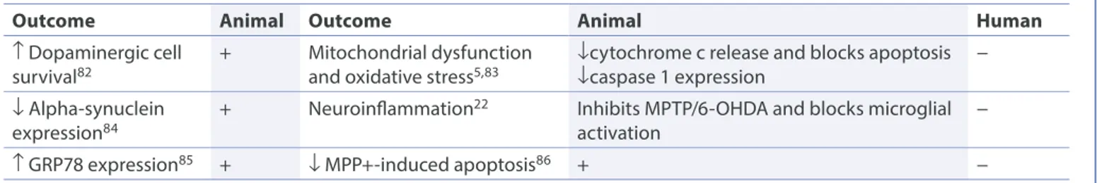
Table 2. Neuroprotective effects of minocycline on pathological features of Parkinson’s disease.81
Outcome Animal Outcome Animal Human
↑ Dopaminergic cell
survival82 + Mitochondrial dysfunction and oxidative stress5,83 ↓cytochrome c release and blocks apoptosis↓caspase 1 expression −
↓ Alpha-synuclein
expression84 + Neuroinflammation
22 Inhibits MPTP/6-OHDA and blocks microglial
activation −
↑ GRP78 expression85 + ↓ MPP+-induced apoptosis86 + −
6-OHDA, 6-hydroxy-dopamine; GRP78, glucose-regulated protein; MPP+, 1-methyl-4-phenylpyridinium, MPTP, 1-methyl-4-phenyl-1,2,3,6-tetrahydropyridine.
the minocycline group nor in the others regarding the primary indicator of disease progression that was defined as the time for starting a symptomatic pharmaceutical treatment.95
The results of that study were in accordance with another study assessing the efficacy of minocycline in patients with Multiple System Atrophy-Parkinsonian subtype (MSA-P) that also revealed no symptomatic beneficial effects of minocycline in MSA-P.96
Nowadays, there is still no randomized clinical minocycline therapy trial conducted on a massive scale.
Possible procognitive effects
of minocycline in clinical trials
of Parkinson’s disease and
Alzheimer's disease
Cognitive dysfunction is a common and significant nonmotor symptom of PD. PD mild cognitive impairment (PD-MCI) was defined in 2012 by the International Parkinson and Movement Disorder Society,97 and it has shown that approximately
one-third of people have PD-MCI.98,99
Longitudinal cohort studies have demonstrated that approximately half of the patients with PD for 10 years develop PD dementia (PDD),100 while the point prevalence of
dementia among those with PD is roughly 30%.101 Although
the underlying mechanisms of cognitive decline or dementia associated with PD remain unclear, it is believed that the presence of Lewy pathology within the limbic system and neocortex play a critical role in the impairment of cognition in PD. Lewy bodies are abnormal alpha-synuclein (α-syn) proteins, which are greatly responsible for the neurotoxicity via different mechanisms, including impairment of axonal transport, oxidative stress, mitochondrial changes, and synaptic dysfunction.102
Despite increased understanding of pathology, neurotransmitter, and genetic drivers, there are no proven pharmacological treatments or protective agents for PD-MCI. Moreover, studies until now have shown that there is an only modest benefit for antidementia drugs that are licensed for PDD. These findings together suggest that there is an emerging need for a mechanism-based clinical treatment strategy for cognitive impairment in PD.103
In this respect, it is worth to notice, that a new study, The minocycline in Alzheimer’s Disease Efficacy (MADE) study, is a multicenter, randomized, controlled trial in patients with very mild AD.104 Recruitment began in January 2014 and the
trial ended in December 2017. It aimed to determine whether minocycline is effective in reducing the rate of cognitive and functional decline over a 2-year period and assess the safety and tolerability of minocycline. This multicenter, randomized double-blind placebo-controlled semifactorial (2×1) designed phase II clinical trial tested the efficacy and tolerability of 400 and 200 mg minocycline in disease modification of AD. There were 480 patients with very mild AD patients recruited hypothesized that minocycline may normalize the activation
of microglia astrocytes and brain-derived neurotrophic factor expression.87
Additionally, in experimental models of LPS-induced neuroinflammation, minocycline reversed the nigral dopaminergic neuronal cell death and the loss of reactive astrocytes that were associated with improved cognitive outcomes.88,89
Consistently, minocycline has been also shown to improve neurobehavioral deficits and cognitive decline in AD animal models that were related to the inhibition of neuroinflammatory markers, Aβ fibril formation, and NMDA toxicity.35,55,56,90 Agreeably, neuroinflammation,
including reactive astrocytes and activated microglia, correlates with a serious cognitive decline and brain atrophy in AD.91,92
Neuroprotective effects
of minocycline in clinical
trials of Parkinson’s disease
and Parkinson plus syndromes
Several mechanisms of action have been proposed to be responsible for the neuroprotective effects of minocycline, which include the attenuation of microglial activation, apoptosis, and ROS production.
Regarding the neuroprotective properties of minocycline, the National Institute of Neurological Disorders and Stroke (NINDS) has aimed to identify potential agents to modify disease progression in PD. Through the Neuroprotective Exploratory Trials in Parkinson’s Disease (NET-PD) program, creatine and minocycline were used for the first study over 12 months (The NET-PD Futility Study 1, NET-PD FS-1).93
Although there was evidence that both agents can protect against the dopaminergic neuronal loss associated with MPTP administration in a mouse model of PD, this study has failed to confirm the clinical neuroprotective effect of minocycline. An additional 6 months of follow up aimed to assess the safety and potential interactions of the study interventions with anti-Parkinsonian therapy (NET-PD FS-2). At the end of following-up for 18 months, this phase II clinical study suggested that there was neither a beneficial effect nor an adverse event from using either creatine or minocycline in the treatment of PD. The authors commented that there was a need for a larger, phase III efficacy trial.94
NET-PD investigators have also conducted a recent cohort-analysis to measure the disease progression in early PD through analyses an evaluation of progression-sensitive scales and questionnaires. Evaluating the effect of minocycline, creatine, Coenzyme Q10 and GPI-1485, a product candidate that belongs to a class of small molecule compounds called neuroimmunophilin ligands, and placebo in different control and patient groups, they have found no difference, neither in
Numerous in vitro and in vivo experimental studies have pointed toward minocycline as a promising agent in the protection of PD; however, clinical studies have failed to confirm the neuroprotective effect of minocycline in PD. Considering all of these promising preclinical data, it may be reasonable to assume that minocycline is a neuroprotective candidate with its well-known effect on different pathological pathways in neurodegenerative diseases.
PD affects about 1 million Americans110 with annual direct
medical care costs of more than $US10,000 per patient.111 If
clinical efficacy were ever proven, then minocycline would be a very cheap therapeutic option. Although we consider our perspective review findings to be significant the confirmation of preclinical results from clinical studies are still lacking. In this respect, further well-designed clinical studies with a larger number of patients with Parkinson’s disease would be the next logical step in this field of clinical neuroprotection research. in England and Scotland. The patients were assessed with
Standardized Mini-Mental State Examination and the Bristol Activities of Daily Living Scale.104 The results of the study have
not been published yet.
Conclusions
Minocycline is a tetracycline that exerts antimicrobial, anti-inflammatory, anti-apoptotic, and antioxidant effects on the CNS.106 There is rapidly growing evidence showing that
neurodegeneration and inflammation are fundamental aspects of many neurological diseases.109 In this respect,
much attention has been focused on the potential use of currently available anti-inflammatory drugs to prevent neurodegeneration. In our perspective review, we have summarized various studies in animals and humans, which have confirmed beneficial effects and safety of minocycline, alone or combined with other drugs in PD.
Contributions: S Cankaya, B Cankaya and B Yulug were responsible for writing the article; E Kilica and U Kilic were responsible for revising
the article; E Kilic and U Kilic carried out a language check of the article; S Cankaya and B Cankaya prepared the data for the article. All named authors meet the International Committee of Medical Journal Editors (ICMJE) criteria for authorship for this article, take responsibility for the integrity of the work as a whole, and have given their approval for this version to be published.
Disclosure and potential conflicts of interest: The authors declare that they have no conflicts of interest. The International Committee
of Medical Journal Editors (ICMJE) Potential Conflicts of Interests form for the authors are available for download at http://www.drugsincontext. com/wp-content/uploads/2019/02/dic.212553-COI.pdf
Acknowledgments: None.
Funding declaration: There is no funding for assisting this article for writing or studying.
Copyright: Copyright © 2019 Cankaya S, Cankaya B, Kilic U, Kilic E, Yulug B. Published by Drugs in Context under Creative Commons License Deed CC BY NC ND 4.0 which allows anyone to copy, distribute, and transmit the article provided it is properly attributed in the manner specified below. No commercial use without permission.
Correct attribution: Copyright © 2019 Cankaya S, Cankaya B, Kilic U, Kilic E, Yulug B. https://doi.org/10.7573/dic.212553. Published by Drugs in Context under Creative Commons License Deed CC BY NC ND 4.0.
Article URL: https://www.drugsincontext.com/the-therapeutic-role-of-minocycline-in-parkinsons-disease
Correspondence: Seyda Cankaya, Department of Neurology, Faculty of Medicine, Alaaddin Keykubat University, Alanya, Turkey.
Provenance: invited; externally peer reviewed.
Submitted: 9 August 2018; Peer review comments to author: 26 November 2018; Revised manuscript received: 20 January 2019; Accepted: 22 January 2019; Publication date: 6 March 2019.
Drugs in Context is published by BioExcel Publishing Ltd. Registered office: Plaza Building, Lee High Road, London, England, SE13 5PT.
BioExcel Publishing Limited is registered in England Number 10038393. VAT GB 252 7720 07.
For all manuscript and submissions enquiries, contact the Editor-in-Chief [email protected]
For all permissions, rights and reprints, contact David Hughes [email protected]
References
1. Alexi T, Borlongan CV, Faull RL, et al. Neuroprotective strategies for basal ganglia degeneration: Parkinson’s and Huntington’s diseases. Prog Neurobiol. 2000;60(5):409–470. https://doi.org/10.1016/s0301-0082(99)00032-5
2. Seabrook TJ, Jiang L, Maier M, Lemere CA. Minocycline affects microglia activation, Abeta deposition, and behavior in APP-tg mice. Glia. 2006;53(7):776–782. https://doi.org/10.1002/glia.20338
3. Chen M, Ona VO, Li M, et al. Minocycline inhibits caspase-1 and caspase-3 expression and delays mortality in a transgenic mouse model of Huntington disease. Nat Med. 2000;6(7):797–801. https://doi.org/10.1038/77528
4. Van Den Bosch L, Tilkin P, Lemmens G, Robberecht W. Minocycline delays disease onset and mortality in a transgenic model of ALS. Neuroreport. 2002;13(8):1067–1070. https://doi.org/10.1097/00001756-200206120-00018
5. Du Y, Ma Z, Lin S, et al. Minocycline prevents nigrostriatal dopaminergic neurodegeneration in the MPTP model of Parkinson’s disease. Proc Natl Acad Sci USA. 2001;98(25):14669–14674. https://doi.org/10.1073/pnas.251341998
6. Lang AE. Clinical trials of disease-modifying therapies for neurodegenerative diseases: the challenges and the future. Nat Med. 2010;16(11):1223–1226. https://doi.org/10.1038/nm.2220
7. Olanow CW, Kieburtz K, Schapira AH. Why have we failed to achieve neuroprotection in Parkinson’s disease? Ann Neurol. 2008;64 Suppl 2(S2):S101–110. https://doi.org/10.1002/ana.21461
8. Olanow CW, Kieburtz K. Defining disease-modifying therapies for PD – a road map for moving forward. Mov Disord. 2010;25(12):1774–1779. https://doi.org/10.1002/mds.23288
9. Aronson AL. Pharmacotherapeutics of the newer tetracyclines. J Am Vet Med Assoc. 1980;176(10 Spec No):1061–1068. PubMed PMID: 7216873
10. Barza M, Brown RB, Shanks C, Gamble C, Weinstein L. Relation between lipophilicity and pharmacological behavior of minocycline, doxycycline, tetracycline, and oxytetracycline in dogs. Antimicrob Agents Chemother. 1975;8(6):713–720. https://doi.org/10.1128/aac.8.6.713
11. Klein NC, Cunha BA. Tetracyclines. Med Clin North Am. 1995;79(4):789–801. PubMed PMID: 7791423
12. Brogden R, Speight T, Avery G. Minocycline: a review of its antibacterial and pharmacokinetic properties and therapeutic use.
Drugs. 1975;9(4):251–291. PubMed PMID: 1173232
13. Yrjanheikki J, Tikka T, Keinanen R, Goldsteins G, Chan PH, Koistinaho J. A tetracycline derivative, minocycline, reduces inflammation and protects against focal cerebral ischemia with a wide therapeutic window. Proc Natl Acad Sci USA. 1999;96(23):13496–13500. https://doi.org/10.1073/pnas.96.23.13496
14. Kielian T, Esen N, Liu S, et al. Minocycline modulates neuroinflammation independently of its antimicrobial activity in staphylococcus aureus-induced brain abscess. Am J Pathol. 2007;171(4):1199–1214. https://doi.org/10.2353/ajpath.2007.070231 15. Saivin S, Houin G. Clinical pharmacokinetics of doxycycline and minocycline. Clin. Pharmacokinet. 1988;15(6):355–366.
https://doi.org/10.2165/00003088-198815060-00001
16. Wang X, Zhu S, Drozda M, et al. Minocycline inhibits caspase-independent and -dependent mitochondrial cell death pathways in models of Huntington’s disease. Proc Natl Acad Sci USA. 2003;100(18):10483–10487. https://doi.org/10.1073/pnas.1832501100 17. Yong VW, Wells J, Giuliani F, Casha S, Power C, Metz LM. The promise of minocycline in neurology. Lancet Neurol.
2004;3(12):744–751. https://doi.org/10.1016/S1474-4422(04)00937-8
18. Colovic M, Caccia S. Liquid chromatographic determination of minocycline in brain-to-plasma distribution studies in the rat. J
Chromatogr B. 2003;791(1–2):337–343. https://doi.org/10.1016/s1570-0232(03)00247-2
19. Smith DL, Woodman B, Mahal A, et al. Minocycline and doxycycline are not beneficial in a model of Huntington’s disease. Ann
Neurol. 2003;54(2):186–196. https://doi.org/10.1002/ana.10614
20. Luccarini I, Ballerini C, Biagioli T, et al. Combined treatment with atorvastatin and minocycline suppresses severity of EAE. Exp
Neurol. 2008;211(1):214–226. https://doi.org/10.1016/j.expneurol.2008.01.022
21. Sapadin AN, Fleischmajer R. Tetracyclines: nonantibiotic properties and their clinical implications. J Am Acad Dermatol. 2006;54(2):258–265. https://doi.org/10.1016/j.jaad.2005.10.004
22. Jackson-Lewis V, Vila M, Tieu K, et al. Blockade of microglial activation is neuroprotective in the 1-methyl-4-phenyl-1, 2, 3, 6-tetrahydropyridine mouse model of Parkinson disease. J Neurosci. 2002;22(5):1763–1771.
https://doi.org/10.1523/JNEUROSCI.22-05-01763.2002
23. Casarejos MJ, Menendez J, Solano RM, Rodriguez-Navarro JA, Garcia de Yebenes J, Mena MA. Susceptibility to rotenone is increased in neurons from parkin null mice and is reduced by minocycline. J Neurochem. 2006;97(4):934–946.
https://doi.org/10.1111/j.1471-4159.2006.03777.x
24. Kim HS, Suh YH. Minocycline and neurodegenerative diseases. Behav Brain Res. 2009;196(2):168–179. https://doi.org/10.1016/j.bbr.2008.09.040
25. Rogers J, Strohmeyer R, Kovelowski C, Li R. Microglia and inflammatory mechanisms in the clearance of amyloid β peptide.
Glia. 2002;40(2):260–269. https://doi.org/10.1002/glia.10153
26. McGeer P, Itagaki S, Boyes B, McGeer E. Reactive microglia are positive for HLA‐DR in the substantia nigra of Parkinson’s and Alzheimer’s disease brains. Neurology. 1988;38(8):1285–1285. PubMed PMID: 3399080
27. Hu X, Li P, Guo Y, et al. Microglia/macrophage polarization dynamics reveal novel mechanism of injury expansion after focal cerebral ischemia. Stroke. 2012;43(11):3063–3070. https://doi.org/10.1161/STROKEAHA.112.659656
28. Cagnin A, Kassiou M, Meikle S, Banati R. In vivo evidence for microglial activation in neurodegenerative dementia. Acta Neurol
Scand. 2006;114:107–114. https://doi.org/10.1111/j.1600-0404.2006.00694.x
29. Wang Q, Liu Y, Zhou J. Neuroinflammation in Parkinson’s disease and its potential as therapeutic target. Transl Neurodegener. 2015;4(1):19. https://doi.org/10.1186/s40035-015-0042-0
30. McCoy MK, Martinez TN, Ruhn KA, et al. Blocking soluble tumor necrosis factor signaling with dominant-negative tumor necrosis factor inhibitor attenuates loss of dopaminergic neurons in models of Parkinson’s disease. J Neurosci. 2006;26(37):9365–9375. https://doi.org/10.1523/JNEUROSCI.1504-06.2006
31. Ferrari CC, Godoy MCP, Tarelli R, Chertoff M, Depino AM, Pitossi FJ. Progressive neurodegeneration and motor disabilities induced by chronic expression of IL-1β in the substantia nigra. Neurobiol Dis. 2006;24(1):183–193. https://doi.org/10.1016/j.nbd.2006.06.013 32. Gabler WL, Smith J, Tsukuda N. Comparison of doxycycline and a chemically modified tetracycline inhibition of leukocyte
functions. Res Commun Chem Pathol Pharmacol. 1992;78(2):151–160. PubMed PMID: 1335592
33. Stirling DP, Koochesfahani KM, Steeves JD, Tetzlaff W. Minocycline as a neuroprotective agent. Neuroscientist. 2005;11(4):308–322. https://doi.org/10.1177/1073858405275175
34. Haines JD, Fragoso G, Hossain S, Mushynski WE, Almazan G. p38 Mitogen-activated protein kinase regulates myelination. J Mol
Neurosci. 2008;35(1):23–33. https://doi.org/10.1007/s12031-007-9011-0
35. Tikka TM, Koistinaho JE. Minocycline provides neuroprotection against N-methyl-D-aspartate neurotoxicity by inhibiting microglia. J Immunol. 2001;166(12):7527–7533. https://doi.org/10.4049/jimmunol.166.12.7527
36. Lin S, Zhang Y, Dodel R, Farlow MR, Paul SM, Du Y. Minocycline blocks nitric oxide-induced neurotoxicity by inhibition p38 MAP kinase in rat cerebellar granule neurons. Neurosci Lett. 2001;315(1–2):61–64.
https://doi.org/10.1016/s0304-3940(01)02324-2
37. Guo G, Bhat NR. p38alpha MAP kinase mediates hypoxia-induced motor neuron cell death: a potential target of minocycline’s neuroprotective action. Neurochem Res. 2007;32(12):2160–2166. https://doi.org/10.1007/s11064-007-9408-8
38. Friedlander RM. Apoptosis and caspases in neurodegenerative diseases. N Engl J Med. 2003;348(14):1365–1375. https://doi.org/10.1056/NEJMra022366
39. Jellinger KA. Cell death mechanisms in neurodegeneration. J Cell Mol Med. 2001;5(1):1–17. https://doi.org/10.1111/j.1582-4934.2001.tb00134.x
40. Tatton NA. Increased caspase 3 and Bax immunoreactivity accompany nuclear GAPDH translocation and neuronal apoptosis in Parkinson’s disease. Exp Neurol. 2000;166(1):29–43. https://doi.org/10.1006/exnr.2000.7489
41. Mochizuki H, Goto K, Mori H, Mizuno Y. Histochemical detection of apoptosis in Parkinson’s disease. J Neurol Sci. 1996;137(2):120– 123. https://doi.org/10.1016/0022-510x(95)00336-z
42. Thomas M, Le WD. Minocycline: neuroprotective mechanisms in Parkinson’s disease. Curr Pharm Des. 2004;10(6):679–686. https://doi.org/10.2174/1381612043453162
43. Choi Y, Kim HS, Shin KY, et al. Minocycline attenuates neuronal cell death and improves cognitive impairment in Alzheimer’s disease models. Neuropsychopharmacology. 2007;32(11):2393–2404. https://doi.org/10.1038/sj.npp.1301377
44. Stirling DP, Khodarahmi K, Liu J, et al. Minocycline treatment reduces delayed oligodendrocyte death, attenuates axonal dieback, and improves functional outcome after spinal cord injury. J Neurosci. 2004;24(9):2182–2190.
https://doi.org/10.1523/JNEUROSCI.5275-03.2004
45. Garcia-Martinez EM, Sanz-Blasco S, Karachitos A, et al. Mitochondria and calcium flux as targets of neuroprotection caused by minocycline in cerebellar granule cells. Biochem Pharmacol. 2010;79(2):239–250. https://doi.org/10.1016/j.bcp.2009.07.028 46. Antonenko YN, Rokitskaya TI, Cooper AJ, Krasnikov BF. Minocycline chelates Ca2+, binds to membranes, and depolarizes
mitochondria by formation of Ca2+-dependent ion channels. J Bioenerg Biomembr. 2010;42(2):151–163. https://doi.org/10.1007/s10863-010-9271-1
47. O Ulgen B, G Field M, Qureshi W, et al. The role of minocycline in ischemia-reperfusion injury: a comprehensive review of an old drug with new implications. Recent Pat Cardiovasc Drug Discov. 2011;6(2):123–132. https://doi.org/10.2174/157489011795933783 48. Filipovic R, Zecevic N. Neuroprotective role of minocycline in co-cultures of human fetal neurons and microglia. Exp Neurol.
2008;211(1):41–51. https://doi.org/10.1016/j.expneurol.2007.12.024
49. Green DR, Kroemer G. The pathophysiology of mitochondrial cell death. Science. 2004;305(5684):626–629. https://doi.org/10.1126/science.1099320
50. Kroemer G. Mitochondrial control of apoptosis: an overview. Biochem. Soc. Symp. 1999;66(32):1–8. https://doi.org/10.1042/bss0660001 51. Wang J, Wei Q, Wang CY, Hill WD, Hess DC, Dong Z. Minocycline up-regulates Bcl-2 and protects against cell death in
mitochondria. J Biol Chem. 2004;279(19):19948–19954. https://doi.org/10.1074/jbc.M313629200
52. Castanares M, Vera Y, Erkkila K, et al. Minocycline up-regulates BCL-2 levels in mitochondria and attenuates male germ cell apoptosis. Biochem Biophys Res Commun. 2005;337(2):663–669. https://doi.org/10.1016/j.bbrc.2005.09.101
53. Yrjanheikki J, Keinanen R, Pellikka M, Hokfelt T, Koistinaho J. Tetracyclines inhibit microglial activation and are neuroprotective in global brain ischemia. Proc Natl Acad Sci USA. 1998;95(26):15769–15774. https://doi.org/10.1073/pnas.95.26.15769
54. Zhu S, Stavrovskaya IG, Drozda M, et al. Minocycline inhibits cytochrome c release and delays progression of amyotrophic lateral sclerosis in mice. Nature. 2002;417(6884):74–78. https://doi.org/10.1038/417074a
55. Familian A, Boshuizen RS, Eikelenboom P, Veerhuis R. Inhibitory effect of minocycline on amyloid β fibril formation and human microglial activation. Glia. 2006;53(3):233–240. https://doi.org/10.1002/glia.20268
56. Fan R, Xu F, Previti ML, et al. Minocycline reduces microglial activation and improves behavioral deficits in a transgenic model of cerebral microvascular amyloid. J Neurosci. 2007;27(12):3057–3063. https://doi.org/10.1523/JNEUROSCI.4371-06.2007
57. Parachikova A, Vasilevko V, Cribbs DH, LaFerla FM, Green KN. Reductions in amyloid-beta-derived neuroinflammation, with minocycline, restore cognition but do not significantly affect tau hyperphosphorylation. J Alzheimers Dis. 2010;21(2):527–542. https://doi.org/10.3233/JAD-2010-100204
58. Wang CX, Yang T, Shuaib A. Effects of minocycline alone and in combination with mild hypothermia in embolic stroke. Brain Res. 2003;963(1–2):327–329. https://doi.org/10.1016/s0006-8993(02)04045-3
59. Stack EC, Smith KM, Ryu H, et al. Combination therapy using minocycline and coenzyme Q10 in R6/2 transgenic Huntington’s disease mice. Biochim Biophys Acta. 2006;1762(3):373–380. https://doi.org/10.1016/j.bbadis.2005.11.002
60. Van Den Bosch L, Tilkin P, Lemmens G, Robberecht W. Minocycline delays disease onset and mortality in a transgenic model of ALS. Neuroreport. 2002;13(8):1067–1070. https://doi.org/10.1097/00001756-200206120-00018
61. Kriz J, Nguyen MD, Julien JP. Minocycline slows disease progression in a mouse model of amyotrophic lateral sclerosis. Neurobiol
Dis. 2002;10(3):268–278. https://doi.org/10.1006/nbdi.2002.0487
62. Traynor BJ, Bruijn L, Conwit R, et al. Neuroprotective agents for clinical trials in ALS: a systematic assessment. Neurology. 2006;67(1):20–27. https://doi.org/10.1212/01.wnl.0000223353.34006.54
63. Garcez ML, Mina F, Bellettini-Santos T, et al. The Involvement of NLRP3 on the Effects of Minocycline in an AD-Like Pathology Induced by β-Amyloid Oligomers Administered to Mice. Mol Neurobiol. 2018:1–12. https://doi.org/10.1007/s12035-018-1211-9 64. Metz LM, Li DK, Traboulsee AL, et al. Trial of minocycline in a clinically isolated syndrome of multiple sclerosis. N Engl J Med.
2017;376(22):2122–2133. https://doi.org/10.1056/NEJMc1708486
65. Kuroda A, Fuchigami T, Fuke S, Koyama N, Ikenaka K, Hitoshi S. Minocycline directly enhances the self-renewal of adult neural precursor cells. Neurochem Res. 2018;43(1):219–226. https://doi.org/10.1007/s11064-017-2422-6
66. Dauer W, Przedborski S. Parkinson’s disease: mechanisms and models. Neuron. 2003;39(6):889–909. https://doi.org/10.1016/S0896-6273(03)00568-3
67. Jiang BP, Le L, Xu LJ, Xiao PG. Minocycline inhibits ICAD degradation and the NF-kappaB activation induced by 6-OHDA in PC12 cells. Brain Res. 2014;1586:1–11. https://doi.org/10.1016/j.brainres.2014.08.001
68. Worlitzer MM, Viel T, Jacobs AH, Schwamborn JC. The majority of newly generated cells in the adult mouse substantia nigra express low levels of D oublecortin, but their proliferation is unaffected by 6‐OHDA‐induced nigral lesion or M inocycline‐ mediated inhibition of neuroinflammation. Eur J Neurosci. 2013;38(5):2684–2692. https://doi.org/10.1111/ejn.12269
69. Radad K, Moldzio R, Rausch WD. Minocycline protects dopaminergic neurons against long-term rotenone toxicity. Can J Neurol
Sci. 2010;37(1):81–85. https://doi.org/10.1017/s0317167100009690
70. Cronin A, Grealy M. Neuroprotective and Neuro-restorative Effects of Minocycline and Rasagiline in a Zebrafish 6-Hydroxydopamine Model of Parkinson’s Disease. Neuroscience. 2017;367:34–46.
https://doi.org/10.1016/j.neuroscience.2017.10.018
71. Meulendyke KA, Pletnikov MV, Engle EL, Tarwater PM, Graham DR, Zink MC. Early minocycline treatment prevents a decrease in striatal dopamine in an SIV model of HIV-associated neurological disease. J Neuroimmune Pharmacol. 2012;7(2):454–464. https://doi.org/10.1007/s11481-011-9332-1
72. Tripathi P, Singh A, Agrawal S, Prakash O, Singh MP. Cypermethrin alters the status of oxidative stress in the peripheral blood: relevance to Parkinsonism. J Physiol Biochem. 2014;70(4):915–924. https://doi.org/10.1007/s13105-014-0359-7
73. Purisai MG, McCormack AL, Cumine S, Li J, Isla MZ, Di Monte DA. Microglial activation as a priming event leading to paraquat-induced dopaminergic cell degeneration. Neurobiol Dis. 2007;25(2):392–400. https://doi.org/10.1016/j.nbd.2006.10.008 74. Yang L, Sugama S, Chirichigno JW, et al. Minocycline enhances MPTP toxicity to dopaminergic neurons. J Neurosci Res.
2003;74(2):278–285. https://doi.org/10.1002/jnr.10709
75. Diguet E, Fernagut PO, Wei X, et al. Deleterious effects of minocycline in animal models of Parkinson’s disease and Huntington’s disease. Eur J Neurosci. 2004;19(12):3266–3276. https://doi.org/10.1111/j.0953-816X.2004.03372.x
76. Noble W, Garwood CJ, Hanger DP. Minocycline as a potential therapeutic agent in neurodegenerative disorders characterized by protein misfolding. Prion. 2009;3(2):78–83. https://doi.org/10.4161/pri.3.2.8820
77. Noble W, Garwood C, Stephenson J, Kinsey AM, Hanger DP, Anderton BH. Minocycline reduces the development of abnormal tau species in models of Alzheimer’s disease. FASEB J. 2009;23(3):739–750. https://doi.org/10.1096/fj.08-113795
78. Ryu JK, Choi HB, McLarnon JG. Combined minocycline plus pyruvate treatment enhances effects of each agent to inhibit inflammation, oxidative damage, and neuronal loss in an excitotoxic animal model of Huntington’s disease. Neuroscience. 2006;141(4):1835–1848. https://doi.org/10.1016/j.neuroscience.2006.05.043
79. Dommergues M-A, Plaisant F, Verney C, Gressens P. Early microglial activation following neonatal excitotoxic brain damage in mice: a potential target for neuroprotection. Neuroscience. 2003;121(3):619–628. https://doi.org/10.1016/s0306-4522(03)00558-x 80. He Y, Appel S, Le W. Minocycline inhibits microglial activation and protects nigral cells after 6-hydroxydopamine injection into
81. Yulug B, Hanoglu L, Kilic E, Schabitz WR. RIFAMPICIN: an antibiotic with brain protective function. Brain Res Bull. 2014;107:37–42. https://doi.org/10.1016/j.brainresbull.2014.05.007
82. Kilic U, Kilic E, Lingor P, Yulug B, Bahr M. Rifampicin inhibits neurodegeneration in the optic nerve transection model in vivo and after 1-methyl-4-phenylpyridinium intoxication in vitro. Acta Neuropathol. 2004;108(1):65–68.
https://doi.org/10.1007/s00401-004-0867-6
83. Teng YD, Choi H, Onario RC, et al. Minocycline inhibits contusion-triggered mitochondrial cytochrome c release and mitigates functional deficits after spinal cord injury. Proc Natl Acad Sci USA. 2004;101(9):3071–3076.
https://doi.org/10.1073/pnas.0306239101
84. Oida Y, Kitaichi K, Nakayama H, et al. Rifampicin attenuates the MPTP-induced neurotoxicity in mouse brain. Brain Res. 2006;1082(1):196–204. https://doi.org/10.1016/j.brainres.2006.01.116
85. Xu J, Wei C, Xu C, et al. Rifampicin protects PC12 cells against MPP+-induced apoptosis and inhibits the expression of an alpha-Synuclein multimer. Brain Res. 2007;1139:220–225. https://doi.org/10.1016/j.brainres.2006.12.074
86. Li J, Zhu M, Rajamani S, Uversky VN, Fink AL. Rifampicin inhibits alpha-synuclein fibrillation and disaggregates fibrils. Chem Biol. 2004;11(11):1513–1521. https://doi.org/10.1016/j.chembiol.2004.08.025
87. Hou Y, Xie G, Liu X, et al. Minocycline protects against lipopolysaccharide-induced cognitive impairment in mice.
Psychopharmacology (Berl.). 2016;233(5):905–916. https://doi.org/10.1007/s00213-015-4169-6
88. Kim WG, Mohney RP, Wilson B, Jeohn GH, Liu B, Hong JS. Regional difference in susceptibility to lipopolysaccharide-induced neurotoxicity in the rat brain: role of microglia. J Neurosci. 2000;20(16):6309–6316.
https://doi.org/10.1523/JNEUROSCI.20-16-06309.2000
89. Tomas-Camardiel M, Rite I, Herrera AJ, et al. Minocycline reduces the lipopolysaccharide-induced inflammatory reaction, peroxynitrite-mediated nitration of proteins, disruption of the blood-brain barrier, and damage in the nigral dopaminergic system. Neurobiol Dis. 2004;16(1):190–201. https://doi.org/10.1016/j.nbd.2004.01.010
90. Biscaro B, Lindvall O, Tesco G, Ekdahl CT, Nitsch RM. Inhibition of microglial activation protects hippocampal neurogenesis and improves cognitive deficits in a transgenic mouse model for Alzheimer’s disease. Neurodegener Dis. 2012;9(4):187–198.
https://doi.org/10.1159/000330363
91. McGeer PL, McGeer EG. Anti-inflammatory drugs in the fight against Alzheimer’s disease. Ann NY Acad Sci. 1996;777(1):213–220. https://doi.org/10.1111/j.1749-6632.1996.tb34421.x
92. Sokolova A, Hill MD, Rahimi F, Warden LA, Halliday GM, Shepherd CE. Monocyte chemoattractant protein-1 plays a dominant role in the chronic inflammation observed in Alzheimer’s disease. Brain Pathol. 2009;19(3):392–398.
https://doi.org/10.1111/j.1750-3639.2008.00188.x
93. Investigators NN-P. A randomized, double-blind, futility clinical trial of creatine and minocycline in early Parkinson disease.
Neurology. 2006;66(5):664–671. https://doi.org/10.1212/01.wnl.0000201252.57661.e1
94. Investigators NN-P. A pilot clinical trial of creatine and minocycline in early Parkinson disease: 18-month results. Clin
Neuropharmacol. 2008;31(3):141–150. https://doi.org/10.1097/WNF.0b013e3181342f32
95. Parashos SA, Luo S, Biglan KM, et al. Measuring disease progression in early Parkinson disease: the National Institutes of Health Exploratory Trials in Parkinson Disease (NET-PD) experience. JAMA Neurol. 2014;71(6):710–716.
https://doi.org/10.1001/jamaneurol.2014.391
96. Dodel R, Spottke A, Gerhard A, et al. Minocycline 1-year therapy in multiple-system-atrophy: effect on clinical symptoms and [(11)C] (R)-PK11195 PET (MEMSA-trial). Mov Disord. 2010;25(1):97–107. https://doi.org/10.1002/mds.22732
97. Litvan I, Goldman JG, Tröster AI, et al. Diagnostic criteria for mild cognitive impairment in Parkinson’s disease: Movement Disorder Society Task Force guidelines. Mov Disord. 2012;27(3):349–356. https://doi.org/10.1159/000433421
98. Broeders M, De Bie R, Velseboer D, Speelman J, Muslimovic D, Schmand B. Evolution of mild cognitive impairment in Parkinson disease. Neurology. 2013;81:346–352. https://doi.org/10.1212/wnl.0b013e31829c5c86
99. Yarnall AJ, Breen DP, Duncan GW, et al. Characterizing mild cognitive impairment in incident Parkinson disease: the ICICLE-PD study. Neurology. 2014;82(4):308–316. https://doi.org/10.1212/WNL.0000000000000066
100. Williams-Gray CH, Mason SL, Evans JR, et al. The CamPaIGN study of Parkinson’s disease: 10-year outlook in an incident population-based cohort. J Neurol Neurosurg Psychiatry. 2013;84(11):1258–1264. https://doi.org/10.1136/jnnp-2013-305277 101. Aarsland D, Zaccai J, Brayne C. A systematic review of prevalence studies of dementia in Parkinson’s disease. Mov Disord.
2005;20(10):1255–1263. https://doi.org/10.1002/mds.20527
102. Irwin DJ, Lee VM, Trojanowski JQ. Parkinson’s disease dementia: convergence of alpha-synuclein, tau and amyloid-beta pathologies. Nat Rev Neurosci. 2013;14(9):626–636. https://doi.org/10.1038/nrn3549
103. Cosgrove J, Alty JE. Cognitive deficits in Parkinson’s disease: current perspectives. J Parkinsonism Restless Legs Syndrome. 2018;8:1–11. https://doi.org/10.2147/jprls.s125064
104. Howard R. Minocycline in Alzheimer’s disease efficacy trial: the MADE trial. https://gtr.ukri.org/projects?ref=MC_PC_13091. Accessed January 30, 2019.
105. Lin S, Wei X, Xu Y, et al. Minocycline blocks 6-hydroxydopamine-induced neurotoxicity and free radical production in rat cerebellar granule neurons. Life Sci. 2003;72(14):1635–1641. https://doi.org/10.1016/s0024-3205(02)02442-6
106. Quintero EM, Willis L, Singleton R, et al. Behavioral and morphological effects of minocycline in the 6-hydroxydopamine rat model of Parkinson’s disease. Brain Res. 2006;1093(1):198–207. https://doi.org/10.1016/j.brainres.2006.03.104
107. Peng J, Xie L, Stevenson FF, Melov S, Di Monte DA, Andersen JK. Nigrostriatal dopaminergic neurodegeneration in the weaver mouse is mediated via neuroinflammation and alleviated by minocycline administration. J Neurosci. 2006;26(45):11644–11651. https://doi.org/10.1523/JNEUROSCI.3447-06.2006
108. Faust K, Gehrke S, Yang Y, Yang L, Beal MF, Lu B. Neuroprotective effects of compounds with antioxidant and anti-inflammatory properties in a Drosophila model of Parkinson’s disease. BMC Neurosci. 2009;10(1):109. https://doi.org/10.1186/1471-2202-10-109 109. Lidman O, Swanberg M, Horvath L, Broman KW, Olsson T, Piehl F. Discrete gene loci regulate neurodegeneration, lymphocyte
infiltration, and major histocompatibility complex class II expression in the CNS. J Neurosci. 2003;23(30):9817–9823. https://doi.org/10.1523/jneurosci.23-30-09817.2003
110. Schapira AH, Olanow CW. Neuroprotection in Parkinson disease: mysteries, myths, and misconceptions. JAMA. 2004;291(3):358–364. https://doi.org/10.1001/jama.291.3.358
111. Noyes K, Liu H, Li Y, Holloway R, Dick AW. Economic burden associated with Parkinson’s disease on elderly Medicare beneficiaries. Mov Disord. 2006;21(3):362–372. https://doi.org/10.1002/mds.20727