www.mme-journal.de
Novel Making of Bacterial Cellulose Blended Polymeric
Fiber Bandages
Esra Altun, Mehmet Onur Aydogdu, Fatma Koc, Maryam Crabbe-Mann, Francis Brako,
Rupy Kaur-Matharu, Gunes Ozen, Serap Erdem Kuruca, Ursula Edirisinghe,
Oguzhan Gunduz, and Mohan Edirisinghe*
E. Altun, M. O. Aydogdu, Dr. O. Gunduz
Department of Metallurgical and Materials Engineering Marmara University
Goztepe Campus, 34722 Istanbul, Turkey F. Koc
Department of Medical Microbiology Medipol University
Beykoz, 34810 Istanbul, Turkey
M. Crabbe-Mann, Dr. F. Brako, R. Kaur-Matharu, Prof. M. Edirisinghe Department of Mechanical Engineering
University College London (UCL) Torrington Place, London WC1E 7JE, UK E-mail: [email protected] G. Ozen
Department of Molecular Medicine Istanbul University
Capa, 34393 Istanbul, Turkey Prof. S. E. Kuruca
Department of Physiology Istanbul University Capa, 34393 Istanbul, Turkey Dr. U. Edirisinghe
Accident and Emergency Department Chelsea and Westminster Hospital
Fulham Road, Chelsea, London SW10 9NH, UK
DOI: 10.1002/mame.201700607
factors and pathogenic invaders.[1]
Com-promised integrity of the skin would jeop-ardize this naturally impenetrable barrier and any kind of skin trauma needs to be repaired as soon as possible. The nature of the human body is designed to withstand skin injuries by a physiological phenom-enon that aims to re-establish the unique formation for each skin layer. However, wound healing processes of the skin con-sists of a complex mechanism of biological regeneration, which is challenging to com-pletely mimic and involves many steps such as cell proliferation, migration, and epithelialization.[2] Additionally, burn wounds can require
spe-cial treatment to prevent fluid loss and infections.[3] Therefore,
wound dressing studies are being pursued by many in recent years with the purpose of finding an alternative way to support self-regeneration of skin.[4–6] Using polymeric fibers to create
the appropriate environment for cells to attach has signifi-cant importance and encouraging results have been achieved according to the previous studies.[7,8] With respect to the
prepa-ration of wound dressings, electrospinning is frequently used because of its fiber production capability and simplicity.[9,10]
However, weaving a wound dressing matt using electrospin-ning can be complicated by the fact of low yield and efficiency of the single-nozzle process and process control complica-tions of the multiple needle process. In this respect, a novel method, pressurized gyration, which is based on production of polymeric nanofibers using application of centrifugal force and dynamic fluid flow offer a viable alternative.[11] This process and
its sister-processes[11–14] are more amenable to the generation
of sizeable mats (wound bandages) and their mass production. Poly(methylmethacrylate) (PMMA) is a synthetic and a hydrophobic polymer, which is also easy to process. How-ever, PMMA fibers bring some limitations such as brittleness and low strength, which restricts its efficient use in engi-neering.[15,16] These limitations can be overcome by blending it
with a natural polymer.[17] Bacterial cellulose (BC) is a natural
polymer which is very promising for making wound healing materials due to its exceptional features such as high strength, durability, thermal stability, biocompatibility, and low cost.[18–21]
Also, various materials based on nongenotoxic and noncyto-toxic bacterial cellulose have been commercialized[22] and
pro-motes it as a major wound healing material. However, it is difficult to process. Thus, using BC together with PMMA can
Bandage Manufacture
Bacterial cellulose (BC) is a very promising biological material. However, at present its utilization is limited by difficulties in shape forming it. In this Communication, it is shown how this can be overcome by blending it with poly(methylmethacrylate) (PMMA) polymer. BC:PMMA fibers are produced by pressurized gyration of blended BC:PMMA solutions. Subsequently, BC:PMMA bandage-like scaffolds are generated with different blends. The products are investigated to determine their morphological and chemical fea-tures. Cell culture and proliferation tests are performed to obtain information on biocompatibility of the scaffolds.
1. Introduction
Skin is a crucial component of the human body and its primary objective is to create a protective shield against environmental
© 2018 The Authors. Published by WILEY-VCH Verlag GmbH & Co. KGaA, Weinheim. This is an open access article under the terms of the Creative Commons Attribution License, which permits use, distribution and re-production in any medium, provided the original work is properly cited.
www.advancedsciencenews.com www.mme-journal.de
help in generating biodegradable and biocompatible bandages to support wound healing on the skin.
In this communication, we show that BC:PMMA blends can be processed by pressurized gyration to obtain fibrous band-ages. Characterization of the chemical, morphological, and most importantly, in vitro properties of the products were used to assess its potential, and set the scene for a more detailed investigation on gyrospun BC containing bandage materials.
2. Experimental Section
2.1. Materials
BC was provided by the Department of Medical Microbi-ology, Medipol University (Istanbul, Turkey). PMMA (Mw ≈
120 000 g mol−1), dimethylformamide (DMF), and tetrahydro-furan (THF) were purchased from Sigma-Aldrich. Saos-2 cell line was purchased from ATCC HTB-85. DMEM, 10% fetal bovine serum, penicillin (100 units mL−1), streptomycin (100 g mL−1),
and dimethylsulfoxide (DMSO) were obtained from Sigma-Aldrich. ELISA plate reader was supplied from Rayto, China. All reagents were used without further purification.
2.2. Preparation of Blended Polymer Solutions
5 wt% and 10 wt% bacterial cellulose were sonicated in DMF:THF (50:50 wt ratio) using a sonifier (Branson Sonifier 250, BRANSON Ultrasonics Corporation, USA) at a power output of 100% for 1 h. PMMA solutions (20, 30, 40, and 50 wt%) were prepared using DMF:THF (50:50 wt ratio) in an air tight vial and magnetically stirred for 3 h at 50 °C. Solutions were blended in different concentrations with previously pre-pared 5 and 10 wt% BC solutions (Table 1). Final blends were magnetically stirred for 1 h at the ambient temperature (23 °C).
2.3. Pressurized Gyration
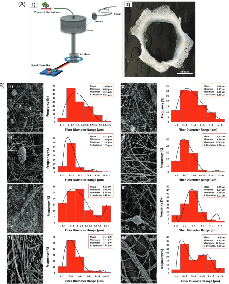
Gyration system used consists of a rotary aluminum cylin-drical vessel, ≈60 mm in diameter and ≈35 mm in height containing 20 round orifices on its surface (Figure 1A1). The
size of one orifice is 0.5 mm. Top of the vessel is connected to pressurized gas with a rotary joint. Flow of the N2 into the
vessel is controlled by a regulator. N2 pressure, can be varied
up to 3 × 10⁵ Pa. Bottom end of the vessel is connected to a DC
motor, which can generate speeds up to 36 000 rpm. High speed of the rotating vessel formed a polymer jet, which subsequently stretched into fibers through the orifices and it generated a fiber mesh (video provided in the Supporting Information). While forming the polymer jet, solvents evaporate from polymer solu-tion to generate the fibers. The stretching of polymer jet can be enhanced by blowing the gas under pressure into the vessel. To facilitate the collection of fibers, a stationary collector made of aluminum foil sheet was placed around the spinning vessel (on the transparent plastic protector surrounding the gyrator). Bracelet-like bandages were made from the BC:PMMA blends (Figure 1A2) and used in characterization tests. All experiments were conducted at a fixed rotating speed of 36 000 rpm, at a working pressure of 3 × 10⁵ Pa, at ambient temperature (23 °C)
and 42% relative humidity.
2.4. Characterization
Viscosity, density, and surface tension of all BC:PMMA blend solutions were determined. Viscosity was measured using a Brookfield DV-III ULTRA viscometer (Brookfield Viscometers Ltd, Harlow, UK). Density was measured using a standard 5 mL density bottle. Surface tension of the solutions was evalu-ated using a Kruss digital tensiometer (K9, Kruss GmbH, Ham-burg, Germany) by the Wilhelmy’s plate method. Equipment was calibrated before use and each test was carried out five times at ambient temperature (23 °C) and a relative humidity of 40–50%. Measurements are shown in Table 1.
A scanning electron microscope (SEM, JSM-6301F, JEOL) was used to characterize the surface morphology of the formed fibers at an accelerating voltage of 5 kV. Before imaging, sam-ples were coated with gold using a Quorum Q1500R ES sputter coater for 300 s. Fiber diameters were calculated using ImageJ analysis software. Fibers from 100 different locations of each image were measured and average, maximum, and min-imum diameters of fibers and standard deviation values were calculated.
Infrared spectra of fibers were recorded using a Fourier transform infrared spectroscope (FTIR, JASCO 6600, Japan) at the ambient temperature between 4400 and 400 cm−1 with a resolution of 4 cm−1.
In this preliminary work, Saos-2 cell line was used for cell viability assays. More pertinent cellular interactions are being investigated in follow-up studies. The scaffold samples were cut to 1 cm2 to fit into the 96-well cell culture polystyrene
plates. They were sterilized using UV rays for 15 min. Prior to the seeding, samples were maintained in DMEM (Dulbec-co’s modified Eagle’s medium) supplemented with 10% fetal bovine serum and penicillin (100 units mL−1), streptomycin
(100 g mL−1) at 37 °C in a humidified 5% CO2 atmosphere.
MTT assay (a colorimetric assay using tetrazolium dye MTT 3-(4,5-dimethylthiazol-2-yl)-2, 5-diphenyltetrazolium bromide) was performed on samples to determine any cytotoxic effect to measure the cell viability at the end of each time point.[23] Table 1. Physical properties of the solutions used in forming
experiments. BC:PMMA [wt ratio] Viscosity [mPa s] Density [kg m−3] Surface tension [mN m−1] 5:20 26.8 ± 1.6 930 ± 0.5 43.2 ± 0.4 10:20 78.6 ± 4.5 950 45.1 ± 0.7 5:30 510.4 ± 10.6 940 51.4 ± 1.3 10:30 1271.2 ± 29.3 976 ± 8.9 55.6 ± 1.3 5:40 11213 ± 95.6 952 ± 0.5 62.2 ± 1.1 10:40 5040.4 ± 27.5 978 ± 0.5 68.3 ± 1.1 5:50 15253 ± 96.2 994 ± 5.5 Too viscous to measure 10:50 77058.8 ± 1475.7 996 ± 5.5 Too viscous to measure
Figure 1. A) (1) Schematic illustrating the experimental setup of pressurized gyration and (2) typical bracelet-like bandages prepared from the 5:50 (wt ratio) BC:PMMA blend). B) Scanning electron images and corresponding fiber diameter distributions of the samples made from (1) 5:20, (2) 5:30, (3) 5:40, (4) 5:50, (5) 10:20, (6) 10:30, (7) 10:40, and (8) 10:50 BC:PMMA solutions.
www.advancedsciencenews.com www.mme-journal.de
For this purpose, cells were cultured in 96-well polystyrene plates with 104 cell/100 µL in each well, uniformly coated with
polymers, for 72 h. After treatment, 10 µL MTT incubation (5 mg mL−1) was performed in the wells for 4 h with
protec-tion from any light. Then, medium was discarded from wells and formazan crystals formed by MTT solution were dissolved with 200 µL DMSO. Finally, absorbance values were read using an ELISA plate reader at 570 nm, according to the 620 refer-ence wavelength. All experiments were repeated at least three times for all samples. Bandages were sterilized overnight with UV. After that, cells were seeded on the surface of the polymers at an approximate density of 106 cells per well in 6-well plates.
Cell cultures were maintained for 24 h. After this period, the cells were fixed with 2.5% glutaraldehyde. Subsequently, the samples were dehydrated in graded series of alcohol (30–100% ethanol in phosphate buffered saline (PBS)) for 15 min each. Following dehydration, they were left dry and stored at −20 °C until SEM imaging. DMEM medium containing the SAOS-2 cells in the well polystyrene plate was used as positive control.
3. Results and Discussion
Solution viscosity and surface tension are fundamental determi-nants of fiber formation such that both parameters depend on polymer concentration, molecular weight and polymer–solvent interactions,[24] and affect the two major process control
param-eters of the pressurized gyration process, i.e., rotation speed and applied pressure as discussed in our previous work.[11] In
this study, an increase in viscosity was observed on increasing BC:PMMA wt ratio. Likewise, a systematic increase in solution surface tension was observed with increasing BC:PMMA concen-tration (Table 1). Another key parameter which affects the genera-tion of fibers from a gyrating pot is solvent evaporagenera-tion. Trial and error experiments showed that the DMF:THF (50:50 wt ratio) mix-ture gave a high fiber yield and was therefore adapted in this work. Figure 1B1–8 shows SEM images of some particle containing fibrous products generated by pressurized gyration. Higher
amounts of particles are observed in Figure 1B5, compared with Figure 1B1. This indicates that particles are caused by increasing BC ratio in BC:PMMA blend solutions. Therefore, it is logical to infer that these particles are caused by the pres-ence of BC. However, as the PMMA concentration increased the mean fiber diameter increased and fiber diameter distribu-tion became wider and this is consistent with previous work[25]
and this increase coincides with changes in the viscosity and surface tension of the feed solutions (Table 1). Consequently, the 5:50 BC:PMMA composition (Figure 1B4) seems to offer the best compromise.
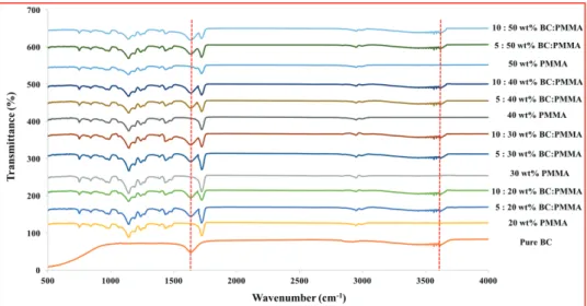
Figure 2 presents the FTIR spectra of pure BC and BC:PMMA
scaffold samples with varying BC:PMMA content. Comparison of the traces conducted on pure BC and BC:PMMA fibers confirmed the presence of BC. The broad absorption band in the region of wave number 3500 cm−1 represents the stretching of hydroxyl
groups,[26] 2900 and 1620 cm−1 shows the CH stretching and
the HOH bending of the absorbed water, respectively.[26]
Absorption band at 1060 cm−1 represents the COC pyranose ring skeletal vibration of BC.[26] Changes in bandwidth for the
absorption centered around 3500 and 1620 cm−1 characteristic of BC also occurred in the blended fiber samples.
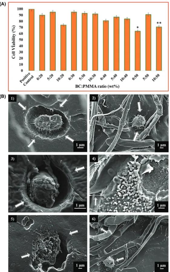
As a primary requirement of biocompatibility, a scaffold mate-rial must be non-toxic and allow cell attachment, proliferation, and differentiation.[27] As can be seen in Figure 3A, BC:PMMA
scaffolds were found to be biocompatible and there was no indi-cation of any toxicity on the Saos-2 cell line. At the end of the 72 h of cell culture, the growth of Saos-2 cells on all the BC:PMMA scaffolds was comparable to positive control group. The cell via-bility of the samples containing 10 wt% BC was lower compared with those containing 5 wt% BC for all compositions. The sig-nificant decrease in viability of the 50 wt% PMMA sample was associated with toxic effects of increased PMMA concentration. Even if significant decrease in viability was observed in 50 wt% PMMA, adding 5 wt% BC to 50 wt% PMMA clearly increased cell viability of Saos-2 cell line. Any wound dressing mate-rial that results in cell viability above 85% can be considered nontoxic for use as a wound dressing.[28] Therefore, BC:PMMA
Figure 2. FTIR spectra of BC:PMMA blend samples together with only PMMA and BC. (Main peak differences are indicated by lines between the spectra.)
Figure 3. A) MTT assay showing cell proliferation with respect to 72 h culture period of Saos-2 cell line. All the tested samples were relative to the control set at 100%. (P value < 0.05 is significant). B) Scanning electron microscope images of scaffold samples with different BC:PMMA ratios after incubation for 72 h: (1) 5:30, (2) 10:20, (3) 5:40, (4) 10:30, (5) 5:50, and (6) 10:40. Arrows in the images shows embedded cells and extension of the cells.
www.advancedsciencenews.com www.mme-journal.de
scaffolds including 5 wt% BC would be considered appropriate based on their effects on the Saos-2 cell line.
Cell behavior on different bandage (scaffold) samples with different BC:PMMA ratios was also observed by SEM imaging. After 72 h of incubation with cells, DAPI stained cells were analyzed. As can be seen in Figure 3B BC:PMMA scaffold sam-ples have demonstrated suitable degrees of cell spreading and proliferation. Compared with controls, all samples exhibited improved metabolic activity. However, 5 wt% BC containing scaffolds showed enhanced proliferation than 10 wt% BC scaf-folds. The slow proliferation rate can be related with insolu-bility of the BC:PMMA scaffolds (containing 10 wt% BC) and their slow deposition on the polystyrene plate.[29] The MTT
assay also indicated that the scaffolds prepared using 5 wt% BC show improved proliferation rate for seeded cells and meta-bolic activity compared with 10 wt% BC. It should be noted that cells show perforation movement and start to embed into the scaffolds at 72 h of incubation. This spontaneously progressing phenomenon can be taken as a positive indicator of biocompat-ibility. Additionally, extensions of the cells were detected, which means they tend to hold on to where they are attached. In fur-ther work, DAPI stained cells, which cannot be contrasted in SEM images, are being further studied using other advanced characterization techniques. These interactions support the proposition of building scaffolds with the purpose of providing a friendly environment for cells while increasing the rate of complete recovery and decreasing the time required.
We have also carried out preliminary mechanical tests on the fibrous products made and these clearly indicate that in the BC:PMMA material the modulus (stiffness) is lower and duc-tility (strain to failure) is higher. For example, the tensile strain at fracture is ≈2.6 times higher in the case of 5:50 BC:PMMA in comparison with electrospun PMMA.[30]
Gelin et al.[31] have shown that only 10% of the 99 wt% water,
which can be present in bacterial cellulose behaves like free bulk water. Also, a study by Meftahi et al.[32] showed that cotton
gauze coating with microbial cellulose increases water absor-bency and wicking ability over 30%, and reduces drying time sig-nificantly. These are very positive facts with respect to making bandages using BC-containing materials. Our own preliminary experiments done at the body temperature and in PBS (pH 7.4) indicate swelling of the 5:50 BC:PMMA products stabilize at a swelling ratio of about 4 after ≈2 h.
The BC and PMMA polymer combination provide a novel and unique, biologically compatible material that could be used to aid the complex process of wound healing. Wound healing poses a significant burden to the healthcare system, not only in terms of acute injury, burns, and post-surgery but also due to chronic ineffective wound healing in patients with pathologies such as chronic skin ulcers caused by pressure, venous stasis or diabetes mellitus. The prevention of acute wounds developing into chronic wounds is also important to consider. There are an estimated 2.2 million wounds managed by the National Health-care Service (NHS) in the UK annually, creating over 10 million community nurse visits, over 7 million GP visits, and over 3 million hospital outpatient visits. The annual NHS cost of managing these wounds is about £4.5 billion. The sheer breadth and scope of current wound healing methods eludes to the gap that exists for an ideal bandage and wound healing aid.
Many of the current methods such as gauze, foam dressings, hydrogels, transparent film dressings, silicon dressings, and alginates face criticism. In fact, there are very high-quality, ran-domized controlled trials evaluating wound dressings and they do not clearly demonstrate superiority of any material. This would be the necessity for BC:PMMA going forward.
In fact, Kwak et al.[33] researched second-degree burn wound
healing treatment of SD rats with BC and authors found that topical application of BC for two weeks was inducing the accel-eration of second-degree burn wound healing, including tissue regeneration, connective formation, and angiogenesis.
Cai and Kim[34] produced a BC:poly(ethylene glycol) (PEG)
composite prepared by freeze drying method. The biocompat-ibility of the composite was estimated by cell adhesion studies using BC:PEG and pure BC. BC/PEG scaffolds showed the potentiality to be used for wound dressing. Biocomposite scaffolds by freeze-drying using poly(3-hydroxubutyrate-co-4-hydroxubutyrate) and BC were produced and established by Zhijiang et al.[35] At the end of the 48 h scaffolds (containing
BC) were capable of forming cell adhesion and prolifera-tion and showed better biocompatibility than scaffolds made without BC. Also, the authors found that the prepared poly(3-hydroxubutyrate-co-4-hydroxubutyrate):BC composite scaffold is bioactive, may be suitable for cell adhesion/attachment and can be used for wound dressing researches. Park et al.[36] used
BC as wound dressing material and compared these with two different commercial dressings, Vaseline gauze and Algisite M, in a rat model and this study showed that BC-dressed animals presented more rapid wound healing on day 14 without any evi-dence of toxicity when compared to other groups. Suzuki and Kuroyanagi[37] studied PMMA-based tissue adhesive (PMMA-ta)
for wound closure. An excellent macroscopic wound appear-ance was observed with PMMA-ta, and found promising for use as a tissue adhesive in wound closure.
The process of wound healing is an intricate one involving an acute inflammatory stage, a proliferation stage, and a remod-eling stage. It is perhaps useful to consider the ideal proper-ties of a dressing that are desired. Key factors include providing protection to peri-wound skin, creating a moist environment but controlling excess exudates, forming an effective bacterial barrier, conforming to wound shape, producing minimal pain during application and removal, being free of toxic or irritant properties, not releasing particles/fibers into the wound, and maintaining the wound at an optimal temperature and pH as well as cost effectiveness and ease of use. By combining a nat-ural substance with a polymer as in this case, a balance could be found to satisfy all these parameters creating a nontoxic, mouldable, biocompatible substance to act as the ideal scaffold of a bandage. What is most important is the ability to manufac-ture these bandages as demonstrated in this work.
4. Conclusions
This preliminary work demonstrates the spinability of BC-loaded PMMA polymer fibers and the formation of bandage-like fibrous structures by pressurized gyration. BC solutions cannot form fibers, but BC:PMMA mixtures do generate continuous fibers with BC particles. At a lower PMMA concentration fibers
were obtained with numerous BC particles. The fiber diameter was decreased by reducing the PMMA concentration in solu-tions. Fibers in the range of 690 nm to 25 µm were formed at various BC:PMMA concentrations. FTIR analysis confirmed the presence of BC and PMMA in the fiber structures. The miscibility of 5 wt% of BC in PMMA solutions was better than the 10 wt% BC in PMMA solutions. In vitro biocompatibility studies show that adding 5 wt% BC to PMMA can increase biocompatibility of PMMA polymer. Preliminary results on the mechanical properties, swelling characteristics indicate that these gyrospun BC:PMMA materials show promising charac-teristics for the manufacturing bandages. Thus, the BC in the PMMA polymer matrices will contribute to a unique reinforced polymer composite for biomedical applications, such as skin wound dressing bandages.
Supporting Information
Supporting Information is available from the Wiley Online Library or from the author.
Acknowledgements
The authors wish to thank UCL for part supporting the visits of Esra Altun and Mehmet Onur Aydogdu. The authors also wish to thank the Engineering and Physical Sciences Research Council UK for supporting gyration spinning work at UCL (Grant Nos. EP/L 023059/1 and EP/N 034228/1).
Conflict of Interest
The authors declare no conflict of interest.
Keywords
bacterial cellulose, bandages, polymer, pressurized gyration
Received: November 29, 2017 Published online: January 17, 2018
[1] A. Zimmerman, L. Bai, D. D. Ginty, Science 2014, 4, 346.
[2] L. Zhang, P. Xu, X. Wang, M. Zhang, Y. Yan, Y. Chen, L. Zhang, L. Zhang, Int. J. Biochem. Cell Biol. 2017, 87, 69.
[3] J. S. Boateng, K. H. Matthews, H. N. E. Stevens, G. M. Eccleston, J. Pharm. Sci. 2008, 97, 2892.
[4] M. Abrigo, S. L. McArthur, P. Kingshott, Macromol. Biosci. 2014, 14, 772.
[5] S. E. Wharram, X. Zhang, D. L. Kaplan, S. P. McCarthy, Macromol. Biosci. 2010, 10, 246.
[6] J. Koehler, L. Wallmeyer, S. Hedtrich, A. M. Goepferich, F. P. Brandl, Macromol. Biosci. 2017, 17, 1600369.
[7] Y. Zhang, C. T. Lim, S. Ramakrishna, Z. M. Huang, J. Mater. Sci. Mater. Med. 2005, 16, 933.
[8] Q. L. Loh, C. Choong, Tissue Eng., Part B 2013, 19, 485.
[9] S. Agarwal, S. Greiner, J. H. Wendorff, Prog. Polym. Sci. 2013, 38, 963. [10] S. R. Bhattarai, N. Bhattarai, H. K. Yi, P. H. Hwang, D. I. Cha,
H. Y. Kim, Biomaterials 2004, 25, 2595.
[11] S. Mahalingam, M. Edirisinghe, Macromol. Rapid Commun. 2013, 34, 1134.
[12] S. Zhang, B. T. Karaca, S. K. VanOosten, E. Yuca, S. Mahalingam, M. Edirisinghe, C. Tamerler, Macromol. Rapid Commun. 2015, 36, 1322.
[13] Z. Xu, S. Mahalingam, P. Basnett, B. Raimi-Abraham, I. Roy, D. Craig, M. Edirisinghe, Macromol. Mater. Eng. 2016, 301, 922. [14] X. Hong, S. Mahalingam, M. Edirisinghe, Macromol. Mater. Eng.
2017, 302, 1600564.
[15] N. Z. Tomic´, D. Veljovic´, K. Trifkovic´, B. Med¯o, M. Rakin, V. Radojevic´, R. Jancˇic´-Heinemann, Int. J. Adhes. Adhes. 2017, 73, 80.
[16] H. Liu, D. Liu, F. Yao, Q. Wu, Bioresour. Technol. 2010, 101, 5685. [17] P. H. Chen, H. C. Liao, S. H. Hsu, R. S. Chen, M. C. Wu, Y. F. Yang,
C. C. Wu, M. H. Chen, W. F. Su, RSC Adv. 2015, 5, 6932.
[18] G. F. Pichetha, C. L. Pirich, M. R. Sierakowski, M. A. Woehl, C. N. Sakakibara, C. F. Souza, A. A. Martina, R. Silva, R. A. Freitas, Int. J. Biol. Macromol. 2017, 104, 97.
[19] V. I. Legeza, V. P. Galenko-Yaroshevskii, E. V. Zinov’ev, B. A. Paramonov, G. S. Kreichman, I. I. Turkovskii, E. S. Gumenyuk, A. G. Karnovichi, A. K. Khripunov, Bull. Exp. Biol. Med. 2004, 138, 311.
[20] W. Lin, C. Lien, H. Yeh, C. Yu, S. Hsu, Carbohydr. Polym. 2013, 94, 603.
[21] K. Tajima, R. Kusumoto, R. Kose, H. Kono, T. Matsushima, T. Isono, T. Yamamoto, T. Satoh, Biomacromolecules 2017, 18, 3432.
[22] R. Jonas, L. F. Farah, Polym. Degrad. Stab. 1998, 59, 101. [23] T. Mosmann, J. Immunol. Methods 1983, 65, 55.
[24] B. T. Raimi-Abraham, S. Mahalingam, P. J. Davies, M. Edirisinghe, D. Q. M. Craig, Mol. Pharmaceutics 2015, 12, 3851.
[25] S. Piperno, L. Lozzi, R. Rastelli, M. Passacantando, S. Santucci, Appl. Surf. Sci. 2006, 252, 5583.
[26] Z. Yang, S. Chen, W. Hu, N. Yin, W. Zhang, C. Xiang, H. Wang, Car-bohydr. Polym. 2012, 88, 173.
[27] R. Kiss, C. A. Brebbia, Modelling in Medicine and Biology X, WIT Press, Southampton, UK 2013, p. 118.
[28] W. Lin, C. Lien, H. Yeh, C. Yu, S. Hsu, Carbohydr. Polym. 2013, 94, 603. [29] S. Moreira, N. B. Silva, J. Almeida-Lima, H. A. Rocha,
S. R. Medeiros, C. Alves Jr., F. M. Gama, Toxicol Lett. 2009, 189, 235.
[30] H. R. Munj, D. L. Tomasko, Polym. Scie., Ser. A 2017, 59, 695. [31] K. Gelin, A. Bodin, P. Gatenholm, A. Mihranyan, K. Edwards,
M. Strømme, Polymer 2007, 48, 7623.
[32] A. Meftahi, R. Khajavi, A. Rashidi, M. Sattari, M. E. Yazdanshenas, M. Torabi, Cellulose 2010, 17, 199.
[33] M. H. Kwak, J. E. Kim, J. Go, E. K. Koh, S. H. Song, H. J. Son, H. S. Kim, Y. H. Yun, Y. J. Jung, D. Y. Hwang, Carbohydr. Polym. 2015, 122, 387.
[34] Z. Cai, J. Kim, Cellulose 2009, 17, 83.
[35] C. Zhijiang, H. Chengwei, Y. Guang, Carbohydr. Polym. 2012, 87, 1073.
[36] S. U. Park, B. K. Lee, M. S. Kim, K. K. Park, W. J. Sung, H. Y. Kim, D. G. Han, J. S. Shim, Y. J. Lee, S. H. Kim, I. H. Kim, D. H. Park, Int. Wound J. 2014, 11, 35.
![Table 1. Physical properties of the solutions used in forming experiments. BC:PMMA [wt ratio] Viscosity [mPa s] Density [kg m−3] Surface tension [mN m−1] 5:20 26.8 ± 1.6 930 ± 0.5 43.2 ± 0.4 10:20 78.6 ± 4.5 950 45.1 ± 0.7 5:30 510.4 ± 10.6 940 51.4](https://thumb-eu.123doks.com/thumbv2/9libnet/5449790.104834/2.892.72.432.877.1072/physical-properties-solutions-experiments-viscosity-density-surface-tension.webp)