Contents lists available atScienceDirect
Redox Biology
journal homepage:www.elsevier.com/locate/redox
Research paper
Particular phosphorylation of PI3K/Akt on Thr308 via PDK-1 and PTEN
mediates melatonin's neuroprotective activity after focal cerebral ischemia
in mice
Ulkan Kilic
a,b, Ahmet Burak Caglayan
a,c, Mustafa Caglar Beker
a,c, Mehmet Yalcin Gunal
a,c,
Berrak Caglayan
a,c, Esra Yalcin
a,c, Taha Kelestemur
a,c, Reyhan Zeynep Gundogdu
a,c,
Burak Yulug
a,d, Bayram Y
ılmaz
f, Bilal Ersen Kerman
a,e, Ertugrul Kilic
a,c,⁎aRegenerative and Restorative Medical Research Center, Istanbul Medipol University, Istanbul, Turkey bDept. of Medical Biology, Istanbul Medipol University, Turkey
cDept. of Physiology, Istanbul Medipol University, Turkey dDept. of Neurology, Istanbul Medipol University, Turkey
eDept. of Histology and Embryology, Istanbul Medipol University, Turkey fDept. of Physiology, Yeditepe University, Istanbul, Turkey
A R T I C L E I N F O
Keywords:
PI3K/Akt signaling pathway PI3K inhibition
Melatonin Brain injury
A B S T R A C T
Apart from its potent antioxidant property, recent studies have revealed that melatonin promotes PI3K/Akt phosphorylation following focal cerebral ischemia (FCI) in mice. However, it is not clear (i) whether increased PI3K/Akt phosphorylation is a concomitant event or it directly contributes to melatonin's neuroprotective effect, and (ii) how melatonin regulates PI3K/Akt signaling pathway after FCI. In this study, we showed that Akt was intensively phosphorylated at the Thr308 activation loop as compared with Ser473 by melatonin after FCI. Melatonin treatment reduced infarct volume, which was reversed by PI3K/Akt inhibition. However, PI3K/Akt inhibition did not inhibit melatonin's positive effect on brain swelling and IgG extravasation. Additionally, phosphorylation of mTOR, PTEN, AMPKα, PDK1 and RSK1 were increased, while phosphorylation of 4E-BP1, GSK-3α/β, S6 ribosomal protein were decreased in melatonin treated animals. In addition, melatonin decreased apoptosis through reduced p53 phosphorylation by the PI3K/Akt pathway. In conclusion, we demonstrated the activation profiles of PI3K/Akt signaling pathway components in the pathophysiological aspect of ischemic stroke and melatonin's neuroprotective activity. Our data suggest that Akt phosphorylation, preferably at the Thr308 site of the activation loop via PDK1 and PTEN, mediates melatonin's neuroprotective activity and increased Akt phosphorylation leads to reduced apoptosis.
1. Introduction
Melatonin is a powerful free radical scavenger with the desirable characteristics of a clinically-reliable antioxidant. It detoxifies oxygen-and nitrogen-based free radicals oxygen-and oxidizing agents, including the highly toxic hydroxyl-and peroxynitrite radicals, initiating lipid perox-idation and neuronal injury [1–3]. It was previously revealed that melatonin reduced brain injury and DNA damage after ischemic stroke in rodents [4,5]. So far, the neuroprotective effect of melatonin has been linked mainly to its direct free radical scavenging and indirect antioxidant activities[3].
Moreover, melatonin regulates cellular signaling pathways through receptor-dependent and independent mechanisms [6–8]. Thus, it is
expected that the downstream signaling molecules also contribute to the neuroprotective effects of melatonin. To date, several melatonin receptors have been characterized in mammalian cells; such as Gi protein-coupled metabotropic membrane receptors MT1 (Mel1a), MT2 (Mel1b), and nuclear binding sites ROR and RZR [6]. Binding of melatonin to these receptors promotes several signaling processes via different Gi protein subtypes such as Gq/G11, Go, Gz, or G16[6,8–10]. It was reported that activation of Gi protein by melatonin, leads to the phosphorylation of extracellular signal–regulated kinases-1/2 (ERK1/2) and c-Jun N-terminal kinases (JNK) through increased activation of phospholipase C (PLC) and protein kinase C (PKC), especially in the case of elevated intracellular Ca+2[6,11,12]. Additionally, melatonin inhibits cAMP response element-binding protein (CREB)
phosphoryla-http://dx.doi.org/10.1016/j.redox.2017.04.006
Received 19 March 2017; Received in revised form 27 March 2017; Accepted 1 April 2017
⁎Correspondence to: Department of Physiology, Istanbul Medipol University, Regenerative and Restorative Medical Research Center, Ekinciler cad. 19, TR-34810 Istanbul, Turkey.
E-mail addresses:[email protected],[email protected](E. Kilic).
Available online 05 April 2017
2213-2317/ © 2017 The Authors. Published by Elsevier B.V. This is an open access article under the CC BY-NC-ND license (http://creativecommons.org/licenses/BY-NC-ND/4.0/).
tion via deactivation of cAMP and protein kinase A (PKA)[8,13]. It was reported that melatonin increases phosphorylation of Akt by the activation of phosphatidylinositol 3-kinase (PI3K)[5,6,14,15]. Yet, the connection between these signaling molecules, especially PI3K/Akt phosphorylation by melatonin is not well understood in the aspect of melatonin's neuroprotective activity. PI3K/Akt mediated signaling is one of the most critical pathways in regulation of cellular survival, proliferation and metabolism in mammals[16]. Indeed, Akt phosphor-ylation is elevated during brain ischemia, especially within the penumbra, possibly as a part of the endogenous protection mechanism [17]. Furthermore, phosphorylated Akt levels are increased after growth factor [18–20] and free radical scavenger treatments [21]. Thus, PI3K/Akt pathway is targeted for the promotion of cell survival in the treatment of the neurodegenerative disorders[17,22].
Akt is mainly regulated by phosphatidylinositol (3, 4, 5)- trispho-sphate (PIP3) pathway and phosphorylation of Akt by PI3K can be inhibited by a potent PIP3 inhibitor, Wortmannin[18]. In addition, Akt is also activated by several other pathways, including Ca+2/calmodulin dependent kinase and cAMP/ PKA complex, however this activation is not blocked by Wortmannin, suggesting PIP3 independent alternative mechanisms of Akt phosphorylation[23].
Consequently, the neuroprotective effect of melatonin may not solely be restricted to its antioxidant property. Instead, it is surmised that the signaling pathways that melatonin activates may also con-tribute to its protective activities. Similarly, PI3K/Akt signaling emerges as a leading candidate with its role in cell metabolism and survival. Therefore, we hypothesized that increased Akt phosphoryla-tion by melatonin administraphosphoryla-tion is the major contributor to the neuroprotective effect of melatonin. To test this hypothesis, melatonin was administrated alone or in combination with an irreversible and specific PIP3/Akt inhibitor Wortmannin to animals subjected to 30 and 90 min of middle cerebral artery occlusion (MCAo). The present report provides evidence that PI3K/Akt inhibition reverses neuroprotective effect of melatonin 30 or 90 min after ischemic stroke and that Akt phosphorylation, preferably on the Thr308 site of the activation loop, via PDK1 mediates this neuroprotective effect. Furthermore, we investigated up-and down-stream components of PI3K/Akt signaling pathways and demonstrated that mTOR, PTEN, AMPKα, PDK1 and RSK1 were activated and 4E-BP1, GSK-3α/β, S6 ribosomal protein were decreased in melatonin treated animals. In conclusion, the data presented here help revealing the molecular mechanisms of melatonin's neuroprotective effect against ischemic stroke.
2. Materials and methods 2.1. Animals
Experiments were performed in accordance to National Institutes of Health (NIH) guidelines for the care and use of laboratory animals and approved by local government authorities (Istanbul Medipol University, Animal Research Ethics Committee). All animals were maintained under a constant 12-h light/dark cycle (lights on at 07:00 daily). A total of two sets of adult male C57BL/6j mice weighing 21–26 g were randomly assigned to one of four groups and treated with intraper-itoneal (i.p.) delivery of (i) vehicle (50μl isotonic saline/5% ethanol), (ii) melatonin (4 mg/kg, dissolved in 0.9% isotonic saline/5% ethanol), (iii) Wortmannin, and (iv) melatonin/Wortmannin immediately after reperfusion.
In the first set, mice were exposed to 30 min of focal cerebral ischemia (FCI) and 72 h reperfusion for the evaluation of disseminate ischemic injury in the striatum, and signaling pathway analysis (n=7 per group). The second group of mice was exposed to 90 min of FCI and 24 h reperfusion for the analysis of infarct development, brain swelling and IgG extravasation (n=7 per group).
2.2. Inhibition of the PI3K/Akt pathway
By means of a glass microelectrode with a tip outer diameter of 50 µm, 2μl of either: 1) 100% dimethyl sulfoxide (DMSO) or 2) the PI3K/Akt inhibitor Wortmannin (0.1 mM; Sigma), dissolved in 100% DMSO, was carefully injected i.c.v. within 5̴min. After 30 min, animals were exposed to FCI[18].
2.3. Transient focal cerebral ischemia
FCI was induced by middle cerebral artery occlusion (MCAo). Animals were anesthetized with 1% isoflurane (30% O2, 70% N2O). Body temperature was maintained between 36.5 and 37.0 °C using a feedback-controlled heating system. During the experiments, cerebral bloodflow was measured using a laser Doppler flowmetry (LDF) with a flexible 0.5 mm fiber optic probe (Perimed), which was attached to the intact skull overlying the middle cerebral artery (MCA) territory (2 mm posterior/6 mm lateral from bregma). LDF changes were monitored up to 30 min after the onset of reperfusion. FCI was induced using an intraluminalfilament technique[5,19]. Briefly, a midline neck incision was made, and the left common and external carotid arteries were isolated and ligated. A microvascular clip (FE691, Aesculap) was temporarily placed on the internal carotid artery. A 8-0 nylon mono-filament (Ethilon; Ethicon) coated with silicon resin (Xantopren; diameter of the coated thread: 180–190 µm) was introduced through a small incision into the common carotid artery and advanced 9 mm distal to the carotid bifurcation for MCAo. Either 30 min or 90 min after MCAo, reperfusion was initiated by withdrawal of the monofilament. Anesthesia was discontinued and animals were placed back into their cages. Twenty-four hours after 90 min of MCAo or 72 h after 30 min of MCAo, animals were deeply re-anesthetized and decapitated. Brains were removed, immediately frozen on dry ice and cut on a cryostat in 18 µm coronal sections and tissue samples were harvested from ischemic hemispheres.
2.4. Analysis of infarct volume and brain swelling
For the evaluation of infarct volume and brain swelling and IgG extravasation, coronal brain sections were collected at four equidistant brain levels, 2 mm apart, from mice exposed to 90 min MCAo, which were stained with cresyl violet according to a standard protocol. For each section, the border between infarcted and non-infarcted areas was outlined using ImageJ (National Institute of Health, Bethesda, MD, USA). Infarct area was calculated by subtracting the area of the non-infarcted ipsilateral hemisphere from that of the contralateral side. Infarct volume was calculated by integration of infarct areas. Edema, or brain swelling, was calculated as the volume difference between the ischemic and the non-ischemic hemispheres and presented as a percentage of the volume of the non-ischemic hemisphere.
2.5. Analysis of serum IgG extravasation
Brain sections at the level of bregma of mice exposed to 90 min MCAo, were rinsed for 10 min at room temperature in 0.1 M PBS to remove intravascular IgG, and werefixed in 4% PFA[5]. Following the blocking of endogenous peroxidase with methanol/0.3% H2O2 and immersion in 0.1 M PBS containing 5% bovine serum albumin (BSA) and normal swine serum (1:1000), sections were incubated for 1 h in biotinylated goat anti-mouse IgG (sc-2013; Santa Cruz Biotechnology), and stained with an avidin peroxidase kit (Vectastain Elite; Vector Labs) and diaminobenzidine (Sigma). All sections were processed in parallel for standardization. Sections were scanned and IgG extravasation in the ischemic striatum and cortex was densitometrically analyzed. For correction of background staining, optical densities in corresponding contralateral non-ischemic tissue were subtracted from those in the ischemic tissue.
2.6. Analysis of DNA fragmentation/apoptosis
For the evaluation of neuronal injury, coronal brain sections at the level of the bregma of mice exposed to 30 min MCAo werefixed with 4% paraformaldehyde (PFA)/0.1 M phosphate buffered saline (PBS) and were labeled using a TUNEL kit (In Situ Cell Death Detection Kit; Roche, Switzerland). Sections were counterstained with 4 ′,6-diamidino-2-phenylindole (DAPI). Stainings were analyzed by quantifying DNA-fragmented cells (which in 30 min MCAo are equivalent to neurons) in twelve adjacent ROI in the striatum, each measuring 62,500 µm2, under a confocal microscope (LSM 780, Carl Zeiss, Jena, Germany). 2.7. Western blot and Planar Surface Immunoassay (PSI)
The western blotting was carried out -as described previously[5]. Briefly, brain tissue samples were harvested from the ischemic striatum of mice exposed to 30 min MCAo. Tissue samples of the same group
were pooled, homogenized, sonicated, and treated with protease inhibitor cocktail and phosphatase inhibitor cocktail. Total protein content was evaluated using Qubit 2.0 Fluorometer according to the manufacturer's protocol (Invitrogen, Life Technologies Corporation, Carlsbad, CA, USA). Equal amounts of protein (20 µg) were size-fractionated using any-kD Mini-Protean TGX gel electrophoresis and then transferred to a nitrocellulose membrane using the Trans-Blot TurboTransfer System (Bio-Rad, Life Sciences Research). Next, mem-branes were blocked in 5% nonfat milk in 50 mMol Tris-buffered saline containing 0.1% Tween (TBS-T; blocking solution) for 1 h at room temperature, were washed in 50 mMol TBS-T, and were incubated overnight with polyclonal rabbit anti-total Akt (9272; Cell Signaling), polyclonal rabbit anti-phosphorylated Akt (Thr308; 2965; Cell Signal-ing), rabbit total p53 (9284; Cell SignalSignal-ing), polyclonal rabbit anti-phosphorylated p53 (Ser15; 9284; Cell Signaling), antibody (all 1:1000). The next day, membranes were washed with 50 mM TBS-T and were incubated with horseradish peroxidase-conjugated goat-anti Fig. 1. The PI3K/Akt pathway mediates neuroprotective effect of melatonin after 90 min of MCAo. (A) Laser Doppler flow (LDF) recordings during and after 90 min of MCA occlusions. (B) Infarct volume and (C) Brain swelling was assessed using cresyl violet–stained brain sections, analyzed 24 h after ischemic-stroke onset. (D) Blood-brain barrier (BBB) integrity was evaluated by serum IgG extravasation on the ischemic striatum and overlying cortex. Data are mean ± SEM (n=7 mice/group). **p < 0.01/*p < 0.05 compared to vehicle,§p < 0.05
rabbit (31460; Thermo Scientific) antibody (1:2500) for 1 h at room temperature. Each blot was performed in 3 or more replicates. Protein loading was controlled with polyclonal rabbit anti-β-actin antibody (4967; Cell Signaling Technology). Blots were developed using Clarity Western ECL Substrate kit (Bio-Rad; Life Sciences Research) and visualized using the ChemiDoc MP System (Bio-Rad; Life Sciences Research). Blot intensities were analyzed densitometrically using Im-ageJ. Protein levels were normalized to the β-actin levels and were presented as percentage of the vehicle treated mice.
For in depth analysis of the Akt signaling pathway, planar surface immunoassay (PSI; PathScan Akt Signaling Antibody Array Kit, 9474; Cell Signaling) was used according to the manufacturer's instructions. PSI allows the simultaneous detection of 16 phosphorylated proteins predominantly belonging to the Akt signaling pathways. Each well in the glass slide was blocked by array blocking buffer for 15 min at room temperature. Next, equal amount of protein (75 µg) was loaded to each well and incubated for overnight at 4 °C on an orbital shaker. The next day, wells were washed with array wash buffer, were incubated in Detection Antibody Cocktail for 1 h at room temperature. -Next, the slide was incubated with horseradish peroxidase-linked streptavidin for 30 min at room temperature. The plates were then covered with LumiGLO/Peroxide reagent and were visualized using the ChemiDoc MP System (Bio-Rad; Life Sciences Research). Protein levels were analyzed densitometrically using ImageJ. The values were normalized via the positive and negative controls on array slides. Four repeated PSI were performed.
3. Statistical analyses
A standard software package (SPSS 18 for Windows; SPSS Inc., Chicago, IL, USA) was used for statistical analysis. All values were presented as mean ± S.E.M. Statistical significance was analyzed by one-way ANOVA, followed by LSD tests. P values < 0.05 were con-sidered statistically significant. The n indicate the number of animals analyzed per group.
4. Results
4.1. Inhibition of PI3K attenuates melatonin's protective effect against ischemic injury
To clarify the role of the PI3K/Akt pathway in melatonin's neuro-protection, we first examined neuronal damage following PI3K/Akt inhibition with Wortmannin in vehicle and melatonin treated mice subjected to transient FCI. Wortmannin was chosen as it is a potent, specific, noncompetitive and irreversible inhibitor of PI3K/Akt, without blocking PI4 kinase, protein kinase C or protein tyrosine kinase[24,25]. FCI was induced by either 30 min or 90 min middle cerebral artery occlusion (MCAo). 30 min of MCAo induced disseminate neuronal injury in the striatum without causing cellular necrosis and brain edema. Thus, it is suited for the analysis of cell-signaling. In contrast, extensive necrotic tissue damage and edema were observed following 90 min of MCAo (Fig. 1).
LDF measurements during ischemia did not reveal differences in cerebral blood flow differences among the vehicle, melatonin, Wortmannin and melatonin/Wortmannin treated groups (Fig. 1A; 4A). Yet, PI3K/Akt inhibition with Wortmannin reversed melatonin's neuroprotection when the infarct volumes were compared (Fig. 1B). Interestingly, neither brain swelling (Fig. 1C) nor blood-brain barrier (BBB) leakage that was measured by IgG extravasation (Fig. 1D) were different between melatonin and melatonin/Wortmannin treated ani-mals, while solely Wortmannin treated animals had the least swollen brains and the minimum IgG extravasation (Fig. 1C–D). This effect may be due to a melatonin independent role of Akt phosphorylation in BBB leakage after ischemic-stroke. It also indicates that activation of Akt may be responsible for the BBB leakage after ischemic stroke.
4.2. Modulation of PI3K/Akt pathway components by melatonin PI3K/Akt signaling pathway is composed of a number of signal transduction molecules, including mechanistic target of rapamycin (mTOR), phosphatase and tensin homolog (PTEN), AMP-activated protein kinaseα (AMPKα), pyruvate dehydrogenase kinase-1 (PDK-1), 90 kDa ribosomal S6 kinase-1 (RSK1), eukaryotic translation initiation factor 4E-binding protein-1 (4E-BP1), glycogen synthase kinase-3α/β (GSK-3α/β), S6 ribosomal proteins, extracellular signal–regulated kinases (ERK-1/2), Bcl-2-associated death promoter (Bad) and protein 53 (p53). These molecules have crucial roles in cell-metabolism, function, death and survival. In order to investigate how melatonin regulates PI3K/Akt signaling pathway, we evaluated the phosphoryla-tion states of these proteins in the brains of animals, which were submitted to 30 min of MCAo and 72 h reperfusion.
Akt can be phosphorylated at Thr308 and Ser473[23]. For the full activation of Akt, phosphorylation of both sites is required. Phosphor-ylation of Thr308 activates PI3K/Akt pathway to a significant extend, while Ser473 phosphorylation alone has a minor effect on Akt activa-tion[23]. We observed that both of these sites were phosphorylated after melatonin treatment. However, only Thr308 phosphorylation levels were significantly altered by melatonin and this increase in Akt phosphorylation was reversed by Wortmannin (Figs. 2A–B;3A).
To elucidate whether Akt phosphorylation was due to activation of PIP3 pathway, we further analyzed the molecules upstream and down-stream of Akt. Increased phosphorylation of PDK-1, an updown-stream regulator of Akt phosphorylation, was observed in melatonin treated animals (Fig. 2G). Furthermore, increased levels of activated PTEN (Fig. 2C), RSK-1 (Fig. 2F), mTOR (Fig. 2D) and AMPKα kinases (Fig. 2E) were detected in melatonin treated animals. ERK-1/2 phosphorylation (Fig. 2M) and Bad phosphorylation (Fig. 2N) were mildly inhibited, while phosphorylations of S6 Ribosomal Protein (Fig. 2K), 4E-BP1 (Fig. 2L), GSK-3α and GSK-3β (Fig. 2I, J) were significantly inhibited. Finally, PRAS40 (Fig. 2H) phosphorylation was slightly increased. In conclusion, melanin treatment altered the components of the PI3K/Akt signaling pathway, resulting in phosphorylation of Akt at Thr308. Additionally, Wortmannin inhibited melatonin's regulatory effects on several of these proteins.
4.3. Reduced p53 phosphorylation results in neuronal survival after melatonin treatment
Phosphorylation of PI3K/Akt pathway stabilizes mitochondrial function under hypoxic conditions and protects neurons via the activation of Bcl-XLand inhibition of caspase-3 and p53 [18,26]. In agreement with that, number of apoptotic neurons were significantly decreased in melatonin treated animals (Fig. 4B). Moreover, this decrease in apoptosis was reversed by Wortmannin, suggesting that melatonin's pro-survival effect is mediated through the PI3K/Akt path-way (Fig. 4B).
Phosphorylation of p53 is inhibited by Akt and mTOR signaling[27] and p53 is also implicated as a mediator of neuronal injury after ischemic stroke[28–30], Hence, we wanted to test whether melatonin decreased apoptosis by inhibiting the phosphorylation of p53. Western blot analysis showed that p53 phosphorylation was decreased signi fi-cantly in melatonin treated animals after FCI and Wortmannin abol-ished this effect (Fig. 3B). Taken together, these observations suggest that melatonin prevents neuronal apoptosis by inhibiting p53 phos-phorylation through the Pi3K/Akt pathway (Fig. 5).
5. Discussion
In a previous study, we demonstrated for the first time that melatonin activated Akt phosphorylation following FCI in mice[14], which was later shown in various tissues, including brain[4,5,15], seminiferous tubules[31]as well as in cancer cells[32].
Fig. 2. Effect of melatonin on intracellular PI3K/Akt signaling pathway after 30 min of MCA occlusion. PI3K/Akt signaling was evaluated using a planar surface immunoassay tool from tissue samples collected 72 h after 30 min MCA occlusion. (A) p-Akt (Thr308), (B) p-Akt(Ser373), (C) p-PTEN (Ser380), (D) p-mTOR (Ser2481), (E) p-AMPKα (Thr172), (F) p-RSK1 (Thr421/Ser424), (G) p-PDK1 (Ser241), (H) p-PRAS40 (Thr246), (I) p-GSK-3α (Ser21), (J) p-GSK-3β (Ser9), (K) p-rpS6 (Ser235/236), (L) p-4E-BP1 (Thr37/46), (M) p-ERK-1/−2 (Thr202/Tyr204), (N) p-Bad (Ser112). Data are mean ± SEM (n=7 mice/group). **p < 0.01/*p < 0.05 compared to vehicle, §§p < 0.01/§p < 0.05 compared to melatonin,
induced apoptosis of cancer cells is intervened by Pi3K/Akt pathway [32]. Thus, PI3K/Akt pathway may also directly contribute to melato-nin's neuroprotective effect or increased PI3K/Akt phosphorylation may simply be a collateral consequence of melatonin treatment after FCI. To clarify how PI3K/Akt pathway contributes to melatonin's neuroprotec-tive effect, we evaluated (i) the significance of Akt signaling in the neuroprotective activity of melatonin and (ii) the effect of melatonin on the molecules both upstream-and downstream of Akt. To this end, C57Bl6/j mice were submitted to 30 or 90 min of intraluminal MCAo. The infarct volume, brain swelling, DNA fragmentation and BBB leakage were evaluated. PI3K/Akt signaling pathway components were evaluated by Western blot and planar surface immunoassay, after 30 min of MCAo, which is a well-accepted animal model for the
analysis of cellular signaling, due to presence of only disseminate neuronal injury in the striatum without necrotic tissue and brain edema. Wortmannin was used to investigate the contribution of PI3K/Akt signaling to the melatonin's neuroprotective effect in vivo due to its potent, specific, noncompetitive and irreversible inhibitory effect on PI3K/Akt[24,25].
Here, we demonstrated that the PI3K/Akt pathway mediated melatonin's neuroprotective activity. In line with this, infarct volume and the number of apoptotic cells were also decreased by melatonin treatment, after 90 and 30 min of MCAo, respectively. Moreover, inhibition of PI3K/Akt signaling reversed the beneficial effects of Fig. 3. Effect of melatonin treatment on phosphorylation of PI3K/Akt and p53, 72 h after
30 min of MCA occlusion. Melatonin (A) significantly increased the phosphorylation of Akt and (B) decreased the phosphorylation of p53 after MCA occlusion. Inhibition of PI3K/Akt with Wortmannin significantly decreased the phosphorylation of Akt. Data are mean ± SEM (n=7 mice/group). **p < 0.01/*p < 0.05 compared to vehicle,
§§p < 0.01/§p < 0.05 compared to melatonin treated group.
Fig. 4. The PI3K/Akt pathway mediates neuroprotective effect of melatonin after 30 min of MCAo. (A) LDF recordings during and after 30 min of MCA occlusions. (B) DNA fragmentation assessed by TUNEL staining, and analyzed 72 h after 30 min of MCA occlusion. Note that the number of TUNEL+(i.e., DNA-fragmented) cells is significantly decreased by melatonin treatment, which is reversed by PI3K/Akt inhibition by Wortmannin. Data are mean ± SEM (n=7 mice/group). *p < 0.05 compared to vehicle,
§p < 0.05 compared to melatonin treated group. Bar, 50 µm.
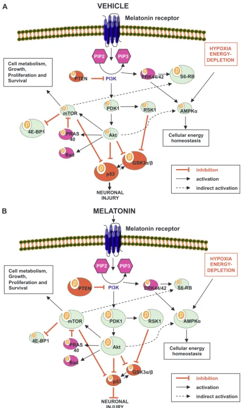
Fig. 5. Regulation of Akt signaling pathways after (A) vehicle, and (B) melatonin treatments following ischemic-stroke. Akt is activated via its upstream kinase PDK1, while PTEN is a major negative regulator of Akt signaling. When phosphorylated, inhibitory effect of PTEN on PI3K/Akt is decreased. Activated Akt stimulates mTOR phosphorylation, which is a central regulator of cell metabolism, growth, proliferation and survival. Both Akt and mTOR are negative regulators of p53 phosphorylation. Here we demonstrated that melatonin activated Akt, PDK1, PTEN, mTOR and AMPKα phosphorylation. Furthermore, AMPKα was demonstrated to be activated via a PI3K independent pathway as it was not deactivated by IP3K inhibition. It is possible that AMPKα is activated by decreased ATP levels due to anabolic reactions stimulated by Akt and hypoxia. Therefore, phosphorylated AMPKα stimulates catabolic pathways to generate ATP from alternative pathways such as fatty acid oxidation. Moreover, melatonin activated RSK1 phosphorylation and reduced 4E-BP1, GSK-3α/β, S6 ribosomal protein and p53 phosphorylation. GSK-3α and GSK-3β are critical players in the activation of apoptosis. Both proteins are deactivated by phosphorylation of Akt and RSK1. GSK-3β is not only inhibited by Akt, but also provides a strong link between Akt and p53. Finally, reduced p53 phosphorylation resulted in decreased apoptosis. In addition, PRAS40 phosphorylation was slightly increased, whereas ERK-1/2 and Bad phosphorylation were decreased after melatonin treatment. Thus, our data provide evidence for how melatonin utilizes PI3K/Akt pathway to protect neurons after ischemia.
melatonin. As expected, even though not significant, the number of apoptotic cells was slightly increased after the inhibition of Akt pathway without melatonin treatment. However, infarct volume was not increased by Wortmannin treatment after 90 min of MCAo, which results in a significant amount of necrosis. Therefore, it is tempting to speculate that the inhibition of Akt signaling diminishes brain edema and IgG extravasation.
In agreement with the recent experimental data indicating increased Akt phosphorylation by melatonin in the ischemic brain[14], we also observed that melatonin stimulated Akt phosphorylation after FCI. PI3K/Akt pathway mediates neuroprotective effects of several growth factors, including vascular endothelial growth factor (VEGF) [18], erythropoietin [20], brain-derived neurotrophic factor (BDNF), and insulin-like growth factor 1 (IGF1) [17]. Furthermore, PI3K/Akt phosphorylation is increased in superoxide dismutase 1 (SOD1) and glutathione peroxidase (GPX) transgenic animals after FCI or exposure to oxidative stress [33,34]. Similarly, treatment with another free radical scavenger agent, NXY-059, attenuates the decrease in PI3K/ Akt after ischemic-stroke[35]. Taken together, these observations point to the significant role that PI3K/Akt pathway plays in the protective effect of free radical scavenger agents, especially under oxidative-stress conditions. Moreover, phosphorylation of PI3K/Akt pathway stabilizes mitochondrial function under hypoxic conditions and protects neurons via activation of Bcl-XLand inhibition of caspase-3 and p53[18,26]. Consistently, we observed decreased phosphorylated p53 levels accom-panying reduction in apoptosis with melatonin treatment after FCI. Reversal of melatonin's effects by Wortmannin indicated the involve-ment of PI3K/Akt signaling.
Our detailed signaling pathway analyses further demonstrated the components of PI3K/Akt pathway that were under the regulation of melatonin (Fig. 5). Akt is a downstream target of PI3K which regulates cellular survival, cell proliferation and protein synthesis in response to various extracellular stimuli [23,36]. Akt is activated by two site-specific phosphorylations at Thr308 and Ser473 [16]. Its upstream kinase PDK1 phosphorylates Akt on Thr308 within its activation loop [23]. For the maximum activation of Akt, Ser473 is phosphorylated possibly by autophosphorylation or by mTOR signaling [37]. In accordance with this mechanism of activation of Akt, we observed that melatonin promoted phosphorylation of both Thr308 and Ser473. However, only the Thr308 phosphorylation was increased statistically significantly as analyzed by both Planar Surface Immunoassay and Western blot. PTEN is a major negative regulator of Akt signaling and catalyzes the dephosphoylation of PIP3 [22]. Interestingly, PDK1, mTOR and PTEN were all activated in the melatonin treated animals. Moreover, Wortmannin treatment decreased activated Akt (Thr308) and PDK1, indicating direct activation of Akt phosphorylation through Thr308 phosphorylation via IP3/PDK1 signaling. While Akt (p-Ser473), PTEN and mTOR phosphorylation was not inhibited by Wortmannin.
Additionally, we observed that melatonin activated AMPKα and RSK1 in an IP3 independent manner following FIC. AMPK responds to intracellular energy level changes. It is activated by decreased ATP levels or by ERK-1/2 phosphorylation and stimulates the catabolic pathways such as fatty acid oxidation to generate ATP. AMPK is also activated indirectly by increased anabolic activity of Akt, resulting in elevated energy consumption[27]. In contrast to Akt, AMPKα inhibits anabolic activity, indicating an interactive mechanism between Akt and AMPKα[38]. Therefore, melatonin may activate alternative collateral survival pathways through AMPKα phosphorylation. Activated AMPKα can drive the cell to a catabolic state that utilizes alternative energy sources, such as fatty acid oxidation, in order to supply ATP for the increased energy demand during ischemic stroke.
RSK1 promotes cell survival via inactivation of several pro-apopto-tic molecules[39]. It is activated primarily by Erk1/2[39]but may also be activated by PDK1[40]. In our present study, although RSK1 was activated, ERK-1/2 was not activated. Thus, it is likely that melatonin promotes phosphorylation of RSK1 via elevated phosphorylation of
PDK1.
In addition, we observed reduced phosphorylation of GSK-3α and GSK-3β, in melatonin treated animals following FIC. Consistently, GSK-3α, GSK-3β are deactivated by Akt and RSK1 phosphorylation[39,41]. Moreover, overexpression of catalytically active GSK-3 promotes apop-totic cell death, while dominant-negative GSK-3 prevents apoptosis [41]. mTOR, a central regulator of cell metabolism, growth, prolifera-tion and survival[36], deactivates 4E-BP1[42]. Active 4e-BP1 levels were decreased in our observations. Furthermore, phosphorylation of GSK-3α, GSK-3β and 4E-BP1 were increased by Wortmannin.
S6 Ribosomal Protein is activated by mTOR[43]. Yet, melatonin treatment decreased phosphorylation of S6 Ribosomal Protein follow-ing FIC. Moreover, this decrease was not reversed by Wortmannin treatment. In addition, PRAS40 phosphorylation was increased while ERK-1/2 and Bad phosphorylation were decreased slightly after mela-tonin treatment following FCI.
In conclusion, phosphorylation of p53 protein was decreased in melatonin treated animals after FCI. This effect was reversed by Wortmannin. The tumor-suppressor transcription factor p53 is impli-cated as a mediator of neuronal injury after ischemic-stroke[28–30]. Its phosphorylation can be activated by extrinsic and intrinsic stress signals, including hypoxia, and phosphorylation of p53 is inhibited by Akt and mTOR signaling [27]. Thus, our observations suggest that melatonin acts through the PI3K/Akt pathway to inhibit p53 phosphor-ylation which results in increased neuronal survival (Figs. 4and5). Declaration of interest
No competingfinancial interests and potential conflict exist. Acknowledgement
This work was supported by The Turkish Academy of Sciences (TUBA), Istanbul Medipol University and Bezmialem Vakif University Scientific Research Project Committee.
References
[1] L.C. Manchester, A. Coto-Montes, J.A. Boga, et al., Melatonin: an ancient molecule that makes oxygen metabolically tolerable, J. Pineal Res. 59 (2015) 403–419. [2] R.J. Reiter, J.C. Mayo, D.X. Tan, R.M. Sainz, M. Alatorre-Jimenez, L. Qin, Melatonin
as an antioxidant: under promises but over delivers, J. Pineal Res. 61 (2016) 253–278.
[3] R.J. Reiter, D.X. Tan, J. Leon, U. Kilic, E. Kilic, When melatonin gets on your nerves: its beneficial actions in experimental models of stroke, Exp. Biol. Med. 230 (2005) 104–117.
[4] U. Kilic, B. Yilmaz, R.J. Reiter, A. Yuksel, E. Kilic, Effects of memantine and melatonin on signal transduction pathways vascular leakage and brain injury after focal cerebral ischemia in mice, Neuroscience 237 (2013) 268–276.
[5] M.C. Beker, A.B. Caglayan, T. Kelestemur, et al., Effects of normobaric oxygen and melatonin on reperfusion injury: role of cerebral microcirculation, Oncotarget 6 (2015) 30604–30614.
[6] R. Hardeland, Melatonin: signaling mechanisms of a pleiotropic agent, BioFactors 35 (2009) 183–192.
[7] F. Luchetti, B. Canonico, M. Betti, et al., Melatonin signaling and cell protection function, FASEB J.: Off. Publ. Fed. Am. Soc. Exp. Biol. 24 (2010) 3603–3624. [8] U. Kilic, B. Yilmaz, M. Ugur, et al., Evidence that membrane-bound G
protein-coupled melatonin receptors MT1 and MT2 are not involved in the neuroprotective effects of melatonin in focal cerebral ischemia, J. Pineal Res. 52 (2012) 228–235. [9] M.L. Dubocovich, M. Markowska, Functional MT1 and MT2 melatonin receptors in
mammals, Endocrine 27 (2005) 101–110.
[10] S.R. Pandi-Perumal, I. Trakht, V. Srinivasan, et al., Physiological effects of melatonin: role of melatonin receptors and signal transduction pathways, Progress. Neurobiol. 85 (2008) 335–353.
[11] A.S. Chan, F.P. Lai, R.K. Lo, T.A. Voyno-Yasenetskaya, E.J. Stanbridge, Y.H. Wong, Melatonin mt1 and MT2 receptors stimulate c-Jun N-terminal kinase via pertussis toxin-sensitive and -insensitive G proteins, Cell. Signal. 14 (2002) 249–257. [12] M.L. Dubocovich, P. Delagrange, D.N. Krause, D. Sugden, D.P. Cardinali, J. Olcese,
International union of basic and clinical pharmacology. LXXV. Nomenclature, classification, and pharmacology of G protein-coupled melatonin receptors, Pharmacol. Rev. 62 (2010) 343–380.
[13] S. McNulty, A.W. Ross, K.Y. Shiu, P.J. Morgan, M.H. Hastings, Phosphorylation of CREB in ovine pars tuberalis is regulated both by cyclic AMP-dependent and cyclic AMP-independent mechanisms, J. Neuroendocrinol. 8 (1996) 635–645.
[14] U. Kilic, E. Kilic, R.J. Reiter, C.L. Bassetti, D.M. Hermann, Signal transduction pathways involved in melatonin-induced neuroprotection after focal cerebral ischemia in mice, J. Pineal Res. 38 (2005) 67–71.
[15] E. Kilic, U. Kilic, R.J. Reiter, C.L. Bassetti, D.M. Hermann, Tissue-plasminogen activator-induced ischemic brain injury is reversed by melatonin: role of iNOS and Akt, J. Pineal Res. 39 (2005) 151–155.
[16] A. Bellacosa, T.O. Chan, N.N. Ahmed, et al., Akt activation by growth factors is a multiple-step process: the role of the PH domain, Oncogene 17 (1998) 313–325. [17] H. Zhao, R.M. Sapolsky, G.K. Steinberg, Phosphoinositide-3-kinase/akt survival
signal pathways are implicated in neuronal survival after stroke, Mol. Neurobiol. 34 (2006) 249–270.
[18] E. Kilic, U. Kilic, Y. Wang, C.L. Bassetti, H.H. Marti, D.M. Hermann, The phosphatidylinositol-3 kinase/Akt pathway mediates VEGF's neuroprotective ac-tivity and induces blood brain barrier permeability after focal cerebral ischemia, FASEB J.: Off. Publ. Fed. Am. Soc. Exp. Biol. 20 (2006) 1185–1187.
[19] G. Jin, N. Omori, F. Li, et al., Protection against ischemic brain damage by GDNF affecting cell survival and death signals, Neurol. Res. 25 (2003) 249–253. [20] E. Kilic, U. Kilic, J. Soliz, C.L. Bassetti, M. Gassmann, D.M. Hermann, Brain-derived
erythropoietin protects from focal cerebral ischemia by dual activation of ERK-1/ −2 and Akt pathways, FASEB J.: Off. Publ. Fed. Am. Soc. Exp. Biol. 19 (2005) 2026–2028.
[21] A. Saito, T. Hayashi, S. Okuno, T. Nishi, P.H. Chan, Oxidative stress affects the integrin-linked kinase signaling pathway after transient focal cerebral ischemia, Stroke 35 (2004) 2560–2565.
[22] P. Liu, H. Cheng, T.M. Roberts, J.J. Zhao, Targeting the phosphoinositide 3-kinase pathway in cancer, Nat. Rev. Drug Discov. 8 (2009) 627–644.
[23] G. Song, G. Ouyang, S. Bao, The activation of Akt/PKB signaling pathway and cell survival, J. Cell. Mol. Med. 9 (2005) 59–71.
[24] A. Arcaro, M.P. Wymann, Wortmannin is a potent phosphatidylinositol 3-kinase inhibitor: the role of phosphatidylinositol 3,4,5-trisphosphate in neutrophil re-sponses, Biochem. J. 296 (Pt 2) (1993) 297–301.
[25] G. Powis, R. Bonjouklian, M.M. Berggren, et al., Wortmannin, a potent and selective inhibitor of phosphatidylinositol-3-kinase, Cancer Res. 54 (1994) 2419–2423. [26] E. Kilic, A. ElAli, U. Kilic, et al., Role of Nogo-A in neuronal survival in the
reperfused ischemic brain, J. Cereb. Blood Flow. Metab.: Off. J. Int. Soc. Cereb. Blood Flow. Metab. 30 (2010) 969–984.
[27] K.H. Vousden, K.M. Ryan, p53 and metabolism, Nat. Rev. Cancer 9 (2009) 691–700. [28] M. Chopp, Y. Li, Z.G. Zhang, S.O. Freytag, p53 expression in brain after middle
cerebral artery occlusion in the rat, Biochem. Biophys. Res. Commun. 182 (1992) 1201–1207.
[29] H. Watanabe, S. Ohta, Y. Kumon, S. Sakaki, M. Sakanaka, Increase in p53 protein expression following cortical infarction in the spontaneously hypertensive rat, Brain Res. 837 (1999) 38–45.
[30] K.C. Ahn, E.M. MacKenzie, C.R. Learman, et al., Inhibition of p53 attenuates ischemic stress-induced activation of astrocytes, Neuroreport 26 (2015) 862–869. [31] B. Niu, B. Li, C. Wu, et al., Melatonin promotes goat spermatogonia stem cells
(SSCs) proliferation by stimulating glial cell line-derived neurotrophic factor (GDNF) production in sertoli cells, Oncotarget 7 (2016) 77532–77542. [32] W. Deng, Y. Zhang, L. Gu, et al., Heat shock protein 27 downstream of P38-PI3K/
Akt signaling antagonizes melatonin-induced apoptosis of SGC-7901 gastric cancer cells, Cancer Cell Int. 16 (2016) 5.
[33] N. Noshita, T. Sugawara, A. Lewen, T. Hayashi, P.H. Chan, Copper-zinc superoxide dismutase affects Akt activation after transient focal cerebral ischemia in mice, Stroke 34 (2003) 1513–1518.
[34] J.M. Taylor, U. Ali, R.C. Iannello, P. Hertzog, P.J. Crack, Diminished Akt phosphorylation in neurons lacking glutathione peroxidase-1 (Gpx1) leads to increased susceptibility to oxidative stress-induced cell death, J. Neurochem. 92 (2005) 283–293.
[35] T. Yoshimoto, P. Kanakaraj, J. Ying Ma, et al., NXY-059 maintains Akt activation and inhibits release of cytochrome C after focal cerebral ischemia, Brain Res. 947 (2002) 191–198.
[36] M. Laplante, D.M. Sabatini, mTOR signaling at a glance, J. Cell Sci. 122 (2009) 3589–3594.
[37] A. Toker, A.C. Newton, Akt/protein kinase B is regulated by autophosphorylation at the hypothetical PDK-2 site, J. Biol. Chem. 275 (2000) 8271–8274.
[38] M.M. Mihaylova, R.J. Shaw, The AMPK signalling pathway coordinates cell growth, autophagy and metabolism, Nat. Cell Biol. 13 (2011) 1016–1023.
[39] A. Carriere, H. Ray, J. Blenis, P.P. Roux, The RSK factors of activating the Ras/ MAPK signaling cascade, Front. Biosci.: J. Virtual Libr. 13 (2008) 4258–4275. [40] C.J. Jensen, M.B. Buch, T.O. Krag, B.A. Hemmings, S. Gammeltoft, M. Frodin,
90-kDa ribosomal S6 kinase is phosphorylated and activated by 3-phosphoinositide-dependent protein kinase-1, J. Biol. Chem. 274 (1999) 27168–27176. [41] M. Pap, G.M. Cooper, Role of glycogen synthase kinase-3 in the
phosphatidylino-sitol 3-Kinase/Akt cell survival pathway, J. Biol. Chem. 273 (1998) 19929–19932. [42] I. Ruvinsky, O. Meyuhas, Ribosomal protein S6 phosphorylation: from protein
synthesis to cell size, Trends Biochem. Sci. 31 (2006) 342–348.
[43] O.H. Iwenofu, R.D. Lackman, A.P. Staddon, D.G. Goodwin, H.M. Haupt, J.S. Brooks, Phospho-S6 ribosomal protein: a potential new predictive sarcoma marker for targeted mTOR therapy, Mod. Pathol.: Off. J. US Can. Acad. Pathol., Inc. 21 (2008) 231–237.