J Exp Clin Med 2021; 38(3): 272-276
doi: 10.52142/omujecm.38.3.12
1. Introduction
Infertility is seen in 10-15% of married couples, and nearly 50% of the cases are male related. One of the main causes of male infertility is azoospermia, which is defined as having no sperm cells in the ejaculate and found in 1% of men (Kumar and Singh, 2015). The quality and quantity of the spermatozoa directly affect the reproduction (Sapanidou et al., 2015). Apoptosis is one of the important processes that provide quality and quantity of germ cells in the testicle (Hikim et al., 2003). Apoptosis may decrease the number of germ cells and their viability (Kang et al., 2017). Accordingly, sufficient number of healthy spermatozoa can’t be produced in the azoospermia and this leads to infertility (Lee and Cheng, 2008). The treatment of these cases is performed by surgical operations including ‘testicular sperm extraction’ in case of sperm cell absence in the tissue (Kumar and Singh, 2015).
The blood-testis barrier (BTB) is located between adjacent Sertoli cells in the testis where it provides a unique microenvironment for the development and maturation of meiotic and post-meiotic germ cells in seminiferous tubules. It compounds junctional proteinous structures, such as tight junctions (TJs), adhesion junctions and gap junctions (GJs).
Some of the junctional proteins function as structural proteins of BTB and some have regulatory roles. Deletion or functional silencing of coding genes of these junctional proteins may disrupt the BTB, and concordantly this may cause immunological or other damages to meiotic and postmeiotic cells, this eventually leads to spermatogenic arrest and infertility (Jiang et al., 2014). The intercellular tight junction dynamics induce the formation of blood-testis barrier which play a crucial role in spermatogenesis (Gao et al., 2017). The tight junctions between cells in the testicular tissue, zonula occludens-1 (ZO-1) and regular actin network affect sperm production (Lee and Cheng, 2003). The aim of this study is to understand the role of tight junctions by comparing ZO-1 expression levels between azoospermia testicular tissue sperm positive and negative samples which are obtained by TESE. 2. Materials and methods
2.1. Ethics committee approval
All related material has been obtained in accordance with the Helsinki declaration and all procedures have been approved by İstanbul Şişli Etfal Training and Research Hospital Ethics committee 18.12.2012 date and 2012/133 number. This study Journal of Experimental and Clinical Medicine
https://dergipark.org.tr/omujecm
Research Article
Ultrastructural examination of testicular tissues and evaluation of ZO-1 protein levels
obtained from azoospermic cases
Ayşe ALTUN1,* , Yasemin E. CANILLIOĞLU2 , Evrim ÜNSAL3 , Seda KARABULUT4 Canan HÜRDAĞ5
1Department of Obstetrics and Gynaecology, Faculty of Medicine, Istanbul University, Istanbul, Turkey 2Department of Histology and Embryology, Faculty of Medicine, Bahçesehir University, Istanbul, Turkey
3Genart Woman Health and Reproductive Biotechnology Center, Ankara, Turkey
4Department of Histology and Embryology, Faculty of Medicine, Istanbul Medipol University, Istanbul, Turkey 5Department of Histology and Embryology, Faculty of Medicine, Demiroğlu Bilim University, Istanbul, Turkey
Received: 20.11.2020 • Accepted/Published Online: 07.02.2021 • Final Version: 23.04.2021
Abstract
Spermatogenesis is a process that involves cellular and biochemical processes. The purpose of this study is to understand the role of tight junctions in spermatogenesis by evaluating Zonula occludens-1 (ZO-1) protein levels of cells from the testicular sperm extraction (TESE) biopsies in patients with azoospermia. The study included 40 infertile men with azoospermia. They were divided into two groups, as spermatozoa positive (n=20) and spermatozoa negative (n=20). Testis tissues were examined morphologically, immunohistochemically and ultrastructurally by using transmission electron microscopy (TEM). The structure of seminiferous tubules was deteriorated, degenerated and hyalinized in spermatozoa negative group. ZO-1 expression level was normal in blood-testis barrier of spermatozoa positive group in comparing to lateral surfaces of Sertoli cells spermatozoa negative group. The ultrastructural results were also supported with light microscopy findings. These findings suggest that decrease of the ZO-1 protein expression level in Sertoli cells might be one of the factors involved in the impairment of spermatogenesis in spermatozoa negative group. Therefore, we suggest that tight junctions may have crucial roles in spermatogenesis.
Altun et al. / J Exp Clin Med
was supported by Istanbul Bilim University withresearch code HE/1122012.
2.2. Testicular sperm extraction
All patients referred for testicular biopsy to infertility center for diagnosis or infertility treatment. TESE was performed under general anesthesia. The scrotal skin, tunica albuginea and one or two pieces of the extruding testicular tissue were excised using a pair of curved scissors. The specimen was then transferred into a Petri dish (Falcon; Becton-Dickinson, Aalst, Belgium) filled with ~2 ml modified HEPES-buffered heparin 0.4% (H 3149; Sigma, St Louis, MO, USA). The biopsy specimen was shredded into small pieces with two sterile glass microscope slides under a stereomicroscope to release spermatozoa from the seminiferous tubules into the medium. The presence of spermatozoa was examined by using inverted microscope. The testicular incision was continued until the spermatozoa could be distinguishable from the debris, immature cells, red blood cells or Sertoli cells.
In the case of spermatozoa absence in the tissue, up to three biopsies were performed on different areas of each testis. The shredded biopsy tissue was gently homogenized by repeated aspiration and spilling out maneuvers into a tuberculin syringe. The effluent and the minced tissue were transferred into a Falcon tube (Becton-Dickinson) and incubated for 2–3 h in 5% CO2 containing atmosphere at 37°C. Then, the fluid was
aspirated, put into another tube and centrifuged at 300 g for 5 min., after the centrifuge the pellet gently re-suspended by following the removal of supernatant. Finally, droplets were aspirated from the suspension as described in the ESP for searching of spermatozoa.
2.3. Histological and immunohistochemical analysis In this study, a total of 40 men ages 25-55 were 2012-2013 between evaluated for persistent azoospermia. Testicular biopsy samples were divided into two groups as spermatozoa positive and spermatozoa negative for azoospermia men. The patients were enrolled to study who have diagnosed with non-obstructive azoospermia, could not have a child after one year of unprotected sexual intercourse in their first TESE, and never receive any medication and hormone replacement therapy before TESE procedure, plus was not treated for chronic disease. Testicular samples were evaluated by morphological, immunohistochemical and ultrastructural analysis.
Testicular samples were fixed in Bouin’s solution for 6h. After fixation, tissues were incubated with 1% lithium carbonate. After carbonate incubation tissues were transferred to the alcohol. Then samples were embedded in paraffin. 3 μm thick sections were prepared and stained with hematoxylin-eosin and Masson’s trichrome. Sections were observed under light microscopy and images were taken (Olympus Bx53, Japan).
2.4. Immunohistochemical analysis
Sections (3µm) were deparaffinized with xylene and rehydrated with decreasing percentages of ethanol and finally
with water. Surroundings of the sections were marked with a PAP pen (Pappen, Dako). The endogenous peroxidase activity was blocked with 3% H2O2 for 20 min at room temperature and
later rinsed with distilled water and phosphate buffered saline (PBS). Antigen retrieval was accomplished by Decloacking chamber (Bicare Medical DC2008) in aEDTA (1/10) for 20 min at 110°C. Blocking reagent (Invitrogen Blocking Solution REF: 859043) was applied to each slide followed by 5 min incubation at room temperature in a humid chamber. Sections were incubated for overnight at 4°C with anti ZO-1 rabbit polyclonal primary antibody (1:100, Millipore, AB2272), ki67 mouse anti rat (1:100 Dako, M7248). Antibodies were diluted in a large volume of UltrAb Diluent (Thermo Scientific, TA-125-UD).
The sections were biotinylated and incubated with goat anti-rabbit antibodies (Invitrogen Broad Spectrum REF: 859043). After slides were washed in PBS, the streptavidin peroxidase label reagent (Invitrogen HRP–Streptavidin REF: 859043) was applied for 30 min at room temperature in a humid chamber. The colored product was developed by incubation with AEC chromogen (Invitrogen, D22187). The slides were counterstained with Mayer’s hematoxylin (LabVision, TA-125-MH) and mounted in glycerol gelatin. ZO-1and Ki67 reactions were observed under light microscopy and images were taken (Olympus Bx53, Japan).
2.5. Electron microscopy
Testis tissues were fixed and dissected cut into smaller blocks of 1 mm3. Tissue samples (1 mm3) were put in the 2.5%
glutaraldehyde with 0.1 M phosphate buffer (pH 7.2) for overnight fixation at +4°C. Then samples were washed again with PBS for 3 times (30 minutes for each wash). Samples were transferred to 1% osmium tetroxide with 0.1 M PBS for 1 h at room temperature (RT) and wash in PBS 3 × 30 min. Samples were dehydrated with increasing concentrations of ethanol (70% Ethanol-5 min. 96% Ethanol 10 min., 100% Ethanol, 2 × 15 min.). Then, dehydrated samples were transferred to the propylene oxide. Tissues were taken in the mixture of propylene oxide and embedding medium (Epon 812) (1:1) at RT for 45 min and in the mixture of propylene oxide and embedding medium (1:3) at RT for 45 min. Then samples were embedded in the fresh medium at +4°C for overnight. Samples were again embedded and polymerized at 60°C for 24–48 h. Ultra-thin sections were taken on ultramicrotome. Semi-thin sections were stained at 60°C with toluidine blue for 1 min. Ultra-thin sections are taken on copper grids and stained with uranyl acetate and Reynolds lead citrate. Tissues were examined in a transmission electron microscope (TEM, JEOL 1200 SX) for more detailed examination and imaging. 3. Results
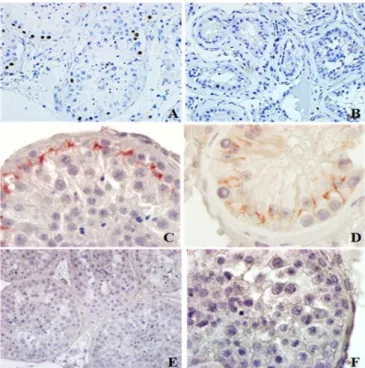
In TESE (+) group, testis tissues were stained with H&E, the seminiferous tubules were associated with basal membrane area. Cells interacted with each other along the tubular wall at different stages of spermatogenesis. Spermatids were clearly
observable. Cell colonies in different spermatogenic stages and cell debris were found in the lumen of seminiferous tubules. Seminiferous tubules were associated with the basal membrane in the areas stained with Masson's trichrome. Also, oedema and hemorrhage where observed in some of these areas (Fig. 1A, Fig.1C).
Fig. 1. (A) TESE+and (B) TESE-seminiferous tubules. x200, H&E staining. (C) TESE+and (D) TESE-seminiferous tubules. x400, Masson’s trichrome staining. (E) TESE+and (F) TESE-intracellular junctions between germ and Sertoli cells. x5000, TEM
In TESE (-) seminiferous tubules were disordered, and absence of cells in other stages of spermatogenesis were observed. Hyalinated seminiferous tubules with thick basal membrane were observed. Connective tissue deteriorated by interstitial tissue thickening enclosing the seminiferous tubule completely and also hemorrhages area were examined (Fig. 1B, 1D).
Basal membrane which is enclosed to the seminiferous tubules and thin collagen fibril network were examined in TESE (+) group by using transmission electron microscope (TEM). Tight junctions between Sertoli cells within seminiferous tubule were observed (Fig. 1E) In TESE (-) group, peritubular wall was thickened because of increased collagen fibers in the testicle tissue. There were mainly Sertoli cells in the tubular wall enclosed by the basal membrane. Vacuolated hyaline material in Sertoli cell and tight junction were detected along with the whole lateral surface of the Sertoli cell (Fig. 1F). The Ki67 protein is present during all active phases of the cell cycle but it is absent only in resting cells. This finding renders this molecule a proper marker. Correspondingly, in TESE + group we detected Ki67 expression around the seminiferous tubules but not in the TESE (-) group (Fig. 2A and Fig. 2B). In spermatozoa containing testis tissue ZO-1 expression was observed in the blood testes barrier (BTB) with normal localization (Fig. 2C).
Spermatozoa-absent group was examined and ZO-1 in the structure of TJs was not only in the basal compartment of the seminiferous epithelium but also observed on the lateral surface covering the adluminal compartment (Fig. 2D). The ultrastructural results were also supported with light microscopy findings.
Fig. 2. (A) TESE+and (B) TESE- germ cells in seminiferous tubules. x400, Ki-67 immunohistochemistry. (C) TESE+and (D) TESE-seminiferous tubules. x1000, ZO-1 immunohistochemistry. (E) Ki67, x400 and (F) ZO-1, x1000 negative control sections
4. Discussion
Infertility is a common disorder and nearly 50% of all cases are caused by male factor(s) (Garg and Gard, 2011). In our study we detected that spermatogenesis can be affected by the tight connections between the Sertoli cells and impairment of tight connections is one of the significant causes for infertility. Description of the blood-tissue barrier (BTB) was based on observations which were reported in the early twentieth century. However, the term blood-testis barrier, also known as the Sertoli cell seminiferous epithelium barrier was firstly used by Chiquoine (Chiquoine, 1964) in a study that they examined the effects of cadmium toxicity as it related to testicular necrosis. The number of sperm is effective in fertilization thus, it is important among the sperm parameters. According to World Health Organization (WHO) standards it is considered normal to have 15x106 cell/ml or more. (WHO, 2010)
Azoospermia is described as no sperm in the ejaculate, and common in 1% of men. In our study, morphological differences of spermatozoa and non-azoospermic testis tissues were evaluated. In addition, the role of tight junctions between cells in the presence and absence of sperm in the cases of azoospermia also were observed. As supported by the literature, the tight junctions between the cells form the blood-testis barrier within the blood-testis structure and the BTB plays an important role in male infertility (Sun et al., 2013).
Altun et al. / J Exp Clin Med
The testis barrier (BTB) is one of the tightest blood-tissue barriers in the mammalian body. It divides the seminiferous epithelium into the basal and the apical (adluminal) compartments. Meiosis I and II, spermiogenesis, and spermiation take place in a specialized microenvironment behind the BTB in the apical compartment but spermatogonial renewal and differentiation and cell cycle progression up to the preleptotene spermatocyte stage normally occur outside of the BTB. However, in the azoospermic cases the BTB is distorted. Besides, unexplained male infertility cases involve 30-40% of men with distorted BTB and abnormal semen parameters (Nieschlag et al., 2011). The causes of spermatogenic defects in infertile patients are multifactorial (Skakkebaek et al., 2001). The immunological barrier provided by the BTB even transiently cannot be compromised during the epithelial cycle to avoid the production of antibodies against meiotic and post meiotic germ cells. Studies have demonstrated that some adhesion protein complexes (e.g., occludin-ZO-1, N-cadherin-β-catenin, claudin-5-ZO-1), steroids (e.g., testosterone, estradiol-17β), non-receptor protein kinases (e.g., focal adhesion kinase, c-Src, c-Yes), polarity proteins (e.g., PAR6, Cdc42, 14-3-3), endocytic vesicle proteins (e.g., clathrin, caveolin, dynamin 2), and actin regulatory proteins (e.g., Eps8, Arp2/3 complex), are associated to each other and collaborative with the cytokines. The findings showed that impairment of BTB was responsible for subsequent damage to germ-cell adhesion, thereby leading to germ-cell loss, reduced sperm count, and male infertility or subfertility (Yan Cheng and Dolores, 2012).
According to our microscopic observation, the ZO-1 protein, which provides tight junctions between Sertoli cells in the spermatozoa group, was more intense than the spermatozoa-free group (n:20). In the spermatozoa-containing group (n:20), the ZO-1 staining localized to the expected region between the Sertoli cells, while in the spermatozoa-free group (n:20), the staining took place along the entire lateral surface of the Sertoli cells. Parallel to this, in the spermatozoa group, the tight connections were between the Sertoli cells within the abdominal and basal compartments, while the tight connections in the spermatozoa-free group were along the entire side wall of Sertoli cells. Therefore, ZO-1 plays an effective role in Sertoli cells for the spermatogenesis, as supported with literature.
ZO-1 is suggested to play a role in the regulation of germ cell development and differentiation. Sertoli cell / germ cells in the epithelium of the testis are responsible to preserve the correct ratio and producing vital and efficient sperm by using blood testis barrier protein. Therefore ZO-1 is important to maintain male fertility and pathogenesis (Jiang et al., 2014). Based on this, our electron microscopy observations showed that the number of germ cells in the non-spermatozoa group decreased and the ZO-1 interaction with blood testes barrier was less significant than the group with spermatozoa.
In addition, Ki67 is a non-histon nuclear protein found in all phases of the cell cycle except the Go phase, it is associated
with tight intercellular connections and effective in cell proliferation. Ki67 protein expression is associated with tight binding proteins (E-cadherin, ZO-1, etc.) and involved in cell proliferation, differentiation and apoptosis (Chen et al., 2012). In our study we detected morphological abnormalities in the connective tissue in both groups, suggesting that these changes may also affect the testicular tissue function. The absence of spermatozoa proves that seminiferous tubule structure deteriorates the tubules and hyalinized the spermatogenesis. Ki67 weren't expressed and the absence of cells in different stages of spermatogenesis proved that spermatogenesis never occurred.
In Vitro Fertilization (IVF) applications have also begun to use in male subfertility except for anovulation, tubal factor and unexplained infertility, when classical in vitro fertilization has shown that there may be a solution in male subfertility if there is a sufficient number of motile sperms. However, it is known that in adequacy of sperm parameters in male subfertile caused decreasing of fertilization rate (Rumbold et al., 2019). Consequently, our study emphasizes that BTB integrity is important for spermatogenesis and tight junctions play effective role in regulation of sperm cells. Germ cell transplantation is not yet ready for the human fertility clinic, but it may be eligible for young cancer patients, who have no any other option to preserve their fertility (Vrankovic et al., 2012). Conflict of interest None to declare. Acknowledgments None to declare. References
1. Chen, W., Hu, J., Zhang, Z., Chen, L., Xie, H., Dong, N., Chen Y., Zuguo Liu, Z., 2012. Localization and expression of Zonula Occludins-1 in the rabbit corneal epithelium following exposure to benzal konium chloride. PLoS ONE. 7(7): e40893.
2. Chiquoine, A.D., 1964. Observations on the early events of cadmium necrosis of the testis. Anat. Rec. 149, 23–35.
3. Garg, V., Gard, S.P., 2011. Role of nitric oxide in male infertility. J Indian Acad. Forensic Med. 33, 65-68.
4. Gao, Y., Mruk, D., Chen, H., Lui, W.Y., Lee, W.M., Cheng, C.Y., 2017. Regulation of the blood-testis barrier by a local axis in the testis: role of laminin α2 in the basement membrane. FASEB J. 31, 584-597.
5. Hikim, A. P., Lue, Y., Yamamoto, C. M., Vera, Y., Rodriguez, S., Yen, P. H., Soeng, K., Wang, C., Swerdloff, R.S., 2003. Key apoptotic pathways for heat-induced programmed germ cell death in the testis. Endocrinology. 144, 3167–3175.
6. Jiang, X.H., Bukhari, I., Zheng, W., Yin, S., Wang, Z., Cooke H.J., Shi, Q.H., 2014.Blood-testis barrier and spermatogenesis: lessons from genetically-modified mice. Asian J. Androl.16, 572-580. 7. Kang Z., Qiao N., Liu G., Chen H., Tang Z., Ying Li Y., 2017.
Copper-induced apoptosis and autophagy through oxidative stress-mediated mitochondrial dysfunction in male germ cells.
Toxicol In Vitro. 61, 104639.
8. Kumar, N., Singh, A.K., 2015. Trends of male factor infertility, an important cause of infertility: A review of literature. J. Hum. Reprod Sci. 8, 191-196.
9. Lee, N.P., Cheng, C.Y., 2008. Nitric oxide and cyclic nucleotides: Their roles injunction dynamics and spermatogenesis. Adv. Exp. Med. Biol. 636, 172-185.
10. Lee, N.P.Y., Cheng, C.Y., 2003. Regulation of sertoli cell tight junction in the rat testis via the nitric oxide synthase/soluble guanylate cyclase/cGMP/Protein Kinase G signaling pathway: an in vitro study. Endocrinology. 144, 3114-3129.
11. Nieschlag, E., Behre, H., Nieschlag, S., Van, Ahlen H., 2011. Male reproductive health and dysfunction. Internistische. Praxis. Andrology. 51, 751.
12. Rumbold, A.R., Sevoyan, A., Oswald, T.K., Fernandez R.C., Davies, M.J., Moore, V.M., 2019. Impact of male factor infertility on offspring health and development. Fertil. Steril. 111, 1047-1053.
13. Sapanidou, V., Taitzoglou, I., Tsakmakidis, I., Kourtzelis, I., Fletouris, D., Theodoridis, A., Zervos, I., Tsantarliotou, M., 2015. Antioxidant effect of crocin on bovine sperm quality and in vitro fertilization. Theriogenology. 84, 1273-1282.
14. Skakkebaek, N.E., Rajpert De Meyts, E., Main, K.M., 2001. Testicular dysgenesis syndrome: an increasingly common developmental disorder with environmental aspects. Hum. Reprod. 16, 972−978.
15. Sun, H., Yang, B., Zhu, C., Liu, R., Wang, H., Li, W., 2013. Presence of metastasis-associated protein 1 in Sertoli cells is required for proper contact between Sertoli cells and adjacent germ cells. Urology.81, 66-73.
16. Yan Cheng, C., Dolores, D.M., 2012. The Blood-Testis Barrier and Its Implications for Male Contraception. Pharmacol. Rev. 64, 16–64.
17. WHO, 2010. World Health organization laboratory manual for the examination and the processing of human semen. 5 th ed; pp 161.