Dosimetric comparison of two different whole brain
radiotherapy techniques in patients with brain
metastases: How to decrease lens dose?
INTRODUCTION
Brain metastases are the most common intracranial tumors in adults, accounting for signi icantly more than one‐half of brain tumors. In patients with systemic malignancies, brain metastases occur in 10 to 30 percent of adults and 6 to 10 percent of children (1, 2). Palliative whole brain radiotherapy (WBRT) has been the
al metastases and is associated with increase in the median survival of these patients to approximately 4 to 6 months (3). In a pooled recursive partitioning analysis of patients treat‐ ed with WBRT on Radiation Therapy Oncology Group (RTOG) trials, the median survivals were 7, 4, and 2 months, respectively, for patients in the most favorable, intermediate, and poor prognostic subgroups (4). A cumulative response
G. Yavas
1*, C. Yavas
2, O.V. Gul
3, H. Acar
3, O. Ata
41Selcuk University, Department of Radiation Oncology, Konya, Turkey 2Konya Training and Research Hospital, Department of Radiation Oncology, Konya, Turkey 3Medipol University, Department of Radiation Oncology, Istanbul, Turkey 4Selcuk University, Department of Medical Oncology, Konya, Turkey
ABSTRACT
Background: Pallia ve whole brain radiotherapy (WBRT) has been the standard treatment for brain metastases. Ionizing radia on is known to be one of the most potent cataractogenic agents. We aimed to evaluate two different radiotherapy techniques with respect to the doses received by the organs at risk (OAR) in pa ents with brain metastasis who undergone WBRT.
Materials and Methods: Ten consecu ve pa ents with brain metastasis were included. For each pa ent, two different treatment plans were created for whole brain. Helmet‐field (HF) (anterior border was 2 cm posterior to lens, inferior border was the bo om of C2 vertebra) and classical technique with collima on (CT) (anterior border was defined as skin fall off, inferior border was the bo om of cranial base) were generated for all pa ents. Two techniques were compared with respect to the doses received by the OAR including bilateral lenses, op c nerves and eye‐balls, the dose homogeneity index (DHI), and the monitor unit counts (MU) required for the treatment. Student‐t test was used for sta s cal analysis. Results: There was no difference between two techniques in terms of both DHI (p: 0.182) and MU counts (p: 0.167). The maximum and mean doses received by the right lens, le lens and right eye‐ball were significantly reduced with CT (p values for maximum doses 0.007, 0.012 and 0.010; for median doses 0.027, 0.046 and 0.002 respec vely). Conclusion: CT was found to be more advantageous, with respect to the lens doses in addi on the dose received by the right eye‐ball during WBRT.
Keywords: Brain metastasis, whole brain radiotherapy, lens dose, cataract.
*Correspondence author: Dr. Guler Yavas, Fax: +90 332 241 60 65 E‐mail: [email protected] Revised: Aug. 2013 Accepted: Jan. 2014
Int. J. Radiat. Res., October 2014; 12(4): 311-317
with WBRT (5).
The lens is an avascular tissue that receives nourishment from its surrounding aqueous and vitreous luids (6). Its anatomy is unique, with a single epithelial cell layer on the anterior, corneal‐facing surface that contains the progenitors of the underlying lens ibre cells (7).
Cataract is opacity of the eye lens that interferes with vision (8). Ionizing radiation is known to be one of the most potent cataractogenic agents (9). From a histopathological perspective, the earliest changes following irradiation involve the dividing cells of the germinative zone of the lens epithelium, and are typi ied by mitotic inhibition, the duration of which is dose and species dependent (10‐12). Resumption of cell division is marked by transient hyperplasia (10, 11) and the appearance of abnormal mitoses characterized by anaphase bridges, collapsed chromatin, and fragmented nuclei. During the weeks following irradiation but well before the appearance of a cataract, the meridional rows of the epithelium become disorganized and lose their typical cytoarchitecture, and lens bow organization is disrupted (10, 13). These events
are followed by the appearance and accumulation of abnormally shaped lens ibre
cells beneath the posterior capsule, which often assume a rounded bladder‐like appearance.
Later, these cells may rupture leaving eosinophilic material and cellular debris strewn
between apparently intact cells (13). As abnormally differentiating lens ibre cells continue to accumulate in the posterior cortex of the lens, deeper, more internal lens ibre cells,
as judged by light microscopy, remain morphologically unaffected (14). Altered anterior
and posterior cortical cytoarchitecture is considered to be the basis for at least the early stages of radiation‐ induced lens opaci ication (15). Radiation cataracts are expressed after latency. The duration of the latency depends inversely on dose: the higher the dose, the more rapidly the cataract develops. For single fraction and fractionated schedules the cataractogenic doses were reported to be 2‐10 Gy and 6‐12 Gy respectively (16).
In the current study, we aimed to compare two different radiotherapy techniques as
Helmet‐ ield (HF) and classical technique with collimation (CT) with respect to the doses re‐ ceived by the organs at risk (OAR) including the bilateral lenses, optic nerves and eye‐balls, dose homogeneity index (DHI), monitor unit (MU) counts required for the treatment.
MATERIAL AND METHODS
PatientsTen consecutive patients with brain metastases were enrolled to the study. The median age of the patients was 51 years (ranging from 37‐62 years). Patients were
scanned in the supine position with thermoplastic mask and computed tomography
data was acquired with adjacent axial slice spacing of 3 mm, covering the entire head. The data obtained from computed tomography was transferred to the treatment planning system (TPS) (Eclipse, version 8.6; Varian Medical Systems Inc, Palo Alto, CA, USA).
Target volumes and organs at risk
The clinical target volume (CTV) was de ined as whole brain and the planning target volume (PTV) was generated by expanding the CTV5mm isotropically. OAR outlined were bilateral optic nerves, eye‐balls and lenses.
Treatment planning
All the treatment plans were created by the same medical physicist. Two parallel opposed
lateral cranial portals were place using an isocentric technique with the isocenter placed in
the patients’ midline. For each patient, two different treatment plans were created for
whole brain. Helmet‐ ield (HF) (anterior border was 2 cm posterior to lens, inferior border was
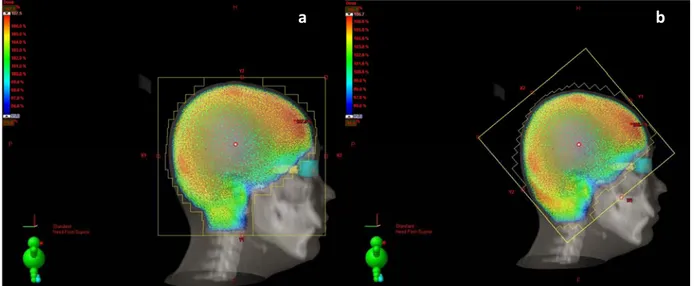
the bottom of C2 vertebra) and classical technique with collimation (CT) (anterior border was de ined as skin fall of, inferior border was the bottom of cranial base) were generated for all patients ( igure 1a and igure 1b). Three‐dimensional treatment planning was performed using 6‐MV photons designed to treat the whole brain for both techniques. Irregular beam portals of the both plans were shaped with a multileaf collimator and manually
optimized using the beam’s eye view technique. The perfect lens superimposition for maximal lens sparing on beam's eye views was achieved.
Dosimetric evaluation
In this study, Varian millennium 80‐leaf colli‐ mators (MLC) (Varian Medical Systems Inc, Palo Alto USA) were used for both HF and CT. The prescribed dose, which was de ined as the mean dose in the PTV, was 30Gy in 10 fractions at 3Gy
per day. The 100% of the planning target volumes covered by 95% of the prescribed dose.
The anisotropic analytical algorithm with tissue
heterogeneity corrections and a 2.5 mm calculation grid was used for dose calculations.
The dose‐volume histograms (DVH) were obtained from both plans.
Dose homogeneity in the PTV for both plans was compared by means of the dose homogenei‐ ty index (DHI, (17)):
In this formula, D98 is the maximum dose absorbed in those 2% of the PTV least irradiat‐ ed, D2 is the minimum dose absorbed in those 2% of the PTV most irradiated.
The maximum, minimum and the mean doses of the bilateral optic nerves, eye‐balls and lenses and MU settings required for each plan were compared.
Statistical analysis
The Statistical Package for Social Sciences (SPSS) v. 11.0 was used for statistical analysis (SPSS Inc. Chicago, II., USA). Paired samples t‐test was used for comparisons. The maximum, minimum and mean doses of OAR (including bilateral optic nerves, eye balls and lenses), were compared. A p value of < 0.05 was consid‐ ered to be signi icant.
RESULTS
The maximum and mean doses to the right lens, left lens and right eye‐ball were signi icant‐ ly decreased with CT (for the maximum doses p values were 0.007, 0.012 and 0.010; and for the mean doses p values were 0.027, 0.046 and 0.002 respectively ) (table 1 and table 2). When the minimum doses to the OAR were compared, CT allowed us signi icantly lower doses in the right optic nerve (p= 0.017) (table 3).
The DHI values were 0.088±0.013 and 0.087±0.013 for HT and CT respectively (p=0.182). The mean MU counts ± SD required for HT and CT were 327.1± 3.9 and 326.0±2.8 respectively. The difference in the average MU values used in the HT and CT was not statistical‐ ly signi icant (p=0.167).
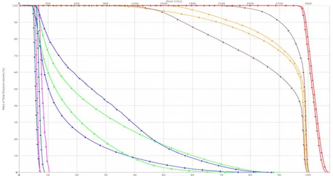
Figure 2 shows the DVH comparison of a patient. For the entire OAR, CT allowed us to obtain lower doses at OAR .
Figure 1. Isodose curves of a sagi al sec on of one representa ve pa ent (a) with Helmet technique and (b) with Collima on technique.
Figure 2. Dose‐volume histogram comparison of a pa ent (a) (Red: PTV, brown: le op c nerve, orange: right op c nerve, green: le eye‐ball, blue: right eye‐ball, magenta: right lens dark blue: le lens; ▲: Collima on technique and ■: Helmet technique).
Table 1. The maximum doses of the OAR.
Parameter Helmetmean±SD‡ (cGy) Collima on mean±SD(cGy) P
Right Lens 421,4±84,8 345,7±89,5 0,007* Le Lens 389,7±104,9 335,1±109,7 0,012* Right Eye‐ball 2972,5±147,8 2861,7±174,5 0,010* Le Eye‐ball 2940,3±139,7 2905,5±256,7 0,312 Right Op c Nerve 3082,1±42,7 3074,3±48,9 0,286 Le Op c Nerve 3082,8±45,6 3072,6±51,2 0,185
¥OAR: Organ at Risk ‡SD: Standard Devia on * p<0.05
Table 2. The mean doses of the OAR.
Parameter Helmet mean±SD‡ (cGy) Collima on mean±SD(cGy) P
Right Lens 286,6±44,2 251,4±34,6 0,027* Le Lens 268,9±53,9 250,0±52,6 0,046* Right Eye‐ball 1087,3±175,4 852,4±175,9 0,002* Le Eye‐ball 941,5±375,5 1074,9±66,5 0,259 Right Op c Nerve 2920,7±115,1 2875,8±104,6 0,229 Le Op c Nerve 2924,4±116,1 2822,4±174,7 0,101
¥OAR: Organ at Risk ‡SD: Standard Devia on * p<0.05
Table 3. The minimum doses of the OAR.
Parameter Helmetmean±SD‡ (cGy) Collima on mean±SD(cGy) P
Right Lens 200,8±36,5 202,1±24,1 0,914 Le Lens 201,5±40,1 202,4±34,0 0,921 Right Eye‐ball 163,7±35,5 167,9±23,7 0,683 Le Eye‐ball 163,0±38,7 149,5±71,1 0,159 Right Op c Nerve 1851,4±591,3 1324,7±639,7 0,017* Le Op c Nerve 1741,5±702,7 1394,9±547,5 0,111
DISCUSSION
Brain metastases occur in 10% to 30% of oncology patients, accounting for approximately 170,000 incident cases per year in the United States (18, 19). The median survival time following diagnosis of metastatic disease in the brain is approximately one year (20). Lung cancer is the most common primary tumor, accounting for 30% to 60% of patients; other common sources include breast cancer, melanoma, renal cell carcinoma, and colorectal cancer (21). The number of cerebral metastases is of pivotal concern in determining appropriate treatment. Palliative WBRT has been the mainstay of therapy for patients with intracranial metasta‐ ses and is associated with increases in the median survival of these patients to approxi‐ mately 4 to 6 months (3).
In our clinic WBRT in patients with brain metastases has been performed with 3‐ dimensional conformal radiotherapy. Therefore we are able to have an idea of the doses received by the entire OAR. However due to the different anatomical structure of different patient’s excess doses in the OAR are unavoidable. Therefore we evaluated the dosimetric bene its of CT com‐ pared with HT in patients with brain metastases who underwent WBRT. Our results suggested that the CT was superior to HT during WBRT in terms of the doses received by OAR, including the bilateral lenses.
Andic and colleagues compared the dosimet‐ ric differences between conventional two‐ dimensional HT WBRT and three‐dimensional conformal radiotherapy techniques in 30 patients (22). Their results suggested that when compared with conventional two‐dimensional radiotherapy planning, three‐dimensional conformal radiotherapy planning signi icantly improved the dose coverage of retro‐orbital areas and the dose homogeneity in WBI while protecting ocular lenses. Gripp and colleagues compared conventional two‐dimensional and
three‐dimensional planning basing on geographic mismatches, without dose distribution information (23). They measured the
minimal distance from the block edge to the contoured organs. The authors concluded that
CT simulation in WBRT is signi icantly superior to conventional simulation with respect to complete coverage of the target volume and protection of the eye lenses. In the current study, different from these two studies, we compared two different three‐dimensional WBRT tech‐ niques. We did not demonstrate any signi icant differences with respect to DHI between two plans. Since the both treatment plans that used in our study were three‐dimensional CT based techniques, we expected to have similar DHI between two plans.
Weiss and colleagues evaluated the doses received by the cribriform plate and the ocular lenses in three‐dimensional isodose distribu‐ tions of 11 patients treated with a standardized HT (24). They found that the average dose received by 95% of the cribriform plate was 85% of the prescribed dose and that suf icient lens shielding was usually not compatible with safe irradiation of the frontobasis. Ugurluer and colleagues evaluated the effects on target volume and lens doses with seven different treatment planning (25). The aim of the study by Ugurluer and colleagues was to evaluate the effects on tar‐ get volume (brain) and lens doses with different treatment plannings in patients who received WBRT. In this study, seven different treatment plannings were done for each patient. Plan I: Angled blocked plan; Plan II: Non‐angled blocked plan; Plan III: Helmet plan; Plan IV: Blocked plan with the inferior border at the inferior orbital ridge; Plan V: Angled unblocked plan; Plan VI: Non‐angled unblocked plan; Plan VII: Convential‐ ly planned non‐angled unblocked plan. When the plans were compared according to the lens doses, the minimum, maximum and mean doses were higher in the unblocked plans (p < 0.05). It was seen that with the angling of beams ive degrees to the posterior the lens doses decreased (p < 0.05). The authors concluded that
using three‐dimensional treatment planning, brain minimum doses increases and the dose
inhomogeneity decreases, and lens doses decreases using customized blocks and angling
of beams ive degrees posteriorly. In our study the maximum and mean doses to the right lens, left lens and right eye‐ball were signi icantly decreased with CT.
A large amount of work is ongoing, investigating the biological and biophysical mechanisms that may be involved in cataract development (26). For example, the lens of the eye is constantly subject to oxidation from a number of sources and a number of intrinsic mechanisms exist which ensure maintenance of a stable redox state, including gap junction communication, which can themselves be subject to oxidative stress (27). It has been shown that lens ibres have a very high protein content to enable transparency and refractivity (28). One of the mechanisms proposed to explain lens opaci ication is oxidation of crystallins, either by radiation or by reactive oxygen species
(ROS) (9, 29, 30). There is good evidence from epidemiological studies that any threshold for
radiation‐induced cataract is much lower than
the value of 2 Gy acute exposures given by International Commission on Radiological Pro‐
tection (ICRP) in their 2007 recommendations. The available data are consistent with a low threshold of around 0.5 Gy or no threshold. It is possible that cataract induction is a stochastic process and a threshold does not apply (26).
There are some shortcomings of the current study that should be mentioned. First of all the median survivals of the patients with brain metastasis is reported to be between 2‐7 months in RTOG study (4). This duration may be short to observe radiation‐induced cataracts in some patients. However radiation‐induced cataracts are expressed after latency. The duration of the latency depends inversely on dose: the higher the dose, the more rapidly the cataract develops. Moreover the current study is a dosimetric study that evaluates the effects of two different WBRT techniques on lens doses. On the other hand WBRT is not only used for the patients with brain metastasis but also used for primary central nervous system lymphoma, prophylactic cranial irradiation in patients with small cell lung carcinoma and leukemia. Second‐ ly the current study is a dosimetric study and we suggest our indings be veri ied in a prospective clinical trial, preferably in a larger number of patients.
In parallel to the developments in the ield of radiation oncology the survival times of the
patients with metastatic tumors have been increased as well. It is important to note that there are many more long‐term survivors with mild changes that impair quality of life but whose severity do not meet criteria for gradable toxicity. Therefore the term “quality of life” has been increasingly important. Modern radiothera‐ py techniques allowed us to decrease treatment related toxicities.
Recent studies have demonstrated that computed tomography simulation in WBRT is
signi icantly superior to conventional simulation with respect to complete coverage of the target volume and protection of the eye lenses (22, 23). In the current study we compared two different computed tomography based three‐dimensional radiotherapy techniques. Our results suggested
that CT is superior to HT during WBRT with respect to the doses received by bilateral lenses.
However; we speculate that to decrease lens doses beyond the 0.5 Gy as well as the increase
dose homogeneity, more sophisticated techniques including intensity modulated radiotherapy (IMRT) might be an option. There‐ fore further studies comparing more sophisticat‐ ed radiotherapy techniques needed to determine “how to decrease lens doses during WBRT”. The current study was presented as both “oral presentation and poster presentation” in MASCC/ ISOO 2013 International Symposium Con lict of interest: Declared none.
REFERENCES
1. Posner JB (1992) Management of brain metastases. Rev
Neurol, (Paris) 148(6–7): 477–87.
2. Graus F, Walker RW, Allen JC (1983) Brain metastases in children. J Pediatr, 103(4):558–61.
3. Murray KJ, Sco C, Greenberg HM, Emami B, Seider
M, Vora NL, Olson C, Whi on A, Movsas B, Curran W(1997) A randomized phase III study of accelerated hyperfrac on‐ a on versus standard in pa ents with unresected brain metastases: A report of the Radia on Therapy Oncology Group (RTOG) 9104. Int J Radiat Oncol Biol Phys, 39:571–
574.
4. Gaspar L, Sco C, Rotman M, Asbell S, Phillips
T, Wasserman T, McKenna WG, Byhardt R(1997) Recursive par oning analysis (RPA) of prognos c factors in three
Radia on Therapy Oncology Group (RTOG) brain metastases trials. Int J Radiat Oncol Biol Phys, 37(4): 745–
751.
5. Khan AJ and Dicker AP (2013) On the merits and limita‐ ons of whole‐brain radia on therapy. J Clin Oncol, 1;31
(1):11‐3.
6. Harding JJ and Crabbe JC (1984) The lens. In: Davson H. (Ed.), The Eye, 3rd ed. Vol. 1b. Academic Press, New York, NY, pp. 207–492.
7. Horwitz J and Jaffe NS (1992) Lens and cataract. In: Podos, S.M., Yanoff, M. (Eds.), Textbook of Ophthalmology, Vol. 3. Gower Medical Publishing, New York, NY.
8. Bantseev V, Bhardwaj R, Rathbun W, Nagasawa H, Trevith‐ ick JR (1997) An oxidants and cataract: (cataract induc on in space environment and applica on to terrestrial aging cataract). Biochem Mol Biol Int, 42: 1189–1197.
9. Bardak Y, Cekic O, Totan Y, Cengiz M (1998) Effect of vera‐ pamil on len cular calcium, magnesium and iron in radia‐
on exposed rats. Int Ophthalmol, 22:285–288.
10. Von Sallmann L (1951) Experimental studies on early lens
changes a er roentgen irradia on. III. Effect of X‐radia on on mito c ac vity and nuclear fragmenta on of lens epi‐ thelium in normal and cysteine treated rabbits. Arch. Oph‐
thalmol, 47: 305‐320.
11. Von Sallman L (1957) The lens epithelium in the patho‐ genesis of cataract. Am J Ophthalmol, 44: 159– 170. 12. Harding CV, Thayer MN, Eliashof PA, Rugh R (1965)
Effects of X‐irradia on on the injury reac on in lens epi‐ thelium. Radiat Res, 25: 305‐311.
13. Cogan DG, Donaldson DD, Reese AB (1952) Clinical and pathological characteris cs of radia on cataract. Arch
Ophthalmol, 47: 55–70.
14. Worgul BV, Merriam GR, Szechter A, Srinivasan BD (1976) Lens epithelium and radia on cataract. Arch Ophthalmol,
94: 996–999.
15. Cogan DG, Donaldson DD (1951) Experimental radia on cataracts. I. Cataracts in the rabbit following single X‐ray exposure. Arch Ophthalmol, 45: 508–522.
16. Beyzadeoglu M, Ozyigit G, Ebruli C (2010) Clinical Radia on Oncology. Springer‐Verlag Berlin Heidelberg pp:196.
17. Wu Q, Mohan R, Morris M, Lauve A, Schmidt‐Ullrich R
(2003) Simultaneous integrated boost intensity modulated radiotherapy for locally advanced head and neck squa‐ mous cell carcinomas. Dosimetric results. Int J Radiat On‐
col Biol Phys, 56:573‐585.
18. Brown PD, Asher AL, Farace A (2008) Adjuvant whole brain
radiotherapy: strong emo ons decide but ra onal studies
are needed. Int J Radiat Oncol Biol Phys, 70: 1305‐1309. 19. Regine WF, Sco C, Murray K, Curran W (2009) Neurocog‐
ni ve outcome in brain metastases pa ents treated with accelerated‐frac on vs. accelerated‐hyperfra onated radi‐ otherapy: an analysis from Radia on Therapy Oncology Group Study 91‐04. Int J Radiat Oncol Biol Phys, 51(3):711‐
717.
20. Narita Y and Shibui S (2009) Strategy of surgery and radia‐ on therapy for brain metastases. Int J Clin Oncol, 14: 275‐
280.
21. Suh JH (2010) Stereotac c radiosurgery for the manage‐
ment of brain metastases. N Engl J Med, 362: 1119‐1127.
22. Andic F, Ors Y, Niang U, Kuzhan A, Dirier A (2009) Dosimet‐
ric comparison of conven onal helmet‐field whole brain irradia on with three‐dimesional conformal radiotherapy: dose homogeneity and retro‐orbital coverage. The Bri sh J
of Radiol 82:118‐122.
23. Gripp S, Doeker R, Glag M, Vogelsang P, Bannach B, Doll T,
Muskalla K, Schmi G(1999) The role of CT simula on in whole‐brain irradia on. Int J Radiat Oncol Biol Phys, 45
(4):1081‐1088.
24. Weiss E, Krebeck M, Kohler B, Pradier O, Hess CF (2001)
Does the standardized helmet technique lead to adequate coverage of the cribriform plate? An analysis of current prac ce with respect to the ICRU 50 report. Int J Radiat
Oncol Biol Phys 49:1475–80.
25. Ugurluer G, İzmirli M, Palabiyik ZA, Cakir T (2009) The com‐
parison of brain and lens doses in whole brain radiotherapy with different treatment plannnings. UHOD, 19(3):140‐146. 26. Bouffler S, Ainsbury E, Gilvin P, Harrison J (2012) Radia on‐ induced cataracts: the Health Protec on Agency’s response to the ICRP statement on ssue reac ons and recommen‐ da on on the dose limit for the eye lens. J Radiol Prot, 32:
479–488.
27. Berthoud VM and Beyer EC (2009) Oxida ve stress, lens gap junc ons and cataracts An oxid. Redox Signal, 11: 339
–53.
28. Beebe DC, Holekamp NM, Siegfried C, Shui YB (2011) Vitre‐ ore nal influences on lens func on and cataract Phil. Trans
R Soc Lond B, 366: 1293–300.
29. Bardak Y, Ozerturk Y, Ozguner F, Durmus M , Delibas N (2000) Effect of melatonin against oxida ve stress in ultra‐ violet‐B exposed rat lens. Curr Eye Res, 20:225–230. 30. Barros PS, Angelo AC, Nobre F, Morales A, Fantoni DT,
Barros SB (1999) An oxidant profile of cataractous English Cocker Spaniels. Vet Ophthalmol, 2: 83‐86.