INVESTIGATION OF FACTORS RELATED TO PHYSICAL
ACTIVITY LEVEL IN PATIENTS WITH KNEE
OSTEOARTHRITIS
PT. Hasan KILINÇ
Program of Physical Therapy and Rehabilitation MASTER THESIS
ANKARA 2018
REBUPLIC OF TURKEY HACETTEPE UNIVERSITY
GRADUATE SCHOOL OF HEALTH SCIENCES
INVESTIGATION OF FACTORS RELATED TO PHYSICAL
ACTIVITY LEVEL IN PATIENTS WITH KNEE
OSTEOARTHRITIS
PT. Hasan KILINÇ
Program of Physical Therapy and Rehabilitation MASTER THESIS
ADVISOR OF THE THESIS
Gizem İrem KINIKLI, Assoc. Prof. PhD. PT.
ANKARA 2018
INVESTIGATION OF FACTORS RELATED TO PHYSICAL ACTIVITY LEVEL IN PATIENTS WITH KNEE OSTEOARTHRITIS
Hasan KILINÇ
Supervisor: Gizem İrem KINIKLI, Assoc., Prof., PhD., PT.
This thesis study has been approved and accepted as a Master dissertation in Physiotherapy and Rehabilitation Program by the assesment committee, whose members are listed below, on 02/05/2018
Chairman of the Committee : Prof., PhD., PT. Filiz CAN (Signature)
(Hacettepe University)
Advisor of the Dissertation : Assoc, Prof, PT.Gizem İrem KINIKLI
(Signature) (Hacettepe University)
Member: Prof., PhD., PT. Tülin DÜGER
(Signature)
. (Hacettepe University)
Member : Prof., PhD., PT. Zafer ERDEN
(Signature) (Hacettepe University)
Member : Seyit ÇITAKER, Assoc., Prof., PhD., PT.
(Signature) (Gazi University)
This dissertation has been approved by the above committee in conformity to the relatedissues of Hacettepe University Graduate Education and Examination Regulation.
Prof. Diclehan ORHAN, MD, PhD
ETHICAL DECLARATION
In this thesis study, I declare that all the information and documents have been obtained in the base of the academic rules and all audio-visual and written information and results have been presented according to the rules of scientific ethics. I did not do any distortion in data set. In case of using other works, related studies have been fully cited in accordance with the scientific standards. I also declare that my thesis study is original except cited references. It was produced by myself in consultation with supervisor Gizem İrem KINIKLI, Assoc. Prof. PhD. PT. and written according to the rules of thesis writing of Hacettepe University Institute of Health Sciences .
Hasan KILINÇ
ACKNOWLEDGEMENTS
Most importantly, I would like to thank to my advisor, Gizem İrem KINIKLI, Assoc., Prof., Phd, PT, because without her guidance and patience, this thesis would have not have been possible. She has been an excellent mentor, providing me with the skills required to be an independent researcher.
I would also like to thank all of my friends and colleagues Muhammed ALTIN, thank you for everything, you have been a great friend and colleague. Kadim AKTAŞ, thank you for being such great friends and running partners, I could not have wished to meet better friends.
Besides, I would like to thank Nisa MAKTAY, you've been very helpful in translation period of my thesis and you've always been a good friend and colleagues In addition, I would like to thank my wife, Cansu KILINÇ, who is with me throughout the thesis period, your patient and love make me happy all time.
Last but certainly not least, I would like to thank my family for being so supportive and encouraging. Mom and Dad, thank you for always being there with your words of wisdom and encouragement. Suzan and Nazan, my sisters, your love and support has been amazing, thank you for always being there for me.
ÖZET
Kılınç H., Diz Osteoartritli Hastalarda Fiziksel Aktivite Düzeyi ile İlişkili Faktörlerin İncelenmesi, Hacettepe Üniversitesi Sağlık Bilimleri Enstitüsü, Fizik Tedavi ve Rehabilitasyon Programı, Yüksek Lisans Tezi, 2018. Bu çalışmanın
amacı, diz osteoartritli bireylerde ağrı, alt ekstremite fonksiyonları, fiziksel performans, hareket etme korkusu, anksiyete/depresyon seviyesi ve yaşam kalitesinin fiziksel aktivite düzeyi ile ilişkili faktörlerin incelenmesidir. Çalışma Şubat 2017 – Kasım 2017’de Yeni Mahalle Devlet Hastanesi’ne başvuran 40-65 yaşları arasında diz osteoartrit tanısı alan gönüllü bireyler üzerinde gerçekleştirildi. Normal eklem hareketi gonyometre ile değerlendirildi. Ağrı ve fonksiyonel durumun hasta perspektifinden değerlendirilmesi için Oxford Diz Skalası (ODS), fiziksel performansın değerlendirilmesi için Zamanlı Kalk ve Yürü Testi (ZKYT) ve 6-Dakika Yürüme Testi (6-DYT), hareket etme korkusunun değerlendirilmesi için Tampa Kinezyofobi Ölçeği (TKÖ), anksiyete ve depresyonun değerlendirilmesi için Beck Anksiyete Ölçeği (BAÖ) ve Beck Depresyon Ölçeği (BDÖ) kullanıldı. Fiziksel aktivite Uluslararası Fiziksel Aktivite Değerlendirme Anketi - Kısa Formu
(IPAQ-SF) ile değerlendirildi. Son olarak yaşam kalitesinin değerlendirilmesinde Kısa
Form-12 (KF-12) anketinden yararlanıldı. Çalışmanın sonucunda ODS’nin tüm puanları ile IPAQ-SF arasında negatif yönde anlamlı bir ilişki bulundu (r=-0,550; p<0,001). TKÖ puanları ile IPAQ-SF puanları arasında negatif yönde iyi derecede korelasyon bulunurken (r=-0,693; p<0,001); BAÖ ve BDÖ puanları ile IPAQ-SF puanları arasında negatif yönde orta dereceli bir ilişki tespit edildi (0,970; r=-0,429; p<0,001). KF-12 fiziksel, mental ve toplam puanları ile IPAQ-SF arasında düşük bir korelasyon vardı (r=0,217; p<0,001). Doğrusal regresyon analizi, 6-DYT, TKÖ, BDÖ ve ODS parametrelerinin, diz OA’li bireylerde, IPAQ-SF ile değerlendirilen fiziksel aktivite düzeyini tahmin etmede en güçlü belirteçler olduğunu ortaya koydu. Sonuç olarak; diz OA’li hastaların yetersiz fiziksel aktivite seviyesine sahip olduğu göz önünde bulundurulduğunda, tedavi süresince kinezyofobi, depresyon seviyesi ve fonksiyonel seviyenin geliştirilmesine yönelik uygulamaların kullanılması fiziksel aktivite düzeyinin geliştirilmesi için önemlidir.
Anahtar Kelimeler: Diz, Osteoartrit, Fiziksel Aktivite, Kinezyofobi, Fiziksel
ABSTRACT
Kılınç H., Investigation of Factors Related to Physical Activity Level in Patients with Knee Osteoarthritis, Hacettepe University, Institute of Health Sciences, Physiotherapy and Rehabilitation Programme, Master Thesis, 2018. The aim of
this study was to examine the relationship between pain, lower extremity functions, physical performance, fear of movement, level of anxiety/depression and quality of life in physical activity levels in individuals with knee osteoarthritis. The study was conducted on voluntary individuals who were diagnosed with knee osteoarthritis between the ages of 40 and 65 who applied to Yeni Mahalle State Hospital between February 2017 and November 2017. A goniometer was used to evaluate normal range of motion. The Oxford Knee Scale (OKS) was used to assess pain and functional status from the patient perspective. The Timed Up and Go Test (TUG) and the 6-Minute Walk Test (6-MWT) were used to assess physical performance. The Tampa Kinesiophobia Scale (TSK) was used to assess fear of movement. Beck Anxiety and Beck Depression Inventory (BAI/BDI) were used to assess anxiety and depression. The International Physical Activity Assessment Questionnaire-Short Form (IPAQ-SF) was used to assess physical activity. Finally, Short Form-12 (SF-12) was used to assess the quality of life. As a result of the study, all the scores of OKS and IPAQ-SF showed a significant negative correlation (r=-0,550; p<0,001). There was a good negative correlation between the TSK scores and the IPAQ-SF scores (r=-0,693; p<0,001)while a moderate negative correlation was found between the BAI/BDI scores and the IPAQ-SF scores (r=-0,970; r=-0,429, p<0,001). There was a low correlation between SF-12 physical, mental and total scores and IPAQ-SF (r=0,217; p<0,001).. Finally, the results of linear regression analysis revealed that the total score parameters of 6-MWT, TSK, BDI and OKS were the strongest predictors of knee OA individuals that affected the physical activity level assessed by IPAQ-SF. As a result; considering that patients with knee OA have inadequate physical activity levels, the use of practices for the improvement of kinesiophobia, depression level and function level during the treatment process may be recommended for improving the level of phyical activity.
Keywords: Knee, Osteoarthritis, Physical Activity, Kinesophobia, Physical
CONTENTS ACKNOWLEDGEMENTS ABSTRACT CONTENTS SIMILARS FIGURES TABLES 1. INTRODUCTION 1 2. GENERAL INFORMATION 3 2.1. Knee Anatomy 3 2.1.1. Joint Capsule 3 2.1.2. Bone Constructions 4 2.1.3 Ligament 5 2.1.4. Meniscuses 6 2.1.5. Bursae 2.1.6. Muscles 7
2.1.7. Biomechanics of Knee Joint 9
2.2. osteoarthritis 10
2.2.1. Diagnostic Criteria of Osteoarthritis 2.2.2. Classification of Osteoarthritis 10
2.2.3. Etiology and Pathology of Osteoarthritis 11
2.2.4. Articular cartilage degeneration 14
2.2.5. Bone Marrow Oedema 14
2.2.6. Decrease in Physical Activity in Osteoarthritis 15
2.2.7. Treatment in Osteoarthritis 15
2.2.8. Osteoarthritis and Exercise 17
2.2.9. Invasive Treatment Approaches in the Treatment of Osteoarthritis 18
2.2.10. Osteoarthritis and Physical Activity 18
2.2.11. Relation of Physical Activity to Articular Cartilage 19
2.3.1. Definition of Fear, Anxiety and Phobia 20
2.3.2. Physiological Results of Kinesiophobia Behavior 21
3. INDIVIDUALS AND METHOD 22
3.1. Individuals 23 3.2. methods 24 3.2.1. Reviews 3.3. Statistical Analysis: 30 4. FINDINGS 31 4.1. Descriptive Findings 31
4.2. Pain and Functional Status Findings 32
4.3. Findings of Physical Performance Evaluations 34
4.4. Findings of Tampa Kinesiophobia Scale 36
4.5. Findings of Assessment of Anxiety and Depression Status 37
4.6. Findings of Assessment of Quality of Life 39
4.7. Findings of Assessment of Physical Activity Level 41
8.4. Findings of Evaluating the Parameters Affecting the Level of Physical Activity 42
5. DISCUSSION 49
5.1. Physical Properties 49
5.2. Range of motion 50
5.3. Pain and Functional Status 50
5.4. Physical Performance 51
5.5. Fear of Movement 52
5.6. Anxiety and Depression Level 53
5.7. Quality of Life 55
5.8. Physical Activity Level 56
5.9. Limitations 58
6. CONCLUSION 59 7. REFERENCES 8. APPENDIX
APPENDIX 1: Authorization Letter
APPENDIX 2: Ethics Committee Approval Certificate APPENDIX 3: Informed Consent Form
APPENDIX 4: Evaluation Form
APPENDIX 5: Poster Presentation from the Thesis APPENDIX 6: Verbal Presentation from Thesis 9. CURRICULUM
SIMILARS 6-MWT: 6 Minute Walking Test
Avg: Average
BAI: Beck Anxiety İnventory BDI: Beck Depression İnventory cm: Centimeters
IPAQ-SF: International Physical Activity Questionnaire Short Form M: Meter
Max: Max Min: Minimum
n: Number of individuals OA: Osteoarthritis, OKS: Oxford Knee Score p: Statistical significance value Sd: Standard Deviation
SF-12: Short Form-12
SPSS: Statistical Package for the Social Sciences TSK: Tampa Scale for Kinesiophobia
FIGURES
Figure Page 3.1. Application of Timed Up and Go Test 26 4.1. Daily Exercise Ratios of Individuals 32
TABLES
Table Page 4.1. Physical properties of individuals 31 4.2. Individuals' OKS averages 33 4.3. Relation of OKS scores to anxiety / depression,
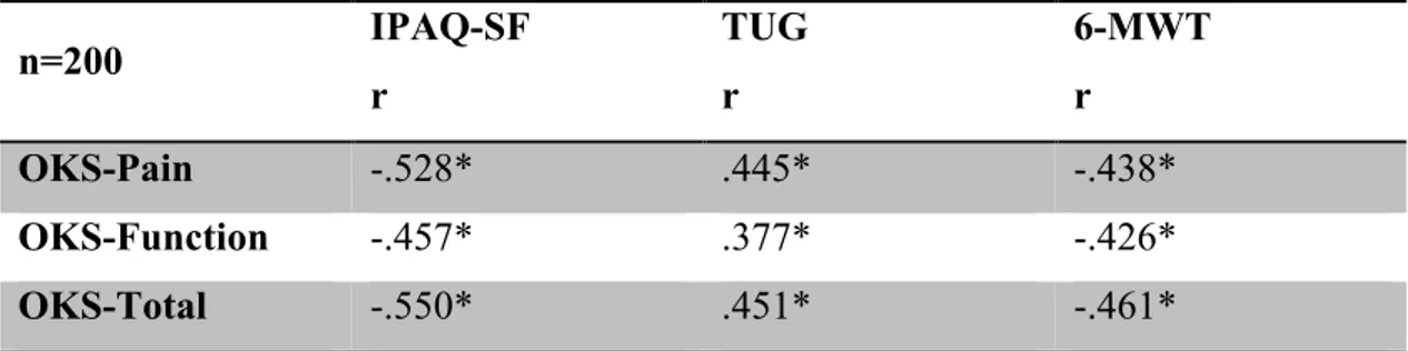
quality of life and kinesophobia scores 33 4.4. The relationship between ODS scores and IPAQ-SF, TUG and
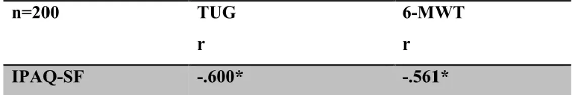
6-MWT scores 34 4.5. Average scores of physical performance tests 35 4.6. The relationship between TUG and 6-MWT scores and
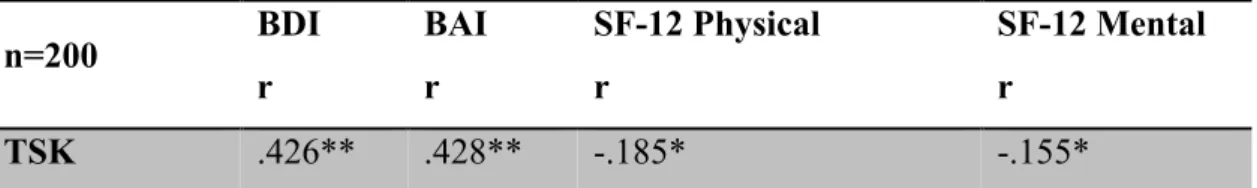
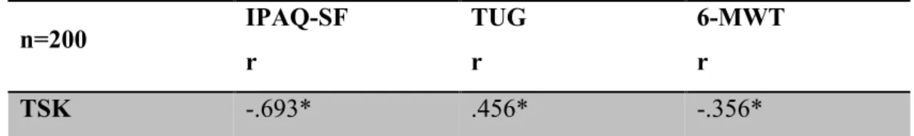
physical activity level IPAQ-SF 35 4.7. The relationship between TSK scores and physical and
mental scores of BAI, BDI, SF-12 36 4.8. The relationship between TSK and IPAQ SF, TUG and
6-MWT scores 37 4.9. Averages of BAI and BDI scores of the subjects 37 4:10. Relationship between BAI and BDI scores and
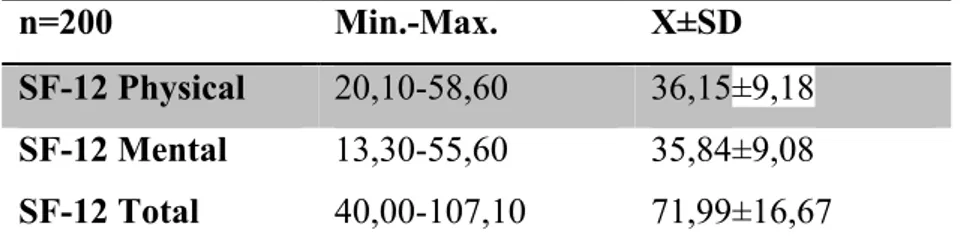
the physical and mental scores of TSK and SF-12 38 4:11. Relationship between BAI and BDI scores and IPAQ-SF scores 38 4.12. SF-12 physical, mental and total point average of individuals 39
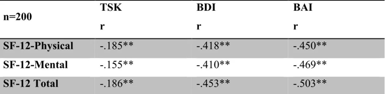
4:13. The relationship between SF-12 physical, mental and
4:14. The relationship between SF-12 physical, mental and total scores and
IPAQ-SF, TUG and 6-MWT 40
4:15. Categorical classification of IPAQ-SF scores 41
4.16. Individuals' levels of physical activity by gender 42
4:17. The relationship between IPAQ-SF and OKS, TUG, 6-MWT, BAI, BDI and SF-12 scores. 43
4:18. Variables entering the model in Linear Regression Analysis 44
4:19. Significant independent variables in regression analysis 45
4:20. Regression model with independent variables BDI, 6-MWT, TSK 46
4:21. Significant variables in Model 2 in Linear Regression Analysis 47
4:22. Summary of Models according to Physical Activity Levels 48
INTRODUCTION
Physical activity involves complex human movements of all ages (1). To examplify physical activity; professional activities, housework (eg self-care, general cleaning), transportation (eg walking, cycling) and leisure activities (eg swimming, dancing) can be shown. Activities such as sleeping, sitting, lying, watching television and using computer, which do not significantly increase rest and energy expenditure, are called sedantary behaviors.
Hip and knee osteoarthritis (OA) is one of the most important causes of pain and physical disability associated with older ages. Primary OA is usually associated with aging and heredity while Secondary OA can be caused by factors such as obesity, joint trauma, or recurrent excessive joint loads (2). In knee OA, fear of movement due to pain and reduced function affect quality of life negatively with balance and loss of proprioception (3). At the same time, daily life activities (DLA) such as going up and down stairs, walking and standing are affected negatively and their dependence gets increased (4).
The aim of physiotherapy and rehabilitation applications in knee OA treatment is to maintain and improve function with muscle strengthening exercises, to control pain, to maintain joint, and to increase quality of life by keeping the damage to a minimum level (5, 6). Regular, mild and moderate physical activity is recommended for beneficial effects such as improved cardiovascular fitness, reduced risk of diabetes and obesity (7). Federation of Exercise and Physical Activity recommends moderate physical activity for at least 3 days a week for 30 minutes (≥3MET) for individuals with knee OA. According to the Chronic Diseases Risk Factor Study, 87% of women and 77% of men do not have enough physical activity (8).
Individuals with knee OA are usually recommended to avoid activities like climbing up stairs, going up and long-term walks which can possibly impose burden on knees (5). In addition, it is seen that there are many parameters affecting the level of physical activity in the literature but there are few studies evaluating these parameters together.
Furthermore, these studies have been found to be made with subjective methods. To this end, for a disease which comes out due to a number of factors such as OA ; it is thought that not only the physical activity level of the patient should be adhered to but also the parameters affecting this physical activity level should be considered.
The difficulty of the costs and availability of methods that directly measure the level of physical activity, such as accelerometers, for individuals with OA is a well known fact. For this reason, studies evaluating the physical activity level in individuals with OA in the clinical setting by subjective methods and examining the parameters that may affect the level of physical activity are inadequate (5).
Hypothesis 1: In individuals with knee OA, pain is associated with lower
extremity functions and physical performance level of physical activity.
Hypothesis 2: Fear of moving in individuals with knee OA is associated with
anxiety / depression level, quality of life and physical activity level.
The aim of this study is to examine the relationship between pain, lower extremity functions, physical performance, fear of movement, level of anxiety / depression, and quality of life, and find out the most convenient indicators of physical activity level in individuals with OA in the knee.
With the results obtained from the study, the most effective parameters on physical activity level and physical activity level in knee OA individuals were revealed. It is thought that the determination of the level of physical activity and the factors affecting it in the knee OA may be useful in evaluating and developing physiotherapy and rehabilitation programs, thus leading to physiotherapists and other health professionals working in this patient group.
2. GENERAL INFORMATION
Osteoarthritis (OA) is the most common joint disease in the world and is the most important cause of chronic musculoskeletal pain. OA is a degenerative joint disease characterized by joint pain, local tenderness, limitation of movement, crepitation, sometimes effusion, and local inflammation at various degrees without clinically systemic symptoms. We can describe the knee OA more closely by examining the anatomy of the knee (5,6).
2. 1. Knee Anatomy
Knee anatomy is very important for the movements of the body, especially for the movements in the standing position. This is why knee stability and painless range of motion is important in maintaining daily work. Most commonly, overuse, age and traumatic injuries cause structural damage that can limit the function of knee. For this reason, a thorough understanding of the anatomy of the knee is important for the accurate diagnosis and treatment of knee pathology (9).
2.1.1. Joint Capsule
The inside of the fibrous capsule is covered with synovium producing synovial fluid which is an important component of the synovial assembly. The joint capsule not only serves for lubrication and shock absorption, but also serves as a conduit for nutrients. It may also have hormonal and messenger functions. Nociceptors buried in the synovial fluid are also protected by the fluid. Normally the amount of synovial fluid is very small and is only a few centimeters. However, flexion and extension movements of the knee joint allow the articular surfaces to be continually washed by fresh synovial; thus allowing the cartilage to feed properly and lubricate the joint surfaces. The capsule encases the proximal part of the head of the two gastrocnemius and the tendon of the popliteus muscle. The joint capsule is folded on itself to form a suprapatellar bursa in the anterosuperior part of the femoral condyle (10, 11).
2.1.2. Bone Constructions
The convex face of the knee joint belongs to the femur condyle, and the concave face belongs to the upper end of the tibia. As the third bone, the frontal patella is also added to the joint (12).
Femur: It is the longest, the heaviest and the strongest bone in the whole
human body. The femur is structurally classified as long bone and is an important component of the appendicular skeleton. The entire weight of the body is supported by femurs during many activities such as running, jumping, walking and standing. The distal femur extends significantly over the knee to form the medial and lateral condyles (12).
The medial and lateral condyles of the femur meet with the medial and lateral condyles of the tibia to form the articular surfaces of the knee joint. Between the condyles, there is a pit called the intercondylar fossa which provides space for anterior cruciate ligament and posterior cruciate ligaments cruciate that stabilizes the knee along the anterior / posterior axis (12).
Tibia: Tibia is bigger and stronger than the two bones in the lower leg. Tibia
forms the knee joint with the femur. Many strong muscles that move the foot and lower leg are attached to the tibia. Tibia's support and movement is necessary for many activities performed by the legs, including standing, walking, running, jumping and supporting body weight. The smooth concave medial and lateral condyles, which form the femur and knee joint, namely the proximal end of the tibia are roughly plane. Between the condyles, there is an intercondylar region that contains tibial object and provides points for the knee to bind meniscus, anterior and posterior cruciate ligaments. At the lower edge of the lateral condyle, there is a small facet formed by the tibia for the proximal tibiofibular joint. This joint is a plane that allows the tibia and fibula to pass slightly and adjust the position of the lower leg (12).
2.1.3 Ligament
There are four main ligaments to prevent excessive movement; anterior cruciate ligament, posterior cruciate ligament, medial and lateral collateral ligaments.
Medial collateral ligament; medial to the knee joint and extends from the
medial femoral epicondyle to the tibia. This prevents the tibia from displacing excessively laterally on the femur.
Lateral collateral ligament;is on the lateral part of the knee joint. It extends
from the lateral femoral epicondyle to the beginning of the fibula. It prevents the Tibia from displacing excessively medially on the femur.
Anterior cruciate ligament; extends from the anteromedial aspect of the tibial plateau to the medial region of the lateral femur condyle. The task of this ligament is to prevent the tibia from moving too far forward on the femur.
The anterior fibrils of the ligament are loose in the knee extension, stretched in flexion. The back fibers are stretched in the extension. It provides posterior stability and helps the flexion of knee. In addition to providing mechanical stabilization, the anterior and posterior cruciate ligaments also play an important role in providing proprioception sensation through the mechanoreceptors in their structures. This suggests that damage to these ligaments has an adverse effect on the proprioceptive sensation (13).
Posterior cruciate ligament; stays from femoral condyle to posterolateral
tibia platosum. It opposes the forces that want to push Tibia posteriorly considering the femur.
There are also three more ligaments in the knee. Patellar, oblique and arcuate popliteal ligaments.
Patellar ligament; It is a continuation of the quadriceps tendon and wraps the
Oblique popliteal ligament; connects to the upper edge of the intercondylar
fossa and to the rear edge of the tibia head.
Arcuate popliteal ligament; is a large fibrous band (11, 12, 14) attached to the
lateral condyle of the femur and blended with the posterior part of the capsule, which passes through the inferomedial route.
2.1.4. Meniscuses
Meniscuses are the shock absorbers of the knee. They are located horizontally between femur and tibia. In addition, these tissues are fibrous cartilaginous structures that synchronize with the anterior cruciate ligament, and play a crucial role in the functional integrity of the knee and are located on the proximal side of the tibia (12). The meniscus fills the gap between the flattened ends of the tibial bone, which the femur condyles sit on. The two meniscuses differ in shape and mobility.
Lateral meniscus; It is more oval shaped and quite mobile. It can slide
forward and backward with the knee motion. The Popliteus tendon continues along an edge through the joint capsule and contributes to the mobility of this meniscus (12).
Medial meniscus; It is larger and more C-shaped and firmly attached to the
capsule structure and to the medial collateral ligament. It moves slightly with the effect of knee movements. Therefore, the medial meniscus is more frequently torn than the lateral meniscus (11, 12, 14).
2.1.5. Bursae
The bursae are collected in three parts: anterior, lateral and medial. There are 4 bursae in each section. Bursa, which is located between the medial part of M. Gastrocnemius, M. Semimembranosus and the medial condyle of the tibia, is the most common form of effusion and inflammation.
2.1.6. Muscles
M. Quadriceps Femoris: Quadriceps femoris, the base of the knee joint, is the
strongest and largest muscle of the human body. It is a 4-headed muscle of M. rectus femoris, Vastus medialis, Vastus lateralis and Vastus intermedialis (16).
M. Rectus Femoris: This muscle located on the anterior and middle parts of
the M. quadriceps Femoris muscle begins by holding on to the upper side of the spina iliaca anterior inferiora and acetabulum with the upper extremity. It moves downward and joins the other heads and ends in the patella with the lower tip (17).
M. Vastus Lateralis: This muscle, located on the outer side of the M.
quadriceps femoris muscle, begins with the upper part of the linea intertrochanterican, the outer part of the trochanter major and the upper part of the labium laterals of the linea aspera. As it goes down and forward, it merges with other heads and ends in the patella (17, 18).
Vastus Intermedius: Located in the middle of the M. quadriceps femoris
muscle and behind the M. Rectus femoris, this muscle starts from under the linea intertrochanterican and from the anterior and lateral femur sides. As it moves downward, it merges with other heads. The common beam ends in the patella (17).
Vastus medialis: This fragment, located on the inner side of the M.
Quadriceps femoris muscle, begins with a long line from the top of the trochanter to the lower part of the trochanter and to the labium mediales of the linea aspera. As it goes downwards it merges with other heads and ends in the patella (17).
M. Quadriceps Femoris which functions for the extension of leg is the only extensor muscle of leg. In addition, M. Rectus femoris, as it holds spina iliaca anterior superior, enables the thigh for flexion movement (17).
M. Hamstrings: It is composed of M. Semitendinosus, M. Semimembranosus
M. Biceps femoris: This muscle located on the outer side of the rear group has
two heads, the long head and the short head. The short head begins by holding the labium laterales of the linea asperan with the upper tip while the long head begins from the tuber ischiadicum and merges with the short head as it moves downward. Both heads attach to the fibula after meeting in the lower parts of the thigh and moving towards the outer side of the thigh (17). M. biceps femoris, a biarticular muscle, has hip extension (when the leg is fixed), leg flexion and brings the knee to external rotation while the leg is in flexion position (16, 18).
M. Semitendinosus: The femoris muscle, semimembranosus muscle, adductor
magnus muscle, and gracilis muscle are located on the outer side of this muscle located on the inner side of the back of the thigh.
This muscle carries down the tube with the tuber ischiadicuma and joins together with the sartorius muscle and the gracilis muscle to form 'Pes anserinus' (17) which is also called the goose foot. The semitendinosus causes the hip to undergo extension, biceps flexion and brings the knee to internal rotation while in the leg flexion position (16).
M. Semimembranosus: It is located on the inner side of the back of the thigh
and there is M. Biceps Femoris on the outer side, M. Adductor Magnus on the back, M. Semitendinosus on the back, and M. Gracilis on the inner side. This muscle begins with the upper end of the tuber ischiadicuma in a widespread beam and ends in the tibial medial condyle (17). It exhibits a function similar to that of M. Semitendinosus, but it is a much more powerful convent. It enables hip extension, leg flexion, and when the leg flexes, it brings the leg to internal rotation (16).
M. Gastrocnemius: It has two heads as lateral and medial. External lateral
head which is called as lateral head; begins from the lateral condyle of the femur, joins the lateral head which is called as the inner lateral head starting from the medial epicondyle of femur and sticks to calcaneus via the tendon calcaneus (17).
The gastrocnemius, which is an important part of the plantar flexion of the foot, also has a knee joint flexion as a biarticular muscle in addition to its role as a supinator (16).
M. Sartorius: A superficial caste of approximately 5 cm wide, 50 cm long.
Spina iliaca begins anterior superior and ends in pes anserinus. M. Sartorius, the longest muscle in the human body, is flexor, abductor and external rotator of the hip, flexor in the knee (16).
M. Gracilis: The gracilis muscle, a thin and long muscle located on the
innermost side of the leg, starts with pubis and ends in pes anserinus (17). It is a two-jointed cast and enables hip joint flexion and internal rotation and flexion of the knee joint (16).
M. Popliteus: It is a superficial muscle. Starting from the lateral epicondyleof
the femur attaches to the back of the tibia. It gives the leg the ability of internal rotation movement (17).
M. Tensor Fascia Latae: It begins from the Spina iliaca anterior superior.
This muscle, expanding downward, becomes chirped under the trochanter major and ends up participating in the fascia latae structure (17). The swing phase brings the leg forward and abduction (16).
2.1.7. Biomechanics of Knee Joint
The movements of the knee joint take place in three planes rather than in a single plane. The knee joint is characterized by the presence of a trochoid (pivot) joint, even though it is a hinge (ginglymus) type joint. Flexion and extension movements take place in the frontal plane while adduction and abduction in the sagittal plane, and outward and inward rotation take place in the transverse plane (19). The knee joint can flex up to 140 ° active and passive 160 °. When knee is 90 ° in flexion; approximately 30 ° active, 30-35 ° passive internal rotation; 40 ° active and 45-50 ° passive external rotation can be seen. Abduction and adduction occurs passively after 30 ° of flexion and is less than 5 ° (18, 20).
Loads coming from the direction of string flexion-extension and varus-valgus are met by capsules and ligaments, contraction of agonist and antagonist muscles and geometry of joint faces. In the case of rotational loads, the role of muscles is very small and are covered by other structures. For this reason, rotational loads carry a greater risk of injury to the knee joint (21)
The movement of the patella in the knee flexion and extension is vertically displaced up and down. As the joint surface of the patella moves upwards in the extension, it tilts 35 ° downwards in the flexion. Patella increases the effectiveness of the M. quadriceps femoris muscle by extending the lever arm. It also provides a contact surface against the trochlea, enhances functional stability during loading and protects the femur condyles while the knee is in the flex. While the patella is moving freely in the knee extension position; the fact that the patella has shifted to the laterale, medial, or distant indicates that the joint is not fully functioning (22).
Q angle: An imaginary line drawn from the patella at the center of the tibial
tubercle to the imaginary line drawn from the center of the patella to the spina iliaca anterior superior is called the Q angle. These two lines can also be measured when the knee is at full extension, as measured at 90 ° (23). The mean is 10-14 ° in males and 15-17 ° in females due to the wider pelvis (22, 24).
2.2. Osteoarthritis
2.2.1. Diagnostic Criteria of Osteoarthritis
The diversity of the etiology of OA has led to the establishment of separate diagnostic criteria for disease in different joint regions. The most commonly used one is proposed by the American College of Rheumatology (ACR) (25).
ACR Knee Osteoarthritis Diagnostic Criteria: Clinically,
1. Knee pain on most days of previous month 2. Crepitation in active joint motion
3. ≤ 30 minutes morning malfunction. 4. Age ≥ 38
5. Bone growth in the tested knee
For OA diagnosis; The presence of 1, 2, 3, 4 or 1, 2, 5 or 1, 4.5 criteria is required.
Clinically and radiographically,
1. Knee pain on most days of the previous month, , 2. Radiographic osteophytes on the sides of joints,
3. The synovial fluid should have at least two of the following findings; clear, viscous, leukocyte count <2000 cells / ml
4. Age ≥ 40
5. Morning malfunction for ≤ 30 minutes in the knee 6. Crepitation in active joint motion
For OA diagnosis; The presence of 1, 2 or 1, 3, 5, 6 or 1, 4, 5, 6 criteria is required (25).
2.2.2. Classification of Osteoarthritis
Radiography is the most useful and important imaging method in OA. Common findings in OA are asymmetric narrowing of the joint space, sclerosis of the subchondral bone, subchondral cysts and osteophytes near the joint. Deformities, subluxations and joint rats are more common in advanced cases. Generally, there is poor correlation between radiological findings and symptoms in OA.
Kellgren and Lawrence rate the most commonly used rating in OA assessment (26).
Kellgren and Lawrence Rating: Stage 0: Normal
Stage 1: Suspicious narrowing of joint space, possible osteophyte Stage 2: Definite osteophyte, possible narrowing of joint space
Stage 3: Reasonable Multiple osteophytes, definite narrowing of the joint space, the onset of sclerosis
Stage 4: Wide osteophyte, severe narrowing of the joint space, severe sclerosis
2.2.3. Etiology and Pathology of Osteoarthritis
Knee OA has a multi-factor etiology such as obesity, aging, and injury (27). Other less established risk factors include genetic, bone density metabolism, and biomechanical effects (28).
In the study of Neame et al., It was found that when people whose siblings had a knee OA and those whose siblings did not have it were compared, the former group had twice higher risk than the latter group, and 62% of the disease variants were genetically determined (29). Similarly, the study of Chitnavis et al. showed that persons with a family history of symptomatic knee OA and those with a knee OA in their siblings would normally require two or five times more total knee arthroplasty as they approach the final stage of OA. The study also concluded that 1/3 of the OA variance was genetically determined (30).
Finally, it was reported in the study of Spector et al. in which they explored the genetic variance of hand and knee OA in twins with or without single-twin twins, that genetics explains the 39% to 65% of the variance of hand and knee OA (31).
In a study by Sowers et al., It was determined that foods may also affect knee OA. Oxidative damage, effects of inflammatory responses, cellular differentiation, problems with bone and collagen synthesis all may be affected by inadequate intake of nutrients (32). In a study by McAlindon and Felson, a decrease in radiographic OA was detected in those who intake continuous and high amounts of vitamin C. The progression of OA can also be reduced to a minimum by taking sufficient amounts of beta-carotene and vitamin E. These nutrients are reactive oxygen species and provide antioxidant effects that react against the oxidative damage in the knee. The study also suggests that vitamin E acts as an inhibitor of synovial inflammation associated with OA by forming arachidic acid from phospholipids and inhibits lipoxygenase activity (33). Sowers et al. suggested that OA pathology was associated with skeletal calcification and that individuals with OA showed higher bone mineral density levels than non-OA patients (32). Similarly, Hart et al. showed that increases in the mean bone density of individuals with early stage knee OA were inversely related to each other (34).
Recognizing that the bone mineral density in the lumbar spine is higher and increases the risk of developing knee OA, Hochberg et al. confirmed that high-level mineral density leads to knee OA (35).
Changing biomechanical alignment due to knee injury may also be a risk factor for knee OA. Chaudhari et al., In a study of the biomechanics of the knee in patients with anterior cruciate ligament (ACL) injury, reported that the failure of an ACL was altering biomechanical properties, leading to the onset of OA (36).
In the case of ACL injuries, different tibiofemoral contact patterns, anterior tibial translation and varying tibial internal and external rotations can lead to the formation of knee OA. These changes lead to a kinematic shift in the knee where degenerative forces are placed on the cartilage. Lohmander et al reported in a survey of female footballers living with ACL injuries that they found radiographic knee OA in 51% of patients 12 years after injury (37).
2.2.4. Joint Cartilage Degeneration
Knee OA is characterized by articular cartilage degeneration (38). Early OA often shows changes in the superficial regions of the joint cartilage. These changes are accompanied by changes in collagen orientation and proteoglycan content. Joint cartilage deformities include the medial and lateral tibiofemoral and patellofemoral regions, which are usually the main areas of the knee. Knee OA is characterized by articular cartilage degeneration (21). Early OA often changes in the superficial regions of the joint cartilage. These changes are seen with changes in collagen orientation and proteoglycan content. In places where the collagen content changes, as the OA progresses, the arthritis advances to the deeper regions of the articular cartilage (23). Joint cartilage deformities may involve the medial / lateral tibiofemoral and patellofemoral regions, which are the main areas of the knee (24).
2.2.5. Bone Marrow Oedema
Subchondral trabecular bone marrow oedema is a consequence of knee OA. In some studies, subchondral trabecular bone marrow oedema lesions were shown in 57% of the osteoarthritic knees evaluated. Osteophytes, synovitis, subchondral cysts, and the presence of subchondral sclerosis have been described as physical markers of OA (38, 39). In a study that studied subchondral bone abnormalities and bone marrow oedema for one year, these changes have been shown to trigger the formation of chondropathy (40). In addition, although the exact source of pain seen in OA individuals is uncertain, it is suggested that bone and bone marrow oedema may be the main cause of OA because these structures are rich in nociceptive fibrils and nerve of cartilaginous tissue is not present (40). Finally, another study showed that the pain exhibited by subchondral trabecular bone marrow oedema was site-specific and that the pain was medial or lateral tibiofemoral (38).
The weakness of M. quadriceps femoris muscle is the primary clinical manifestation of knee OA. This muscle strength is crucial for performance in daily living activities, and daily activities such as walking and stair climbing are also difficult to achieve in case of its weakness (41). Another study reported that M. quadriceps femoris weakness was frequently seen in patients with OA, and that muscle weakness reflects itself to the clinic in the form of muscle atrophy or joint pain (27).
2.2.6. Decrease in Physical Activity in Osteoarthritis
Individuals with OA have lower levels of physical activity than their healthy counterparts. Interestingly, however, no significant relationship was found between the pain and the decrease in the level of physical activity (42). In another study, early stage knee OA patients were compared with accelerometer activity levels, and it was observed that only 30% of these patients passed the physical activity criteria (43). It has been determined that individuals with knee and hip OA in the final stage of the disease have a very low level of physical activity.
2.2.7. Treatment in Osteoarthritis
Since OA has many different treatment approaches, several guidelines have been prepared. OA guidelines published by the Osteoarthritis Research Society International (OARSI) in 2007, 2008, 2010 and 2014 are the most recent reports based on evidence (22, 44).
Current treatments to alleviate OA symptoms include both conservative and surgical precautions. Conservative treatment includes drug-free treatments such as weight loss, exercise and education, as well as medicated treatments such as oral pain relievers, anti-inflammatory drugs and corticosteroids. In surgical applications, the final stage is unilateral or bilateral knee arthroplasty. Treatment options with the most potent evidence level of knee OA are addressed on the basis of OARSI 2010 guidelines.
Evidence-based treatment approaches are as follows; 1-Drug Therapy § Glucosamine Hydrochloride, § The diacerein, § Lavage / Debridement § Acetaminophen,
Non-Steroid Anti-Inflammatory Drugs, § opioids,
Intraarticular corticosteroids, § Intrarticular Hyaluronic Acid, § Glucosamine Sulfate, § Chondroitin Sulphate, 2-Physical Therapy § Self management, § Training, § Information, § Communication,
§ Muscle strengthening exercises, § Balneotherapy
§ Weight loss,
§ Laser, § Ultrasound,
§ Hot / Cold Applications, § Acupuncture,
§ Insoles, § Bracing,
§ Electromagnetic therapy, § Spa / Sauna, Massage 3-Surgical Treatment
§ It has been identified as osteotomy
Treatment goals in knee OA are determined as follows; • To reduce joint pain and stiffness,
• To protect and increase joint mobility, • Reduce physical inadequacy and disability, • To increase the quality of life,
• Limiting the progression of joint damage,
• To inform patients about the nature and management of the disease (45). All of the above-mentioned treatment modalities are tailored to the specific needs of the patient and the OA level.
2.2.8. Osteoarthritis and Exercise
Exercise options for OA treatment include; aerobic exercises, resistance exercises, and a combination of different exercise methods. Patients may have patient-specific conditions such as pain, significant disability, difficulty in walking, etc., and the exercise type should be carefully selected accordingly. A systematic review of the efficacy of home strength training has shown that force training targeting especially the muscles of M. quadriceps femoris may be beneficial in reducing pain and disability in patients with knee OA (45). In another study, the OA patients were treated with a 12-week home exercise program followed by a 12-week empowerment exercise.
As a result; When the control group and the exercise group were compared, it was shown that the joint space of the patients participating in the exercise group was more open and pain decreased (31). There are studies in OA patients that suggest that implementing a quantitative and progressive exercise and rehabilitation program in terms of functional performance and muscle function is beneficial (45).
2.2.9. Invasive Treatment Approaches in the Treatment of Osteoarthritis
If conservative and pharmacological treatments fail, invasive techniques may be needed. In a questionnaire on orthopedic surgeons, as a general view; pain, functional restriction, and narrowing of the joint space are the main indications of total knee arthroplasty indications (46). However, in order to consider surgical intervention, the consensus on which phase of OA should be expected has not yet been reached. In surgical procedures, it is intended to replace the damaged bone and cartilage, or to support the surface with metal or plastic implants. Surgical procedures appear to improve indirectly by increasing mental health and self-perception and quality of life, usually by reducing the pain of patients, by providing mobility.
2.2.10. Osteoarthritis and Physical Activity
Physical activity is any bodily movement that is produced by the skeletal muscle and requires energy expenditure (World Health Organization, WHO, 2012). Although physical activity is recommended by more than one health care provider, most people do not get serious about their recommendations. According to WHO, inactivity is the 4th biggest cause of deaths.
Physical activity may be both protective and harmful to cartilage, but physical activity in sufficient quantity and intensity affects cartilage health in the positive direction, but also positively affects both healthy adults and adults at risk of OA. A large proportion of individuals with knee OA do not meet the recommended level of physical activity (43).
This is partly due to the fact that individuals with OA avoid pain or exercise, as they believe that cartilage will suffer more. However, being physically active has many benefits, such as reducing the risk of hypertension, cardiovascular disease, diabetes and obesity, as well as improving bone health and joint function (WHO, 2012). Physical activity can be considered as one of the most important parameters in reducing knee OA risk. Physical activity is also thought to help cartilage healing.
2.2.11. Relation of Physical Activity to Joint Cartilage
Physical activity has positive effects on joint health, especially in the knees. Exercise may increase cartilage volume; but does not increase the incidence or progression rate of OA (47). For example; in the cohort study performed by Framingham et al., it was shown that the rate of knee OA incidence was not increased by regular recreational physical activity (48). 1279 healthy participants participating in the study were radiographically examined and their physical activity levels were questioned. Tests were conducted at baseline and nine years later and the incidence of OA was not associated with the amount of physical activity performed. Participants who regularly exercise as part of their lifestyle do not increase the risk of progression of the disease. Regular exercises, however, have helped to reduce pain and improve physical function. Physically active children also have a 24.8% greater
joint cartilage as compared to sedentary children of the same age (49). In other words, the thickness of the cartilage is directly proportional to the exposure to the physical activity loaded up to the threshold. Many studies suggest that regular and moderate physical activity will increase cartilage health. In a cross-sectional study of individuals without OA, knee pain, or knee injury in the last 5 years, the medial tibial cartilage volume of healthy women exercising for at least 20 minutes daily was higher when compared to those of sedentary ones (47). In another review, magnetic resonance images of 297 participants without knee trauma or disease were looked at and high physical activity such as swimming and cycling at high intensity to cause dyspnea was reported to be associated with cartilage quality (50). Studies have shown that people who perform strong, difficult sports during the youth period have the highest volume of cartilage; indicating the importance of physical activity from the young age (51).
Conversely, some studies have focused on the consequence that physical activity damages cartilage because of the heavy burden of cartilage. These studies indicate that excessive use of the knee joint causes wear and damage to the cartilage due to this wear. In a study by McAlindon et al., the overuse of the cartilage, which leads to the deterioration of cartilage, has been evaluated. Physical activity has been looked at in many subcategories. As a result of eight years of research, 473 participants with radiographs of their knees showed greater risk of knee OA in intensive and heavy physical activity (52).
2.3. Kinesiophobia
The concept of kinesiophobia is described by Kori et al. As "excessive and unreasonable fear of physical activity, which in the end makes the activity vulnerable to injury or re-injury" (53). Vlaeyen et al. later turned this concept into a theoretical model (54). In literature, kinesophobia, fear of movement and fear of movement associated with pain are mostly used synonymously (55). However, there are psychological differences between these concepts (53). Many researchers such as Asmundson and Taylor (1996) and Crombez et al. (1999) nevertheless used these concepts synonymously. There are many substitutions in the literature due to the difficulty of this distinction and the many similarities of these three concepts (55).
Phobia in fear of movement is an unexplained and disproportionate reaction. Moreover, this reaction is completely involuntary. Later, people living with phobia change their behavior and lives to avoid threatening, even if they know they are overreacting. This anxiety state is found to be the primary emotional component of the phobia and has a high correlation with kinesophobia (53, 55).
2.3.1. Definition of Fear, Anxiety and Phobia
Concepts of fear associated with fear of movement and pain are seen as syndromes by researchers. While these concepts are used, it is possible to switch between the concepts when it is known for what purpose it is used. Also, knowing depression and anxiety conditions is necessary to define the relationship between fear, anxiety, and phobia.
Fear is one of our pure and simple feelings. Fear is often described as feeling an unpleasant sensation of real danger (56). Feelings like fear are reaction syndromes that are not defined by any single feeling or behavior; but can be recognized from multiple stimuli and response models (57). Anxiety is similar to a sense of fear, but comes out without having any source of danger (56, 58). Although there are distinctive differences between fear and anxiety, they are often used interchangeably (59, 60). Phobia on the other hand is the emergence of fear without being bound to danger. There is no explanation or valid reason. A large portion is involuntary and causes the avoidance of fear (56). According to the American Psychiatric Association, certain phobia is a persistent and irrational fear that arises against a particular object or situation (58).
Vlaeyen et al. Compared specific phobia (a particular object or situation-related fear) in the case of chronic pain with the underlying features of fear associated with pain and found many similarities parallel to Kori's original theory (61). The difference between certain phobia and fear related to pain is that many people with pain have convinced themselves that it is a protective function of pain, and that they do not think there is any excess (42), as the fear of people with phobia is awful and unreasonable.
2.3.2. Physiological Results of Kinesophobia Behavior
A consequence of avoidance behavior is to avoid physical activity. The negative consequences of physical inactivity have been known since ancient times. Since the 17th century, physicians have introduced resting principle to patients as a treatment principle. Although bed rest is considered to have some problems; the number of people who believe in this method is also increasing.
It is the first change in the last century that Jones and Lovett offered a contrary view to this situation in 1926. Jones and Lovett first put forward the view that "the patient should be encouraged to act as soon as possible and bed rest should be prohibited" (62). Unfortunately, this recommendation was not widely accepted at that time and continued to be recommended for patients with musculoskeletal pain. There is no definite description of physical inactivity in the literature. However, physical activity advice for an adult is at least 30 minutes per day. A physical activity less than 30 minutes a day can be expressed as a physical inactivity (63).
Fear of movement; has a great effect on the formation of physical inactivity (56, 58-60). Physical inactivity causes; cardiovascular weakness, obesity, musculoskeletal system fragility, depression and premature aging. However, it is not clear where obstacles stop in various fear avoidance models. A more detailed definition has been given by the World Health Organization (2001) with the title "International Functioning, Disability and Health Classification (ICF)".
As a result; knee OA is a common disease that causes many problems and is difficult to treat. Patients with OA suffer from pain, depression and anxiety, physical performance and quality of life negatively. So it is very important to know what these changes are in order to be able to treat OA individuals and to prepare appropriate treatment programs. In our study, the relationship between OA and fear of movement, which is rarely seen in the literature, has been evaluated. It also assessed many important parameters that could have an impact on the level of physical activity, which has great significance for OA individuals.
3. PATIENTS AND METHODS
This study was conducted to investigate the relationship between pain, lower extremity functions, physical performance, fear of movement, level of anxiety / depression and quality of life in physical activity level in patients with knee OA who consulted to Yeni Mahalle State Hospital, between February 2017 and November 2017. It was carried out on voluntary individuals who received OA diagnosis (Annex 1-Authorization Letters).
Hacettepe University Ethics Committee of Non-Interventional Clinical Investigations approved the decision of GO 17 / 85-05 on 31.01.2017 (Annex 2-Ethics Board Approval Certificate) for the purpose of our work.
3.1. Individuals
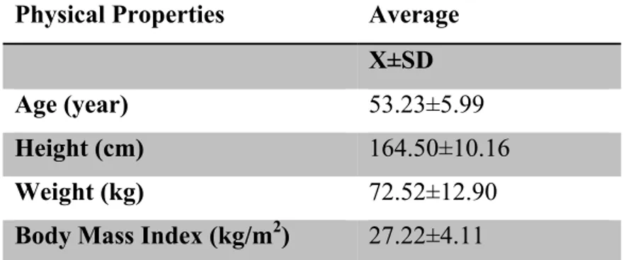
A total of 220 volunteers were enrolled, 130 of which were female and 90 were male, with an average age of 53.23 ± 5.99 years, and who were diagnosed as kne OA (bilateral) according to American Rheumatology Association criteria. However, during the tests, 13 people could not complete the physical performance tests and 7 people did not want to answer some questions in the tests. So the remaining 200 people continued to work.
Inclusion Criteria:
• Be between the ages of 40-65
• Kellgren Lawrence Classification: Level: II-IV • Volunteering to participate in the work
Exclusion Criteria: • Previously knee surgery
• Those with sensory impairment • Those with a broken story
• Those with circulation problems
• Those with infection in areas near the knee joint • Those with severe hearing loss or visual impairment • Individuals with cognitive problems
• Uncontrolled high blood pressure
• Those with vestibular problems such as vertigo and balance problems
• Individuals who have a condition to be considered as an obstacle to the knee joint
At the beginning of the workshop, participants were informed in writing and verbally about the purpose, duration, evaluations to be done, inquiry forms to be used. An informed consent form was signed to the volunteers of the individuals involved in the study (Annex 3: Informed Consent Form).
3.2. Methodology
In this study, which is planned in a cross-sectional study in a single patient group, while the sample size is determined because of the use of linear regression analysis as independent variables (pain, lower extremity functions, physical performance, fear of movement, level of anxiety / depression, and life expectancy) affect the level of physical activity (dependent variable), quality (n = 200) of at least 5 times the independent variables were included in the study (64). The evaluations in the study were done day-to-day to avoid taking patients' time and to make the tests more objective.
3.2.1. Evaluations
The following evaluations were made for all the participants in the study: (a) Physical and Sociodemographic Evaluation
(c) Assessment of Pain and Functional Status (d) Physical Performance Evaluation
(e) Assessment of the fear of movement (f) Evaluation of anxiety and depression (g) Assessment of Physical Activity Level (h) Evaluation of Quality of Life
3.2.1.1. Physical Characteristics and Sociodemographic Evaluation: A
patient assessment questionnaire was used to question the physical characteristics and sociodemographic knowledge of the individuals participating in the study (Annex 4-Evaluation Form). In this survey; age, height, weight, body mass index (BMI), the knee joint on the dominant side, the duration of all exercises performed by sitting or standing outside the daily activities of the individual were questioned.
3.2.1.2. Range Of Motion Assessment: Goniometric measurement, which is
used objectively in the evaluation of range of motion (R.O.M.) in clinic, is also used to determine the treatment program, to determine the functional capacity and to determine the effectiveness of the treatment, in addition to assessing the joint motion limit. Goniometer is a universal, clinical, gravitational, graphical and electronic type that is easy to carry, simple and durable tool that can be easily used on every joint. In our study, all measurements were performed using a "universal goniometer" to evaluate range of motion. The goniometric measurement of index flexion and extension was performed while the patient was in a prone position; the pivot point of the goniometer was placed in the lateral condyle of the femur, parallel to the lateral midline of the fixed arm femur, with the moving arm following the fibula. In the measurements, each measurement was repeated 3 times and their arithmetic mean values were recorded in degrees (65)
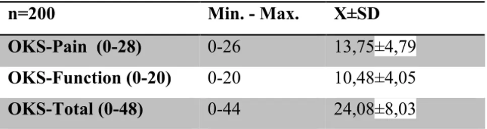
3.2.1.3. Assessment of pain and functional status: The Oxford Knee Score
(OKS) is a valid and reliable questionnaire that evaluates the pain and functional status of individuals with knee OA from their own perspectives (66, 67). It is scored between 0 (no) and 4 (severe) in the Likert system (0-48). In this questionnaire consisting of 12 questions, 2, 3, 7, 11, and 12 questions evaluated the functional status; Questions 1, 4, 5, 6, 8, 9, 10 assess the pain-related condition. OKS pain scores range from 0 to 28 points. The points to be taken from the OKS function dimension range from 0 to 20 points. Higher scores indicate worse pain and functional status.
3.2.1.4. Physical Performance Evaluation:
Timed Up and Go Test (TUG)This test, originally called "Timed Up & Go
Test" (TUG), is frequently used in the measurement of functional status in OA. This test is also investigating the ability of cases to maintain their balance during transfers and walking.
In this test, it was requested that the feet should sit in contact with the floor in a standard armchair, 3 meters walk, 3 meters back from the marked place, walk back to the chair and sit on a chair (Figure 3-1). The time elapsed for the performance of the events was recorded with the stopwatch in seconds. The test was repeated 3 times and averaged (68).
Figure 3.1. Timed up and go test application
6 Minute Walking Test: The functional capacities of the individuals studied
were evaluated with the 6 Minute Walking Test (6-MWT). 6-MWT is a simple test that measures the distance that an individual can walk on a flat, hard surface in 6 minutes. People were asked to walk as long as possible for 6 minutes in their own rhythm. The distance covered in six minutes was recorded in "meters" (69, 70).
3.2.1.5. Assessment of Fear of Movement: The Tampa Kinesophobia Scale
(TSK), consisting of 17 questions, is a measure evaluating the fear of movement / re-injury from 17 to 68 points. In this scale, in which Turkish validity and reliability were performed, high scores indicate that fear of movement is also at a high level (54, 71).
3.2.1.6. Assessment of anxiety and depression: The Beck Depression and
Anxiety Inventory (BDI-BAI) is a valid and reliable test and has also a used Turkish version that measures the anxiety and depression-related symptoms of people aged 13 years and older, with a 21-question. The questionnaire is scored between 0 and 63 separately for anxiety and depression dimensions. High scores indicate severe anxiety and depression (72).
3.2.1.7. Assessment of Quality of Life: Short-Form 12 (SF-12) SF-12 is a
short version of SF-36 containing 12 questions. There are two subtitles: Physical Component Scale (PCS) and mental component (Mental Component Scale, MCS). SF-12 Physical Score includes subscales of physical function, physical role, body pain and general health while SF-12 Mental Score consists of vitality, social function, emotional role and mental health subscales. The Turkish validity study was conducted by Koçyiğit et al. in 1999. The scores on the scale are between 0-60 for the physical and mental component, and the quality of life increases as the score increases (73).
3.2.1.8. Assessment of Physical Activity Level: The International Physical
Activity Questionnaire-Short Form (IPAQ-SF) whose Turkish version was also tested in terms of validity and relevance, was used to determine the level of physical activity of the participants in the study (74, 75). This short questionnaire, consisting of seven questions, assesses the amount of walking in the last week and the amount of moderate and challenging physical activity in work, transportation, home work, garden work and leisure activities. The sitting time is recorded separately on weekdays and weekends. The IPAQ is organized in two forms: special (work, transportation, home-garden work, leisure time) and activity specific (walking, moderate activity, severe activity)
- Calculation on activity specific scoring : Calculation is done by the addition
of walking, moderate intense activity, intense activity in itself.The following values are used for the analysis of IPAQ data:
• Walk = 3,3 MET
• Moderate physical activity = 4,0 MET • Rigorous physical activity = 8.0 MET
For example; MET-min / week score walking in a person walking 5 days 20 minutes a week is calculated as; 3,3x20x5 = 330 MET-min / week. The total walking score is obtained by gathering the scores related to walking within each area. In this way continuous data is obtained.
There are three levels of physical activity that are determined when categorized: 'inactive', 'minimally active' and 'very active' (physical activity that increases fitness). Categorical classification:
Inactive (Category 1): It is the lowest level of physical activity. Situations
that do not meet the criteria for Category 2 or 3 are considered 'inactive'.
Minimal Active (Category 2): Any of the following 3 criteria can be classified
as "minimal active":
a) Difficult activity to be done for 3 or more days, at least 20 minutes a day b) moderate intensity activity for 5 or more days or at least 30 minutes per day for walking,
c) Combination of 5 or more days walking, moderate intensity and challenging activity providing a minimum of 600 MET- min / week.
It is stated that the individual who meets any of the above criteria achieves a minimal level of physical activity.
Active (Category 3): Minimum public health is calculated as a 'very active'
separate category for people who have passed physical activity recommendations. This measurement equals a moderate activity of at least about one hour per day or more. This category is the level of activity required to provide health benefits. Category 3 is at the higher threshold of activity and it is useful to distinguish the difference from the other two groups.
There are two criteria for classifying as 'very active':
a) At least 3 days of tough activity providing a minimum of 1500 MET-min / week or,
b) 7 days walking, moderate intensity or at least 3000 MET-min/week or the combination of challenging activity.
IPAQ Sitting Question:
The IPAQ sitting question is an additional determinant. It is not part of the scoring of physical activity. There is little data on sedentary (sitting) behaviors and there is no accepted threshold value as a categorical level.
The categorical classification of IPAQ-SF, which is used to assess the subjective physical activity level of OA individuals participating in the current study is as follows:
0-599 MET-min/ week: Inactive
600-2999 MET-min/ week: Minimal Active > 3000 MET-min/ week: Active
3.3. Statistical Analysis: Statistical analysis was performed by using IBM
SPSS 22.0 (Statistical Package for the Social Sciences 22.0) package program. The normal distribution of variables was examined visually (histogram and probability graphs) and analytical methods (Kolmogorov-Smirnov / Shapiro-Wilk tests). Descriptive analyzes were expressed as mean and standard deviation for numerical variables, frequency tables for ordinal variables (n) and ratios (%).The relationship between pain, lower extremity functions, physical performance, fear of movement, level of anxiety / depression and quality of life scores were examined by Pearson Correlation Analysis. Before the Regression Analysis, Spearman Correlation Analysis was used to determine the variables associated with the level of physical activity. Correlation coefficients between 0.05 and 0.30 are low or insignificant correlation; Values between 0,30 and 0,40 are low middle correlations; The values between 0.40-0.60 are median correlation ; values between 0.60-0.70 are Good correlation and values between 0.70-1.0 are interpreted as perfect correlation (64). Independent variables (pain, lower extremity functions, physical performance, fear of movement, level of anxiety / depression and quality of life) affecting the level of physical activity (dependent variable) were calculated using the Multiple Linear Regression Analysis Model with stepwise-backward elimination. Those that do not provide linear regression assumptions are removed from the model candidate parameters. For the remaining parameters, Linear Regression Analysis was performed again and the inappropriate parameters were removed from the model for reasons such as not being able to contribute to the model. Four independent variables (fear of movement, pain and function total score, Beck depression score, and 6 minute walking test score) were entered into the final analysis and again as the best predictors of the level of physical activity in knee OA individuals in the final model. The probability of error was accepted as p <0.05.