HPLC ANALYSIS OF OLEUROPEIN IN OLEA EUROPAEA L
OLEA EUROPAEA L.’DAKİ OLEUROPEIN’İN YBSK ANALİZİ
Çiğdem ALTINYAY, M. Levent ALTUN*
Ankara University, Faculty of Pharmacy, Department of Pharmacognosy, 06100, Tandoğan - Ankara, TURKEY
ABSTRACT
In this study a simple and sensitive HPLC method for separation and quantitative determination of oleuropein in the leaves and the branches of the natural (Olea europaea L. var. sylvestris) and cultivated (Olea europaea L. var. europaea) varieties of Olea europaea L. has been developed. Oleuropein was determined on a reversed phase C18 column , by using water-acetonitrile-formic acid (84.6:15:0.4) (v/v/v) as a mobile phase . The oleuropein content of cultivated O.europaea (var. europaea) ( Balıkesir-Edremit samples) was found to be 3.506 % for leaves and 1.438 % for branches. For natural O.europaea (var. sylvestris) (Osmaniye samples) , these values were found as 5.197 % and 1.462 %, respectively. The oleuropein content of cultivated O.europaea (var. europaea) ( Konya samples) was determined as 4.020 % for leaves and 1.097 % for branches.
Key Words: Oleuropein, O.europaea,Oleaceae, HPLC, leaves, branches, cultivars ÖZET
Bu çalışmada, doğal ( Olea europaea L. var. sylvestris ) ve kültüre alınmış ( Olea europaea L. var. europaea ) zeytin ağaçlarının yaprak ve dallarında bulunan oleuropein’in kantitatif analizi için basit ve duyarlı bir yöntem geliştirilmiştir. Oleuropein miktar tayini, ters faz YBSK ile C18 kolonunda su: asetonitril: formik asit (84.6:15:0.4 ) mobil fazı kullanılarak yapılmıştır. Oleuropein miktarı, Olea europaea L. var. europaea bitkisi için Balıkesir-Edremit’ten toplanan örneklerin yapraklarında % 3.506, dallarında % 1.438, Konya’dan toplanan örneklerin yapraklarında % 4.020, dallarında % 1.097 olarak saptanırken Olea europaea L. var. sylvestris bitkisi için Osmaniye örneklerinin yapraklarında % 5.197, dallarında ise % 1.462 olarak bulunmuştur..
Anahtar Kelimeler : Oleuropein, O.europaea, Oleaceae, YBSK, yapraklar, dallar, kültür bitkisi
INTRODUCTION
The olive tree ( Olea europaea L., Oleaceae ) is one of the most important trees in Mediterranean countries. It grows throughout the entire Mediterranean region and in most of the Southern European countries. Moreover, it is cultivated for its edible fruits and to obtain oil from them. In the Mediterranean area, there are nearly eight million hectares of cultivated olive trees (1). Leaves of the tree became important when olive leaf extract was reported to be potent in treating fever and malaria in 1854 (2). Since then, several researchers demonstrated hypotensive (3,4,5), hypoglycemic, coronary dilatatory, antiarrhythmic, antiuricaemic (6), antioxidant (7), anti-complementary (8), antimicrobial (9), thyroid stimulatory (10), antiviral (11) and anti-HIV (2) activities of olive leaf extract.
The leaves of the tree consist of secoiridoids (12), phenolic compounds, flavonoids and volatiles (13).
The active constituent of olive leaf is oleuropein, a secoiridoid, which has reported to be a potent antioxidant endowed with antiinflammatory properties (14). It has pharmacologic and health promoting properties including hypoglycemic, antioxidant (15), antimicrobial (16), antimycoplasmal (17), antiviral (11), anti-tumor and angiogenic (18) activities. It was found to inhibit androstenedione 6β-hydroxylase activity, a cytochrome P450 3A marker in human liver microsomes (19), prevent lipid peroxidation on rat liver microsomes (20). Oleuropein has been also shown to inhibit LDL (Low Density Lipoprotein) oxidation and possess vascular protection activity by inhibiting platelet aggregation induced by platelet-activating factor (21).
In Turkey, species Olea europaea L. is represented by two varieties var.europaea and var. sylvestris and cultivated through the Western and Southern of the country (22). In Turkish traditional medicine 5 % infusion of the leaf is taken orally as appetizing, diuretic, constipate and antipyretic . It is used externally to clean festering sores (23). However, the branches do not have any traditional uses.
In the previous studies, oleuropein content of the olive leaf ethanolic extract, methanolic extract, 60 % aqueous methanolic extract and 100 % aqueous extract was found to be 24.54 % (w/w) (14), 19 % (w/w) (24), 9.04 – 14.16 % (w/w) (25) and 12.8 % (w/w) (2) respectively.
In our research, the amount of oleuropein was determined both in the leaves and the branches of the natural ( Olea europaea L. var. sylvestris (Miller) Lehr. ) and cultivated (Olea europaea L. var. europaea Zhukovsky) varieties by RP-HPLC to find out whether they are good sources of oleuropein isolation or not.
MATERIALS AND METHODS Chemicals
Oleuropein (Extrasynthese; 32619) used as the standard chemical was obtained from Extrasynthese. Chromatographic grade-double distilled water, HPLC grade acetonitrile (Merck - 100030) and analytical grade formic acid (Merck 100264) were used.
Plant material
Natural and cultivated plant materials were collected from Balıkesir-Edremit, Konya and Osmaniye. Plant materials were dried at room temperature. Voucher specimens were deposited at Herbarium of the Faculty of Pharmacy, Ankara University, Ankara, Turkey (AEF 23334, AEF 23547, AEF 23600)
Samples for HPLC analysis
5 grams of the dried powdered materials were macerated with 50 ml methanol for 2 hours at room temperature using a magnetic stirrer. The extracts were filtered and evaporated to dryness under a temperature not exceeding 400C.
The residues were dissolved in 50 ml of HPLC grade Merck methanol. Solutions were passed through a 0.45 µm filter and 20 µl extracts were directly injected into the HPLC column. The results were obtained as a mean value of three separate injections.
Apparatus
The method was performed with a LC system consisting of a Jasco model PU-980 pump and JASCO UV-975 UV/ VIS detector. Samples were injected with a 7725 Rheodyne injector system with a 20 µl sample loop. The detector was set at 240 nm and peak areas were integrated automatically by computer using BORWIN software programme.
Separation was carried out using a Nucleosil 100-5 C18 column (5 µm, 250 x 4.6 mm; GL
Sciences Inc.). All the calculations concerning the quantitative analysis were performed with external standardization by measurement of peak areas.
Stock and standard solution
Oleuropein (25.00 mg) was accurately weighed into a 25 ml volumetric flask and dissolved in the methanol and filled up to volume with methanol.
Standard working solution
Standard working solution was prepared individually in methanol for oleuropein. Aliquots from each working solution was combined and diluted with methanol to obtain a standard solution containing 1000 µg/ ml oleuropein.
Chromatographic conditions
HPLC analysis was performed by isocratic elution with flow rate 1.0 ml/min. The mobile phase composition was Water-Acetonitrile-Formic acid (84.6:15:0.4) (v/v/v). All solvents were filtered through a 0.45 µm Millipore filter before use and degassed in an ultrasonic bath. Volumes of 20 µl of each prepared solutions and samples were injected into the column. Quantification was effected by measuring at the 240 nm. The chromatographic run time was 40 min.
Calibration
Concentrations of 0.1-0.4 mg/ml of standard solutions were prepared in methanol.Triplicate 20 µl injections were made for each standard solution to see the reproducibility of the detector at each concentration level. The peak area of each concentration was plotted against the concentrations to obtain the calibration graph. Finally calibration equation and correlation coefficients were calculated .
RESULTS
Method Development
In our study, several chromatographic conditions were tested for the separation and determination of oleuropein in samples. Good separation and determination of Olea europaea L. (natural and cultivated samples) in leaves and branches were performed by using the mobile phase consisting of Water-Acetonitrile-Formic acid (84.6:15:0.4) (v/v/v) a Nucleosil 100-5 C18 (5 µm,
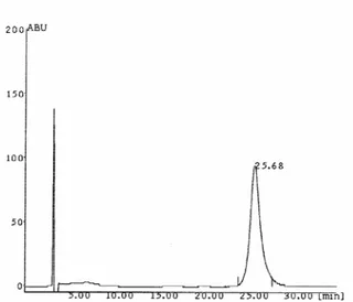
250 x 4.6 mm) column by isocratic elution with flow rate 1.0 ml/min. Detector responses were measured as peak areas. The injection volume was 20 µl and triplicate injections were used for each sample. At the flow rate of 1.0 ml/min the retention time for oleuropein was observed as 25.68 min.(Figure 1).
Figure 1: Chromatogram of oleuropein Linearity
Table 1 presents the equation of the regression line, correlation coefficient (r2), relative standard deviation (RSD) values of the slope and intercept for oleuropein. Excellent linearity was obtained for oleuropein between peak areas and concentrations of 100-400 µg/ ml with r2 = 0.9989.
Limits of Detection and Quantification
Limits of detection (LOD) were established at a signal-to-noise ratio (S/N) of 3. Limits of quantification (LOQ) were established at signal-to-noise ratio (S/N) of 9. LOD and LOQ were experimentally verified by six injections of oleuropein at the LOD and LOQ concentrations.The LOD was calculated to be 30 µg/ml and the LOQ was calculated to be 100 µg/ml for oleuropein (Table 1).
Table 1: Linearity Results, Limit of Detection (LOD) and Limit of Quantification (LOQ)
Compound λ Equation r2 Slope
(RSD %) Intercept (RSD %) LOQ (µg/ml) LOD (µg/ml) Oleuropein 240 Y=19.961X-105.25 0.9989 2.045 10.931 100 30 Precision
The precision of the method (within- day variations of replicate determinations) was checked by injecting nine times of oleuropein at the LOQ level. The precision of the method, expressed as the RSD % at the LOQ level was 2.915 % for oleuropein (Table 2).
Table 2: Precision of the Developed Method at the LOQ Level (n=9)
Compound λ Peak Area (mean) RSD %
Oleuropein 240 1782.75 2.915
Oleuropein analysis of Olea europaea L.
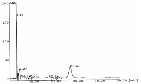
Quantitative determination of oleuropein in the leaves (tR= 26.89 min) and branches (tR=
27.41 min) of Olea europaea L. was carried out by RP-HPLC using external standard method (Figure 2 and Figure 3).
Figure 2: Chromatogram of oleuropein (tR= 26.89 min) in the leaves of Olea europaea var. europaea
Figure 3: Chromatogram of oleuropein (tR= 27.41 min) in the branches of Olea europaea var. europaea The assay results of natural and cultivated Olea europaea L. are shown in Table 3. The standard solution of oleuropein was added respectively to plant extracts and injected at each time.
The area of peaks corresponding to standard were increased to prove the presence of this compound. Its percent means and standard deviation values are summarized in the same table (Table 3).
Table 3: Contents of Oleuropein in Olea europaea L. Samples
Samples Oleuropein %
(n= 3, mean) mean ±SD Leaves of cultivated O.europaea
(var. europaea) (Balıkesir-Edremit samples)
3.506 ±0.0732
Branches of cultivated
O.europaea (var. europaea)
(Balıkesir-Edremit samples)
1.438 ± 0.0418
Leaves of natural O.europaea (var. sylvestris) (Osmaniye samples)
5.197 ± 0.0071
Branches of natural O.europaea (var. sylvestris) (Osmaniye samples)
1.462 ± 0.0868
Leaves of cultivated O.europaea (var. europaea) ( Konya samples)
4.020± 0.0813 Branches of cultivated
O.europaea (var. europaea)
(Konya samples)
DISCUSSION
The present HPLC method was applied to the Olea europaea L. extracts and allowed the efficient separation of oleuropein in the extracts.
HPLC analysis results of oleuropein contents in leaves and branches of Olea europaea L. are given in Table 3.
In the previous studies, oleuropein content of the olive leaf ethanolic extract, methanolic extract, 60 % aqueous methanolic extract and 100 % aqueous extract was found to be 24.54 % (w/w) (14), 19 % (w/w) (24), 9.04 – 14.16 % (w/w) (25) and 12.8 % (w/w) (2), respectively by Benavente-Garcia et al. (14), Bernard et al. (24), Savournin et al. (25) and Lee-Huang et al (2).
In our study, we determined the oleuropein contents of methanolic extracts of cultivated O.europaea L. (var. europaea) ( Balıkesir-Edremit samples) as 3.506 % for leaves and 1.438 % for branches. For natural O.europaea L. (var. sylvestris) (Osmaniye samples) , these values were found as 5.197 % and 1.462 %, respectively. The oleuropein content of cultivated O.europaea L. (var. europaea) ( Konya samples) was found to be 4.020 % for leaves and 1.097 % for branches. As it can be seen from the results, oleuropein content of the leaves of natural O.europaea L. (var. sylvestris) (Osmaniye samples) is higher than other plant samples. When the branches are considered, we see that the quantity of the oleuropein is lower than the leaves for all samples.
To our knowledge this is the first study that reports the oleuropein contents of the methanolic extracts of both the leaves and the branches of some Turkish olive varieties. If the oleuropein contents of Turkish olive varieties are compared with those that are mentioned above, it can be seen that the leaves and the branches of Turkish olive varieties have lower oleuropein contents. Even though, the leaves of both natural and cultivated varieties can be considered as good oleuropein sources. Whereas, the results show that the branches have lower oleuropein contents to be used in oleuropein isolation.
REFERENCES
1. Guinda A., Albi T., Perez-Camino M.C. and Lanzon A. “Supplementation of oils with oleanolic acid from the Olive leaf ( Olea europaea )” Eur. J. Lipid Sci. Technol., 106. 22-26 (2004).
2. Lee-Huang S., Zhang L., Lin Huang P., Chang Y.and Huang P. “Anti-HIV activity of olive leaf extract ( OLE ) and modulation of host cell gene expression by HIV-1 infection and OLE treatment” Biochemical and Biophysical Research Communications, 307. 1029-1037 (2003).
3. Cherif S., Rahal N., Haouala M., Hizaoui B., Dargouth F., Gueddiche M., Kallel Z., Balansard G. and Boukef K. “Essai clinique d’un extrait titre de feuilles d’olivier dans le traitement de l’hypertension arterielle essentielle” J. Pharm. Belg., 51(2). 69-71 (1996) 4. Khayyal M., El-Ghazaly M., Abdallah D., Nassar N., Okpanyi S. and Kreuter M-H.
“Blood pressure lowering effect of an olive leaf extract ( Olea europaea ) in L-NAME induced hypertension in rats” Arzneim.-Forsch. / Drug Res., 52(11). 797-802 (2002)
5. Rauwald H. W., Brehm O. and Odenthal K. P. ″Screening of nine vasoactive medicinal plants for their possible calcium antagonistic activity. Strategy of selection and isolation for the active principles of Olea europaea and Peucedanum ostruthium″ Phytotherapy Research, 8. 135-140 (1994).
6. Fehri B., Mrad S., Aiache J-M. and Lamaison J-L. “Effects of Olea europaea L. extract on the rat isolated ileum and trachea” Phytotherapy Research, 9. 435-439 (1995).
7. Briante R., Ratumi M., Terenziani S., Bismuto E., Febbraio F. and Nucci R. “Olea europaea L. leaf extract and derivatives : Antioxidant properties” J. Agric. Food Chem., 50. 4934-4940 (2002).
8. Pieroni A., Heimler D., Pieters L., Van Poel B. and Vlietinck A.J. “In vitro anti-complementary activity of flavonoids from olive ( Olea europaea L. ) leaves” Pharmazie, 51. 10 (1996)
9. Markin D., Duek L. and Berdicevsky L. “In vitro antimicrobial activity of olive leaves” Mycoses, 46. 132-136 (2003).
10. Al-Qarawi A.A., Al-Damegh M.A. and ElMougy S.A. “Effect of freeze dried extract of Olea europaea on pituitary-thyroid axis in rats” Phytotherapy Research, 16. 286-287 (2002).
11. Micol V., Caturla N., Perez-Fons L., Mas V. and Perez L. “Espeta A. The olive leaf extract exhibits antiviral activity against viral haemorrhagic septicaemia rhabdovirus (VHSV )” Antiviral Research, 66. 129-136 (2005).
12. Skerget M., Kotnik P., Hadolin M., Hras A. R., Simonic M. And Knez Z. ″Phenols, proanthocyanidins, flavones and flavonols in some plant materials and their antioxidant activities″ Food Chemistry, 89. 191-198 (2005).
13. Altarejos J., Salido S., Perez-Bonilla M., Linares-Palomino P. J., Beek T. A., Nogueras M. and Sanchez A. ″Preliminary assay on the radical scavenging activity of olive wood extracts″ Fitoterapia, 76. 348-351 (2005).
14. Benavente-Garcia O., Castillo J., Lorente J., Ortuno A. and Del Rio J.A. “Antioxidant activity of phenolics extracted from Olea europaea L. leaves” Food Chemistry, 68. 457-462 (2000).
15. Al-Azzawie H.F. and Alhamdani M-S S. “Hypoglycemic and antioxidant effect of oleuropein in alloxan-diabetic rabbits” Life Sciences, 78 ( 12 ). 1371-1377 (2006).
16. Bisignano G., Tomaino A., Cascio R., Crisafi G., Uncella N. and Saija A. “On the in vitro antimicrobial activity of oleuropein and hydroxytyrosol” J. Pharm. Pharmacol, , 51. 971-974 (1999).
17. Furneri P.M., Marino A., Sarja A., Uccella N. and Bisignano G. “In vitro antimycoplasmal activity of oleuropein” International Journal of Antimicrobial Agents, 20. 293-296 (2002).
18. Hamdi H.K. and Castellon R. “Oleuropein, a non-toxic olive iridoid, is an anti-tumor agent and cytoskeleton distruptor” Biochemical and Biophysical Research Communications, 334. 769-778 (2005).
19. Stupans I., Murray M., Kirlich A., Tuck K.L. and Hayball P.J. “Inactivation of cytochrome P450 by the food-derived complex phenol oleuropein” Food and Chemical Toxicology, 39. 1119-1124 (2001).
20. Gutierrez V.R., Puerta R. and Catala A. “The effect of tyrosol, hydroxytyrosol and oleuropein on the non-enzymatic lipid peroxidation of rat liver microsomes” Molecular and Cellular Biochemistry, 217. 35-41 (2001).
21. Andrikopoulos N., Antonopoulou S. and Kaliora A. “Oleuropein inhibits LDL oxidation induced by cooking oil frying by-products and platelet aggregation induced by platelet-activating factor” Lebensmittel-Wissenschaft und Technologie, 35. 479-484 (2002).
22. Davis, P. H. Flora of Turkey and the East Aegean Islands, Vol.6, Edinburgh University Press, Edinburgh, p. 155-156 (1978).
23. Baytop, T. Therapy with Medicinal Plants in Turkey, Nobel Tıp Kitabevleri, İstanbul, p. 369 (2nd edn.) (1999).
24. Bernard Le Tutour. and Guedon D. “Antioxidative activities of Olea europaea leaves and related phenolic compounds” Phytochemistry, 31(4). 1173-1178 (1992).
25. Savournin C., Baghdikian B., Elias R., Dargouth-Kesraoui F., Boukef K. and Balansard G. “Rapid high-performance liquid chromatography analysis for the quantitative determination of oleuropein in Olea europaea leaves” J. Agric. Food Chem., 49. 618-621 (2001).
Received: 18.05.2006
![Metil 6-Asetamid-2-Amino-4,5,6,7-Tetrahidrobenzo[b]tiyofen-3-Karboksilat İçeren Schiff Bazı ve Geçiş Metal Kompleksleri: Sentez, Karakterizasyon ve Biyolojik Aktiviteler](data:image/gif;base64,R0lGODlhAQABAIAAAP///wAAACH5BAEAAAAALAAAAAABAAEAAAICRAEAOw==)