42
Acta Haematologica Polonica
Introduction
Extranodal marginal zone lymphomas of mucosa-associated lymphoid tissue (MALTomas) originate from mucosa-associated lymphoid tissue (MALT) and present as low-grade, localized lymphomas, forming 8% of all non-Hodgkin lymphomas (NHLs). The most common localization for MALTomas is the stomach [1]. Follicular lymphoma (FL), the second most common NHL type in Western countries, is another low-grade lymphoma originating from germinal center B cells [2]. FL generally presents with extensive lymphadenopathy, bone marrow involvement, splenomegaly, and less commonly as extranodal disease.
Herein, we report a case of synchronous MALToma and FL in a patient’s two different extranodal tissues.
Clinical history
A 78-year-old male patient applied to our hospital with dyspeptic symptoms. All the laboratory tests were in normal ranges, no palpable lymph node was found on physical examination, and a gastroduodenoscopy was performed. The laboratory results were normal, in addition to the lactate dehydrogenase (LDH) level of 206 U/L and b2-microglobulin level of 2.3 mg/L. The endoscopic biopsy revealed a slightly diffuse, submucosal lymphoid infiltration of small lymphocytes with no evident lymphoepithelial lesions. The infiltration had no nodular or follicular-like pattern. The neoplastic cells immunostained positively for only cluster of differentiation (CD)20,
CD43, and B-cell lymphoma (bcl)-2. Other low-grade lymphoma markers used in differential diagnosis (bcl-6, cyclin D1, CD10, CD5, and CD23) were all negative, with a Ki67 proliferation rate of 3%-5%. The diagnosis was given as MALT lymphoma. After the diagnosis, a bone marrow biopsy was performed; the bone marrow was not infiltrated, showing normal cellularity.
After the diagnosis, a positron emission tomography/computed tomography (PET/CT) was performed, and this technique revealed a parenchymal lesion in the posterobasal segment of the right lung in the paravertebral region. In addition to this finding, slightly increased 18-fluorodeoxyglucose (FDG) involvement was found in millimetric nodular opacities seen in the left apex, the left upper lingular segment of the left lung, and the middle lobe medial segment of the right lung. Moreover, an increased 18-FDG involvement was revealed in the left palatine tonsil, intraaortacaval region of the abdomen, and multiple bone regions. Depending on this extensive disease, lung cancer was thought to be a secondary focus, and a wedge biopsy of the lung lesion was performed.
During microscopic evaluation of the lung, a small lymphocytic infiltration predominantly with nodular and follicular pattern was observed. The infiltration was thought to be the MALT lymphoma with nodular pattern; however, the neoplastic cells showed diffuse immunoexpression of bcl-2, bcl-6, CD20, and CD10. The diagnosis of the lung biopsy was given as FL grade 3A with numerous centroblasts and the characteristic targetoid pattern of Ki67 with a rate of 25%. Upon obtaining these results, the previous endoscopic gastric biopsy was reexamined, but the diagnosis was the same.
Synchronous presentation of two
extranodal lymphomas: follicular
lymphoma and extranodal marginal zone
lymphoma of the mucosa-associated
lymphoid tissue (MALToma)
Abstract
Synchronous malignancies are rare conditions in oncology practices, generally seen as solid tumors with hematological neoplasms. However, occurrence of two different hematological malignancies in the same patient is extremely rare. Two primary malignancies should be considered especially in patients with extraordinary presentations and treatment resistance.
© 2018 Polish Society of Hematology and Transfusion Medicine, Insitute of Hematology and Transfusion Medicine. All rights reserved.
Keywords:
MALToma, mucosa-associated lymphoid tissue, follicular lymphoma, synchronous malignancy
* Corresponding author at: Muğla Sıtkı Koçman University, Faculty of Medicine, Department of Pathology, Muğla, Turkey Mobile: +905054653198, e-mail:[email protected], [email protected]
Yelda Dere1*, Volkan Karakuş2, Özcan Dere3, Nazan Özsan4 1 Sıtkı Koçman University, Faculty of
Medicine, Department of Pathology, Muğla, Turkey
2 Sıtkı Koçman Training and Research
Hospital, Hematology Clinic, Muğla, Turkey
3 Sıtkı Koçman University, Faculty of
Medicine, Department of Surgery, Muğla, Turkey
4 Ege University, Faculty of Medicine,
Department of Pathology, Izmir, Turkey
Article history:
Received: 27.09.2017 Accepted: 14.02.2018
journal homepage: https://content.sciendo.com/ahp
CASE REPORT/KAZUISTYKA
43
Acta Haematologica Polonica
Just before the diagnosis of the second lymphoma and after the wedge biopsy, the patient was given two cycles of cyclophosphamide, doxorubicin, vincristine, and prednisone (CHOP) because of the progressive clinical symptoms, in addition to antibiotherapy for
Helicobacter pylori. After the diagnosis of FL, the Follicular Lymphoma
International Prognostic Index (FLIPI) prognostic index score was calculated as “3: high risk”, and rituximab was added to the treatment protocol. After four cycles of CHOP plus the monoclonal antibody rituximab (R-CHOP), the patient achieved complete response with no pathological 18-FDG involvement on PET/CT and continued with rituximab maintenance therapy. The patient was disease free after 1-year follow-up with complete response.
Discussion
The frequency of occurrence of synchronous multiple malignancies is very low among the population. After the treatment of primary solid tumors, hematological secondary malignancies due to chemo/
radiotherapy can be seen [3]. The most commonly reported double malignancies are solid tumors with hematological neoplasms, such as gastric cancer with diffuse large B-cell lymphoma (DLBCL), lung adenocarcinoma with chronic myeloid leukemia, colon cancer with myeloma, and lung cancer with myeloma [3]. However, synchronous presentation of two hematologic neoplasms is extremely rare and reported generally as myeloproliferative diseases (MPDs) with lymphoproliferative diseases [3], and the combinations were as follows: concomitant histiocytic sarcoma and DLBCL [4], DLBCL with myeloma [5], mantle cell lymphoma with chronic lymphocytic leukemia (CLL) [6, 7], and DLBCL with Hodgkin’s lymphoma [8, 9], MPDs with CLL, and MPDs with hairy cell leukemia [10].
MALToma originates from the MALT tissue, which is widely localized throughout the whole body, such as in the gastrointestinal tract, salivary glands, lungs, and so on. The most common organ of localization is the stomach, with an incidence of 50% of the gastrointestinal MALTomas [11]. The diagnosis generally depends on the exclusion of other types of low-grade lymphomas with the presence of
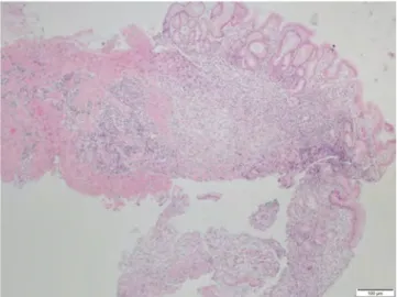
Fig. 1 A. Small lymphocytic infiltration located in the submu- cosal region by gastric endoscopic biopsy (hematoxylin and eosin, ×100)
Fig. 1 B. CD20 positivity (diaminobenzidine, ×100)
44
Acta Haematologica Polonica
characteristic lymphoepithelial lesions (replacement of the epithelial cells with lymphoid cells) [12]. The immunohistochemical features are the same as normal MALT tissue, showing CD20 positivity, as well as CD5, CD10, and cyclin D1 negativity (1). The disease is commonly found to be associated with Helicobacter pylori, and the treatment should also include antibiotherapy for this microorganism.
FL is a lymphoma originating from the germinal center B cells with a bcl2 rearrangement and t(14;18) translocation. The grading depends on the number of centroblasts, and the growth pattern (nodular/diffuse) should be stated in the pathology report (2). The characteristic histological appearance includes a high number of neoplastic follicles with no mantle zones or tingible body macrophages in a back-to-back arrangement showing CD20, CD10, bcl2, and bcl6 positivity and CD5 negativity. The prognosis correlates with the grade and the extent of the disease.
Extranodal FL, although rare, may occur in any area such as the skin, gastrointestinal tract, breast, and testis. Primary pulmonary lymphoma is one of the rare ones, comprising only 4% of all extranodal NHLs [11, 13].
Multiple lymphomas may rarely occur simultaneously and cause diagnostic problems in hematology practice. The firstly diagnosed malignancies may shade the synchronous disease. In addition to the rarity of primary lung FL, the synchronous presentation of two different lymphomas in two different regions made our case more surprising. This is also the first case of synchronous extranodal primary lung FL with gastric MALToma in the literature to our knowledge. As in our case, symptoms may overlap, causing delay in identification of the synchronous neoplasm. But an atypical presentation should remind of a synchronous neoplasm, and the treatment needs to be planned including both diseases.
Fig. 2 A. Nodular/follicular pattern of small lymphocytic infiltration
in the lung wedge resection (hematoxylin and eosin, ×40) Fig. 2 B. CD20 positivity (diaminobenzidine, ×200)
45
Acta Haematologica Polonica
References
[1] Isaacson PG, Chott A, Nakamura S, Muller-Hermelink HK, Harris NL, Swerdlow SH. Extranodal marginal zone lymphoma of mucosa- associated lymphoid tissue (MALT lymphoma) In: Swerdlow SH, Campo E, Harris NL, Jaffe ES, Pileri SA, Stein H, editors. WHO classification of tumours of haematopoietic and lymphoid tissues. Lyon: IARC;2008:214–217.
[2] Harris NL, Swerdlow SH, Jaffe ES, Ott G, Nathwani BN, De Jong D, Yoshino T, Spagnolo D. Follicular lymphoma In: Swerdlow SH, Campo E, Harris NL, Jaffe ES, Pileri SA, Stein H, editors. WHO classification of tumours of haematopoietic and lymphoid tissues. Lyon: IARC; 2008:220–226.
[3] Cui Y, Liu T, Zhou Y, et al. Five Cases report of solid tumor synchronously with hematologic malignancy. Cancer Res Treat 2012;44(1):63–68.
[4] Congyang L, Xinggui W, Hao L, Weihua H. Synchronous Histiocytic sarcoma and diffuse large B cell lymphoma involving the stomach: A case report and review of the literature. Int J Hematol 2011;93(2):247–252.
[5] Sachdev R, Goel S, Gajendra S, Sood N. Synchronous diffuse large B-cell lymphoma and malignant clonal plasma cells in bone marrow as primary presentation: A Diagnostic and Therapeutic Challenge. Journal of Clinical and Diagnostic Research 2015;9(4):12–13. [6] Addada J, Anoop P, Swansbury JG, Wotherspoon A, Thomas JM,
Matutes E. Synchronous mantle cell lymphoma, chronic lymphocytic leukaemia and melanoma in a single lymph node. Acta Haematol 2010;123:194–196.
[7] Kourelis TV, Kahl BS, Benn P, Delach JA, Bilgrami SF. Treatment of synchronous mantle cell lymphoma and small lymphocytic lymphoma with bendamustine and rituximab. Acta Haematol 2011; 126:40–43.
[8] Cheah CY, Spagnolo D, Frost F, Cull G. Synchronous biphenotypic Richter syndrome with Epstein-Barr virus-positive nodal classical Hodgkin lymphoma and bone marrow diffuse large B-cell lymphoma. Histopathology 2016;69:707–710.
[9] Hwang Y-Y, Leung AYH, Lau W-H, et al. Synchronous Epstein–Barr virus-positive diffuse large B-cell lymphoma of the elderly and Epstein–Barr virus-positive classical Hodgkin lymphoma. Histopathology 2011;59:352–355.
[10] Melikian AL, Kolosheinova TI, Goriacheva SR, et al. Synchronous and metachronous myeloid and lymphoid tumors. Ter Arkh 2014;86(7):37–44.
[11] O’Malley DP, Goldstein NS, Banks PM. The recognition and classification of lymphoproliferative disorders of the gut. Hum Pathol 2014;45:899–916.
[12] Bacon CM, Du M-Q, Dogan A. Mucosa‐associated lymphoid tissue (MALT ) lymphoma: a practical guide for pathologists. J Clin Pathol 2007;60.4:361–372.
[13] Freeman C, Berg JW, Cutler SJ. Occurrence and prognosis of extranodal lymphomas. Cancer 1972;29:252–260.
[14] Mian M, Wasle I, Gritsch S, Willenbacher W, Fiegl M. B cell lymphoma with lung involvement: what is it about? Acta Haematol 2015;133:221–225.