SIGNIFICANCE OF QT DISPERSION AS A
DIAGNOSIS TOOL FOR CARDIAC PATIENTS
A THESIS
SUBMITTED TO THE DEPARTMENT OF ELECTRICAL AND ELECTRONICS ENGINEERING
AND THE INSTITUTE OF ENGINEERING AND SCIENCES OF BILKENT UNIVERSITY
IN PARTIAL FULFILLMENT OF THE REQUIREMENTS FOR THE DEGREE OF
MASTER OF SCIENCE
By
Nabil Ben Ahmed Jemel February 2000
I certify that I have read this thesis and that in my opinion it is fully adequate, in scope and in quality, as a thesis for the degree of Master of Science.
Prof. D™ Hayrettin Köyrcşn (Supervisor)
I certify that I have read this thesis and that in my opinion it is fully adequate, in scope and in quality, as a thesis for the degree of Master of Science.
. Dr^Causuf Ziya Ider
I certify that I have read this thesis and that in my opinion it is fully adequate, in scope and in quality, as a thesis for the degree of Master of Science.
v:
''LProf. Dr. Enis Çetin
Approved for the Institute of Engineering and Sciences:
c r j^ ^ t
Prof. Dr. Mehmet
Director of Institute of Engineering and Sciences
ABSTRACT
SIGNIFICANCE OF QT DISPERSION AS A
DIAGNOSIS TOOL FOR CARDIAC PATIENTS
Nabil Ben Ahmed Jemel
M .S. in Electrical and Electronics Engineering Supervisor; Prof. Dr. Hayrettin Köymen
February 2000
Electrocardiogram (EGG) is the recorded electrical potentials generated by the heart during a cardiac cycle. Gardiac abnormalities cause unknown current flows leading to strange waveform morphologies in the recorded EGG. Some of these abnormalities are only visible when the heart is under stress. Exercise EGG is conducted for this reason.
Ischemia is one of the important cardiac abnormalities and is the focus of our study. It occurs when a part of the heart tissue dies or is injured. QT dispersion (QTd) is a proposed method for diagnosing Ischemia. The classical deflnition for QTd is the difference between the maximum and the minimum
QT intervals within the 12 leads.
In this study the effect of exercise on QT dispersion (QTd) is studied and whether QTd could give significant information for diagnosing patients with ischemic heart disease is investigated. A new method for measuring QT interval
is developed and is compared with previous methods. QTd is measured on average beats calculated for 10 seconds intervals throughout the exercise ECG test and a trend curve which we call QTd, is generated. Several decision rules for the diagnosis of cardiac patients are proposed by analyzing these QTd trend curves and the accompanying heart rate curves. It is shown that despite the improvement in QT interval measurements, none of the decision rules proved to be a clinically useful discriminate of cardiac patients, with sufficient confidence.
Keywords-.^xercise ECG, QT interval, QT dispersion. Ischemia.
ÖZET
E G ZE R SİZ T E S T İ SIR ASIN D A Ö LÇ Ü LE N Q T D A Ğ ILIM IN IN K A L P H A STA L IK L A R IN IN TEŞH İSİN D EK İ Ö N EM İ
Nabil Ben Ahmed Jemel
Elektrik ve Elektronik Mühendisliği Bölümü Yüksek Lisans Tez Yöneticisi: Dr. Hayrettin Köymen
Şubat 2000
Elektrokardiyogram(EKG), kalp tarafından bir kalp atımı sırasında oluşturulan
elektriksel potansiyallerin kaydedilmiş halidir. Kalpteki anormallik
ler sıradışı elektrik akımlarının akmasına neder olurlar ki, bu da
kaydedilen EKG’de olağandışı morfolojiler olarak gözlenir. Bu morfolojilerin bazıları sadece kalp stres altındayken gözlenebilir. Egzersiz (Stres) EKG testi bu amaçla uygulanır.
Çalışmamıza konu olan iskemik bozukluklar en önemli kardiyolojik bozuk luklardandır. Bu durum kalp dokusunun bir bölümü öldüğünde veya zarar gördüğünde ortaj^a çıkar. QT dağılımı (QTd) iskemik bozuklukları teşhis et mek için önerilen bir yöntemdir. QTd, en bilinen şekliyle, standart 12 EKG kanalı içinde en uzun ve en kısa QT aralıkları arasındaki fark olarak tanımlanır.
Araştırmamızda egzersiz EKG testinin QTd üzerine etkisini incelenmiş ve bundan iskemik bozuklukların teşhisinde kullanılabilecek önemli bir bilgi elde
edilip edilemediğine bakılmıştır. Bu çalışma sırasında QT aralığını ölçmek için
yeni bir yöntem geliştirilmiş ve eski yöntemlerle karşılaştırılmıştır. QTd her 10
saniyede bir hesaplanan averaj EKG sinyali (bir kalp atımı) üzerinde bu yöntem kullanılarak ölçülmüştür. Bu ölçümlerden zamana bağlı bir QTd eğrisi (QTd trend) çizilmiştir. QTd ve kalp hızı eğrileri kullanılarak, iskemik bozuklukların teşhisi için değişik yöntemler önerilmiştir.
Bu çalışmamız göstermiştir ki, QT aralığı ölçümlerindeki iyileşmeye karşın önerilen yöntemlerden hiçbiriyle, QTd eğrilerinin kardiyolojik bozuklukların teşhisinde klinik değer taşıdığı gösterilememiştir.
Anahtar Kelimeler. QT aralığı, EKG, QT dağılımı. Egzersiz EKG testi. Stres EKG testi, İskemik bozukluklar
A C K N O W L E D G M E N T S
I would like to express my deep gratitude to Dr. Hayrettin Köymen for his su pervision, guidance, suggestions and encouragement through the development of this thesis.
I would like to thank Dr. Enis Çetin and Dr. Yusuf Ziya Ider for reading and commenting on the thesis.
I am intended to Fikret Küçükdeveci and Mustafa Solmaz from ΤΕΡΑ Ltd co. for their assistance in data acquisition. I would like to express my appreciation to Burak Acar, Lutfu Ozcakir and to all my friends inside and outside the department for their continuous support through the development of this thesis.
Finally, I would like to thank my family, especially my parents and my uncle, for their continuous support along my studies.
Contents
1 INTRO D U CTIO N
2 THE M ETH OD 10
2.1 Beat Averaging and QRS D etection ... 11
2.2 Detection of Specific Points on the E G G ... 13
2.2.1 Q point D e t e c t io n ... 14
2.2.2 Isolectric Line Calculation 15
2.2.3 J point’ Detection 16
2.2.4 T-peak Detection 16
2.2.5 Reliability Checks 18
2.2.6 T-end Detection 20
2.3 QTd Trends Computation ... 22
2.3.1 Check for the Reliability of the Detected P o in ts ... 23
2.4 Decision Making ... 31 3 EXPERIM ENTS 33 3.1 Data S e t ... 33 3.2 A n a ly s is ... 34 3.3 R esults... 34 4 DISCUSSION 36
4.1 Significance of the QTd Trends and the Decision Rules as a
Discriminative Factor between Healthy and Cardiac Subjects: . 36
4.2 Comparison between the Least Square Line Fit (LSF) and the
Least Square Parabola Fit (LSPF) Algorithms: ... 37
4.3 Comparison of our QTd Definition with Previous Definitions . . 39
4.4 Effect of TP Fusion on T-end and QTd M easurem ents:... 41
5 CONCLUSION 42
APPENDICES 45
A QTd Trends Plots 45
2.3.2 QTd C o m p u ta tio n ... 27
B D ecision rules evaluation 69
List of Figures
1.1 Standard leads used in EGG recording... 2
1.2 A typical heart beat... 2
1.3 QT interval variation within the 12 leads... 3
1.4 Manual T-end detection
1.5 Algorithms for automatic QT interval measurement... 7
2.1 Block diagram for the algorithm... 12
2.2 Fiducial points... 13
2.3 Q point detection. 15
2.4 T-peak searach interval. 17
2.5 Parabola fitting to the T wave 21
2.6 T-end detection... 23
2.7 Q point elimination... 24
2.8 T-peak elimination... 25
2.9 T-end elimination... 26
2.10 An example of QTd trend... 29
4.1 Comparision between the LSF and the LSPF algorithms... 38
4.2 QTd trends generated by different QT dispersion formulae. . . . 40
A .l QTd, QTpkd and Heart Rate for patient 76 ST = - A = ? Rej=70%. 46 A .2 QTd .QTpkd and Heart Rate for patient 76 ST =- A = ? Rej=0% . 46 A.3 QTd, QTpkd and Heart Rate for patient 3038 ST=- A = ? Rej=70% ... 47
A .4 QTd, QTpkd and Heart Rate for patient 3038 ST =- A = ? Rej=0% . 47 A .5 QTd, QTpkd and Heart Rate for patient 3041 ST = - A = ? Rej=70% ... 48
A.6 QTd, QTpkd and Heart Rate for patient 3041 ST = - A = ? Rej=0% . 48 A .7 QTd, QTpkd and Heart Rate for patient 3048 ST = - A = ? Rej=70% ... 49
A.8 QTd, QTpkd and Heart Rate for patient 3048 ST = - A = ? Rej=0%. 49 A .9 QTd, QTpkd and Heart Rate for patient 3051 ST = - A = ? R,ej=70%... 50
АЛ1 QTd, QTpkd and Heart Rate for patient 3052 S T = - A = ?
R ej=70% ... 51
АЛ2 QTd, QTpkd and Heart Rate for patient 3052 S T = - A = ? Rej=0%. 51
АЛЗ QTd, QTpkd and Heart Rate for patient 3061 S T = - A = ?
Rej=70% ... 52
АЛ4 QTd, QTpkd and Heart Rate for patient 3061 S T = - A = ? Rej=0%. 52
АЛ5 QTd, QTpkd and Heart Rate for patient 3063 S T = - A = ?
Rej=70% ... 53
АЛ6 QTd, QTpkd and Heart Rate for patient 3063 S T = - A = ? Rej=0% . 53
АЛ7 QTd, QTpkd and Heart Rate for patient 3064 ST =- A = ? Rej=70% ... 54
АЛ8 QTd, QTpkd and Heart Rate for patient 3064 S T = - A = ? Rej=0% . 54
АЛ9 QTd, QTpkd and Heart Rate for patient 3074 S T = - A = ?
Rej=70% ... 55
A .20 QTd, QTpkd and Heart Rate for patient 3074 S T = - A = ? R.ej=0%. 55
A .21 QTd, QTpkd and Heart Rate for patient 3076 ST = - A = ?
R e j-7 0 % ... 50
A .22 QTd, QTpkd and Heart Rate for patient 3076 ST =- A = ? Rej=0%. 56 АЛО QTcl, QTpkd and Heart Rate for patient 3051 S T = - A = ? Rej=0%. 50
Rej=70% ... 57
A .24 QTd ,QTpkd and Heart Rate for patient 3068 S T = + A = -R ej=0% ... 57
A .25 QTd, QTpkd and Heart Rate for patient 30 S T = + A = + Rej=70%. 58
A .26 QTd, QTpkd and Heart Rate for patient 30 S T = + A==+ Rej=0% . 58
A .27 QTd ,QTpkd and Heart Rate for patient 3044 S T = + A = + Rej=70% ... 59
A .28 QTd, QTpkd and Heart Rate for patient 3044 S T = + A = + R ej=0% ... 59
A .29 QTd, QTpkd and Heart Rate for patient 16 S T = + A = ? Rej=70%. 60
A .30 QTd ,QTpkd and Heart Rate for patient 16 S T = + A = ? Rej=0% . 60
A .31 QTd ,QTpkd and Heart Rate for patient 19 S T = + A = ? Rej=70%. 61
A .32 QTd, QTpkd and Heart Rate for patient 19 S T = + A = ? Rej=0%. 61
A .33 QTd, QTpkd and Heart Rate for patient 26 ST=4- A = ? Rej=70%. 62
A .34 QTd, QTpkd and Heart Rate for patient 26 S T = + A = ? R.ej=0%. 62
A .35 QTd, QTpkd and Heart Rate for patient 27 S T = + A = ? Rej=70%. 63
A .36 QTd, QTpkd and Heart Rate for patient 27 S T = + A = ? Rej=0%. 63
A .37 QTd, QTpkd and Heart Rate for patient 43 S T = + A = ? Rej=70%. 64 A .23 QTd, QTpkd and Heart Rate for patient 3068 S T = + A =
A .39 QTd ,QTpkd and Heart Rate for patient 47 S T = + A = ? Rej=70%. 65
A .40 QTd, QTpkd and Heart Rate for patient 47 S T = + A = ? Rej=0%. 65
A .41 QTd, QTpkd and Heart Rate for patient 3045 S T = + A = ?
Rej=70% ... 66
A .42 QTd, QTpkd and Heart Rate for patient 3045 S T = + A = ? R ej=0% ... 66
A .43 QTd, QTpkd and Heart Rate for patient 3083 S T = + A = ? Rej=70% ... 67
A .44 QTd, QTpkd and Heart Rate for patient 3083 S T = + A = ? R ej=0% ... 67
A .45 QTd, QTpkd and Heart Rate for patient 3087 S T = + A = ? Rej=70% ... 68
A .46 QTd, QTpkd and Heart Rate for patient 3087 S T = + A = ? R ej=0% ... 68
A .l QTpkd QTd ccc... 69
A .l Heart rate QTd ccc... 70
A .2 Heart rate QTpkd ccc... 70
A.3 CJTd max difference... 71 A .38 QTd, QTpkd and Heart Rate for patient 43 ST==+ A = ? Rej=0%. 64
A .4 QTpkd max difference. 71
A .5 Slope absolute sum... 72
List of Tables
1.1 Published QT dispersion Measurement Methods
3.1 Results of the Specificity-Sensitivit}^ Test ... 35
4.1 Statistical comparision between the LSF and LSPF algorithms . 37
4.2 Comparision between different QTd formulae ... 40
Chapter 1
IN T R O D U C T IO N
The electrical structure of the heart can be divided into three major compo nents: Pacemaker cells, specialized conduction tissue and working muscle of the artria and ventricals.
A cardiac cj^cle starts normally in the pacemaker cells, which are located in the SA node. These cells are self-excitatory and thus provide a regular suc cession of action potentials. These action potentials stimulate the cells in the left and right artria and result in a wave of cellular depolarization, which cause the artria to contract and fill the ventricles with blood. When this excitation reaches the AV node a specialized conduction tissue transmits the excitation to the ventricles. This specialized tissue is characterized by its low conductivity; this results in a delay between ventricular and arterial excitation.
In the ventricular regions the HIS-Purkinje tissue transmits the action poten tial to different parts of the left and right ventricles then the action potential is further transmitted throughout the ventricle muscle on a cell to cell basis leading to the ventricular contraction.
The cardiac electrical activity can be observed in the electrocardiogram
Figure 1.1: Standard leads used in EGG recording.
(EGG). EGG is the recorded electrical potentials on the surface of the body generated by the heart during a cardiac cycle. The EGG is measured using leads connected to the body surface. The standard leads (/, / / , / / / ) were origi nally introduced by Einthoven [3]. These leads are connected to the extremities (wrists and ankles) (Figure 1.1).
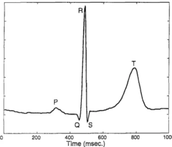
A typical lead voltage as function of time is shown in Figure 1.2. It is
posed of a P wave, a QRS complex and a T wave. The P wave corresponds to the activation of the artria. The repolarization of the artria is usually masked by the QRS complex, which corresponds to the depolarization of the ventri cles. The repolarization of the ventricles is reflected in the T wave. The QT interval gives the total duration of the ventricular systole. It is defined as the time interval between the onset of QRS complex and the end of T wave (Figure 1.2). This interval is widely recognized as an index of ventricular re polarisation. The QT interval varies between the 12 leads (Figure 1.3). This
Figure 1.3: QT interval variation within the 12 leads.
inter-lead variation, known as QT dispersion (QTd) has been proposed as an index of heterogeneity of the repolarization within the ventricles of the heart.
This concept (QTd) was first introduced in 1990 by Day et al. [8] for assessing
patients with long QT syndrome. The definition of QT dispersion varied be tween different studies. The most common definition is the difference between the longest and the shortest QT interval among the 12 leads (also referred to as
12-lead range). Other studies used the standard deviation of the QT interval
in 12 leads , coefficient of variation (standard deviation/mean) ,QT dispersion
ratio (QT dispersion/cycle length) ,and interquartile range(difference between
the upper and lower quartiles of measured QT intervals) . Table 1.1 provides
a set of publications where these formulae have been used.
QT dispersion was a subject of interest by clinical cardiologists because it
Reference Formula Langley et al. [15] Higham et al. [12] Bhullar et al. [11] Davey et al. [13] Hnatkova et al. [14]
Range, Standard deviation. Interquartile range Range, Dispersion ratio
Range, Coefficient of variation
Range, Standard deviation, 10 lead range Range, Standard deviation, 10 lead range Table 1.1; Published QT dispersion Measurement Methods
is hypothesized that QT dispersion is associated with high-risk arrhythmia, infarction, hypertrophic cardiomyopathy and other cardiac diseases. This was based on the idea that QT dispersion could reflect a regional heterogeneity of myocardial recovery. However the signiflcance of QT dispersion as a noninva- sive diagnosis tool for cardiac abnormalities is still a subject of discussion. Previous studies have shown that the measurements of QT dispersion from resting ECG were often not discriminative between patients at high and low risk of cardiac events after myocardial infarction. Stoletniy et al. [4] proposed that exercise-induced QTd may be useful in distinguishing patients with coro nary artery disease from those with angiographically normal coronary arteries.
QTc is derived in order to reduce the effect of heart rate increase and com
pare the measurements, which have been done at different heart rates. The
Heart Study QT interval correction formula(1.2).
QT, = QT + -
^
(
1
,
1
)
V
rU riin t
QTc = OT + 0.154(1 - RIknt) (1.2)
Stoletniy et al. have demonstrated that patients with significant coronary
artery disease had a significant increase in QT^ dispersion compared with pa
tients who had normal exercise test results and low probability of coronary
artery disease. However, in their study they assessed only QTc without the
original QT dispersion.
Gang Yi et al. [5] studied the effect of exercise on QT interval duration and dispersion in patients with sudden cardiac death after myocardial infarction. In their study 26 postinfarction patients were studied, 13 of whom suffered from sudden cardiac death and 13 of them remained event-free. Further 13 patients with chest pain, normal coronary arteriograms and negative exercise test results were studied as controls. QT intervals for this study were measured manually from rest, the end of peak exercise and 3 minutes after exercise. The QT intervals were measured in three consecutive QRS complexes in each lead when ever possible, and the average value was used. Gang Yi et al. found
that exercise induced a prolongation of QTc interval in patients at high risk
of sudden cardiac death [5]. They explained this by the fact that ventricular repolarization fails to adapt normally to increasing heart rate in sudden death patients. However they failed to prove the hypothesis that exercise-induced ischemia may increase the QT dispersion.
Ozcakir et al. [7] studied the effect of exercise on QT dispersion. They have reported that QT dispersion increased in patients with ischemic heart disease. These patients were verified by coronary angiography (invasive diagnostic tool
for ischemic heart disease), however the number of patients with ischemic heart disease was not enough for statistical verification.
The T wave offset is the most important and the most problematic measure ment for QT dispersion analysis. Tend was detected manually by cardiologists by the use of magnifiers, rulers calipers or digitizing tablets. The T-end is detected manually by fitting a line to the falling edge of the T wave. The intersection of this line with the isolectric line is defined as the T-end (Figure 1.4). In addition to its being labor intensive the T-end of T wave is often
Figure 1.4: Manual T-end detection
difficult to determine manually due to the effect of noise on the falling edge of the Twave. Inter-operator differences between 10 and 28 ms have been re
ported [6]. All these make it necessary to develop a reliable automatic QT
measurement technique not only because it is labor saving but also because it will remove subjectivity from the measurement.
Several methods were proposed for automatic T-end measurement. These
methods can l)e classified into two major types. One based on threshold and other on maximum T wave slope (Figure 1.5). In the threshold approach three algorithms have been used:
T h re sh o ld m e t h o d (T H ): It searches for the Tend starting from the max imum T peak. T end is detected as the point at which the T wave amplitude falls below a certain percentage (5 — 20%) of the maximum Tpeak.
D eriva tiv e th resh old m e th o d (D T H ): The derivative of the signal is cal culated. The maximum of the derivative after the Tpeak is determined. The T offset is located as the point at which the derivative falls to a certain percentage (5 — 20%) of its maximum value.
T w ave area m e th o d (T A ): The area under the T wave is calculated. Tend is determined as the point at which the area under the T wave is a certain percentage (90%) of the total area.
Figure 1.5: Algorithms for automatic QT interval measurement.
In the approach based on maximum slope two methods have been used;
M a x im u m slop e m e th o d (S I): The point with maximum derivative on the falling edge of the T wave is determined. A tangential line is fitted to this point. The T offset is determined as the intersection of the maximum slope line with isoelectric line of the TP segment.
Least square fit m e th o d (L S I): A least square line is fitted around the re gion of the maximum slope point on the falling edge of the T wave. The intersection of this line with the isoelctric line is defined as the T-end.
Xue et al. [2] have compared these five algorithms for T oflFset measurements. A reproducibility test was conducted. For comparing the available algorithms test data from 23 normal subjects each of whom had five BCGs recorded was used. The repeated ECGs were obtained 30 minutes, 1 day, I week and one month after the first. QT dispersion was calculated for all five EGG then the median value was determined. Xue et al. defined the individual average of the four absolute differences (IDIFF) as:
4
I D I F F = j ^ \QTdi — median] (1.3)
i=l
According to this definition, a good reproducibility corresponds to a small IDIFF value. Xue et al. have reported that the least-squares fit algorithm achieved better reproducibility of QT dispersion measurements than any other method examined. It was also suggested that the least-squares fit algorithm achieved better reproducibility than the simple slope method because of more stable fitting due to more sample points.
In this study a new algorithm for automated QT measurement (LSPF) is proposed. The fiducial points detected by the algorithm were verified visuallj^ The algorithm is then modified and improved with respect to visual detection. The performance of the algorithm is compared with previously developed al gorithms (mainly with the least-squares fit algorithm)
In this studj^, the QT interval is measured throughout the whole exercise test. The QTd trends are then calculated using the resulting QT intervals. A new approach is presented for calculating the QTd trends. Unlike previous studies where at most three average values (rest, exercise and recovery) of QTd dis persion were calculated, in this study QTd trends are generated for the entire exercise and recovery phases. The efficiency of this new method is discussed and compared with the previously proposed methods for QTd calculation. QTd trends are generated for 24 patients, 12 of which were diagnosed cardiac using ST test. Several decision rules are proposed for classifying the different QTd trends. A sensitivity-specificity test is conducted for evaluating these decision rules and thus the performance of QTd as a diagnosis tool for cardiac patients.
Chapter 2
T H E M E T H O D
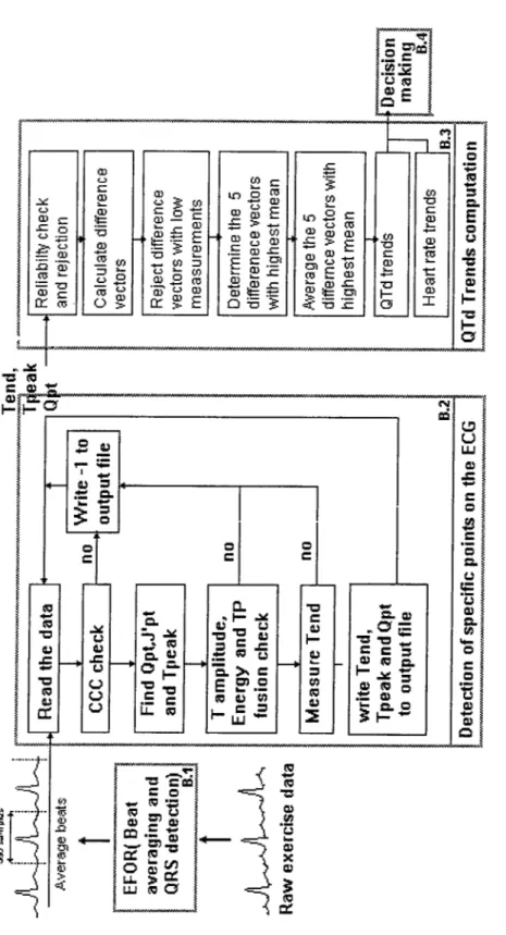
Our algorithm can be divided into four basic blocks. A block diagram for the entire algorithm is shown on Figure 2.1. Each block is explained in detail in the following sections.
Figure 2.1: Block diagram for the algorithm.
2.1
Beat Averaging and QRS Detection
The commerciall}'· available software Efor from ΤΕΡΑ Ltd. is used for beat averaging and noise reduction. The Efor program provides an average beat for
every 10 seconds of raw data.
It is known that the steepest change on the ECG recording occurs at R waves. For this reason a trigger point is defined as the point at which the derivative
of the R wave falls to 80 % of its peak value. Using the rest data, a template,
which represents a normal QRS morphology, is computed separately for each lead. Then a reference channel is determined for all the leads. The reference channel is defined as the channel that has the highest correlation with its tem plate at rest. The calculated templates are used for QRS detection throughout the rest of the exercise data.
The exercise data is divided into 10 seconds segments. After detecting all QRS complex candidates within a segment a correlation test is made. If the corre lation between a QRS complex candidate and the corresponding template is high enough the beat is qualified. The qualified beats are aligned using the ascending edge of the R wave. This results in one average beat for every 10 seconds of data. The average beats for the 12 different leads obtained are time aligned Avith respect to the reference channel. The length of the average beat is 380 samples. The 128th sample corresponds to its trigger point. The beats and its information are then recorded to a file.
2.2
Detection of Specific Points on the E C G
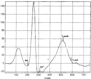
We measure the QT intervals needed for determining the QT trends on the average beats defined in the previous section. We obtain four fiducial points for each lead. They are the Q point (Qpt), J’ point (J p f), T-peak (Tpk) and T-end (T-end) (Figure 2.2).
For determining the specific points we define different search window for each point. We define the search windows according to the characteristics of the exercise ECG. Since the trigger point which corresponds to the falling edge of the R wave is already provided (Section 2.1), it was not necessary to develop an algorithm for determining the R wave peak. In the following the trigger point is used whenever the position of the R wave is needed.
Figure 2.2: Fiducial points.
2.2.1
Q point Detection
The Q point (Qpt) is needed for the calculation of the QT intervals and the calculation of the isoelectric line (baselinel). It is known that the Q wave always proceeds the R wave. For this reason it is reasonable to start searching for the Q point before the R wave peak. In our algorithm we set the search window for the Q point to 50 ms proceeding the trigger point. It has been
shown that the variance of the QRS onset across the 12 leads is very low
compared to that of the T-end [2]. For this reason, knowing that all the leads are time aligned, we detect a global Q point for all 12 leads. We detect it on the reference channel as follows:
We calculate the first derivative of the average beat d{n) as:
|o:(n) — x{n — 2)1
d{n) =
(
2.
1)
We define Qpt as the first point in the search window for which d{n) > 1.
Both the search window and Qpt are shown in Figure 2.3 .
After determining the Q point we make a derivative check. Our aim from this check is to assure that the detected Q point is on the ascending edge of the R wave and not the falling edge of the P wave. Since the Q point is expected to be on the ascending edge of the R wave we expect the derivative of the five sample points after the Q point to be greater than 1. If the derivative check is negative we narrow the search window for the Q point so that to exclude the falling edge of the P wave. We then repeat the search for the Q point on the new search window.
Q poi nt s e a r c h interval
Figure 2.3: Q point detection.
2.2.2
Isolectric Line Calculation
After we determine the correct Q point, we calculate an approximation to the baseline (isoelectric line). We call it ’baselinel’. We first define a search window of length 40 milliseconds proceeding the global Q point. This window corre sponds to the segment between P wave end and QRS onset. On this window we search for the 10 sample points (20 milliseconds) with the lowest standard deviation. We then calculate baselinel as the average of these 10 sample points.
According to [7] the QRS complex returns to its DC level 50 milliseconds after the R wave. We use this physical property of the ECG for defining J point’ (J p f). We define a common J point’ for all leads as follows;
Jpt' : Trigger point + 50 milliseconds .
Jpt’ is not the correct J point but it seemed to be reliable for marking the end of the QRS complex. We use Jpt’ for calculating a second baseline, which is
called baseline2 and defined as:
Baseline2 : The average o f the 20 milliseconds following Jpt'.
The relative values of baselinel and baseline2 can be used to determine whether there is any depression or elevation in the ST segment. Such cases are typical in exercise ECG and complicate the T wave detection considerably.
2.2.3
J point’ Detection
2.2.4
T-peak Detection
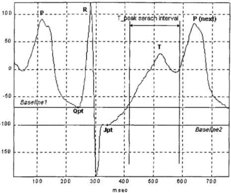
In our study we use a threshold based detection algorithm for determining the T-peak. The advantage of using this algorithm is that it has less computation time and it is able to deal with several complications of exercise ECG such as ST depression ST elevation and TP fusion. TP fusion occurs when the heart rate increases and the R R interval between two consecutive heartbeats shortens. This leads to a fusion between the T wave and the P wave of the next beat(Figure 2.4). The degree of TP fusion depends on the heart rate and the amplitudes of the T and P waves.
We define the search window for the T-peak as:
100 200
300
400
m se c
500
800
70 0
Figure 2.4: T-peak searach interval.
Jpt' + 80ms < t < Qpt + RRint * (2/3) + 20ms
(
2
.
2
)
It was shown in a previous study [7] that this interval excludes the P-peak of the next QRS complex. Since this interval is a function of heart beat the search range for the T-peak decreases as heart rate increases and this avoids detecting the peak of the P wave instead of that of the T wave. A TP fused beat and the corresponding search interval for the T-peak are shown on Figure 2.4 . It is clear how the search interval discards the peak of the P wave.
Once the search window for the T-peak is determined our algorithm searches for two maximums in the previously defined window . It calculates the maximums with respect to the two previously defined baselines (baselinel and Baseline2) as follows.
Tpeakl absolute m,axim,a with respect to baselinel in the interval (2.2.4).
Tpeak2 absolute maxima with respect to baseline2 in the interval (2.2.4).
In the absence of ST depression or elevation Tpeakl and Tpeak2 are the same.
If the two peaks found are different it is more likely that there is an ST depres
sion or an ST elevation. This is checked by comparing the levels of baselinel
and baseline2
We report an ST elevation if baseline2 > baselinel + tol on the other case, if
baseline2 + tol < baselinel, we report an ST depression, tol is a tolerance value which can be changed according to signal level.
For determining the correct T-peak we perform a curvature test on the detected
peaks. We define an index of curvature (Cv) as follows:
Cv =
10 (2.3)
Cv is the absolute average of the derivative on a 20-millisecond window around
the detected T-peak. The value of Cv reflects the behavior of the signal deriva
tive around the T-peak. We select the T-peak with highest Cv value.
2.2.5
Reliability Checks
We make several checks throughout the measurement of the QTd trend. The purpose from these checks is to determine whether the quality of the recording
is good enough for QT anal3'^sis. We reject all beats that fail these checks.
C ross C o rre la tio n C oefficien t C heck We perform the first check on the QRS complex. We obtain 12 templates from rest EGG. These templates are simply the QRS complexes for rest EGG. We set the size of the tem plates to 120 milliseconds centered at the trigger point. We calculate the
cross correlation of every average beat -with the corresponding template as follows.
CCC(n) =
Where T(i) is the template and x(i) the input signal. The cross correla
tion value reflects the quality of the signal. A low SNR signal gives a low correlation between the QRS complex and the corresponding template. We accept a beat only if it has a correlation coefficient bigger than 0.7. If we accept a beat we update the corresponding template by including a fraction of the accepted beat. We perform this update as follows:
New-Temp = 0.7 * old-Temp -P O.Z*new-QRS (2.5)
A m p litu d e C hecks After we determine the T-peak we perform on it an am
plitude check. We set the threshold for T-peak amplitude to 0.1289 mv.
We have determined this value empirically. We have tried to further de crease the value of this threshold for patients with low T wave amplitudes but this lead to a decrease in the measure accuracy. In a study on the in fluence of T wave amplitude on T-end measurement [10] It was reported
that measurement from T waves with amplitude less than 0.25 mvolts
are less reliable.
E n e rg y C heck We perform an energy check to make sure that what we have detected is the peak of the T wave and not a noise spike. This check consists of measuring the energy in a window around the T-peak. The idea is that the energy in a window around a noise spike is much lower than that around the T-peak. We determine the size of the window
empirically and we set to 120 msec. We set the threshold for passing the
energy test to 4.684 If the measured energy under the window is less than this threshold value we reject the detected T-peak.
T P Fusion C h eck We check the behavior of the T wave around the T-peak in order to determine whether we have TP fusion or not. If the second half of the T wave doesn’t fall to 80% of the T-peak maximum amplitude we conclude that there is a TP fusion and the beat is rejected. If the RR interval keeps decreasing (i.e. heart rate is increasing) the T-end is masked by the P wave and it will not be possible to detect it till the heart rate starts to decrease (recovery phase). It is important here to note that TP fusion is critical only for the leads where we have a small T wave compared to the P wave. The reason is that a small amplitude T wave is masked easily by the P wave leading to TP fusion "rejection starting from early stages of exercise.
Whenever a beat fails in any of the above checks, we reject the beat. Oth erwise, we assume that it is good enough for T-end detection.
2.2.6
T-end Detection
The T-end detection algorithm, which we use in this study, is based on a similar idea to the one used by cardiologists. The idea of this algorithm was first introduced by L.Ozcakir [7]
We model the T wave by a second-degree parabola:
y{t) = a*t^ + b * t + c (2.6)
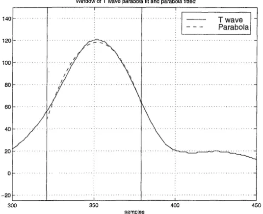
The window to which we fit the parabola is defined as the points at which the T wave amplitude is above the 80% of its maximum around T-peak. In Figure 2.5 the T-end and the fitted parabola are shown.
Window of T wave parabola fit and parabola fitted
140
120
-100
-300 350 400 450
samples
Figure 2.5; Parabola fitting to the T wave
We use the least square approximation technique for fitting the second- degree parabola. This technique minimizes the error between the signal and the fitted parabola as;
min {vi ~ [a * x l + h*Xi + c)Y
a ,b ,c i= .\
(2.7)
Where 1 is the length of the interval to which we fit the parabola and Xi all
sample indexes within this interval. After minimizing the above equation we
obtain the following system of equations:
f
n f c ] ' E v i ''E 4 E ^ f b = E vi^i
E ^ ? ) [ a J \ E Vi^i )
(
2
.
8
)
We use the conjugate gradient algorithm for solving this system of equa tions.
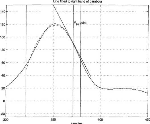
Once we obtain the parameters of the parabola, we determine Idgo as the point
where the fitted parabola falls to 80 % of its absolute maximum on its falling
edge. We then draw the tangential line (Tgo) through (Wo)· Unlike the signal itself the fitted parabola is mathematically well defined and so is the tangential line. Thus the errors due to tangent line fitting to the T wave [l] are avoided. We then determine the T-end as the intersection of Tgo with the baseline voltage
level baselinel.
2.3
QTd Trends Computation
After determining the fiducial points the next step in our algorithm is to calcu late the QT dispersion trends. The first step in this calculation is the elimina tion of suspicious measurements. This elimination is based on the continuity assumption: we assume that in a short interval there can be no abrupt change in the EGG features. In case any occur, we report it as an erroneous measure. Once we eliminate all suspicious measurements we calculate the QTd on the reliable leads and finally we obtain a single QTd trend.
Line fitted to right hand of parabola
Figure 2.6: T-end detection.
2.3.1
Check for the Reliability of the Detected Points
Q p o in t V erifica tion In this part we check for points where we have erro neous detection of Qpt. A derivative check test is made according to the principle of continuitjL We assume that the Q-point can not change by more than 10 milliseconds between two successive average beats. Assume
Qpt{n) is the position of Qpt in the average beat, measured with re
spect to the trigger point, then Qpt{n)' = [Qpt{n) — Qpt{n — l)]/2 is
the first order difference of this discrete signal. We eliminate the average
beats that correspond to Qpt{n)' > 5.
Q lime
beat Index Q time deriuotixe
Figure 2.7: Q point elimination.
Tpeak Verification Similarly we perform a T-peak test for each lead. Unlike the Qpt{n) Tpeaki{n) may show some discontinuity. These are due to the beats where measurements were not possible either due to very low amplitude T wave, or TP fusion.
Figure 2.8 shows a continuous and discontinuous Tpeaki. The disconti
nuity in the second 2"'^ graph is due to biphasic T waves in this example. When a biphasic T wave exists, the T-peak detection algorithm keeps al ternating between the two peaks since both peaks can qualify the deriva
tive check (Cv) (Figure 2.8). Although the second peak of the T wa.A'^e
can be detected at the earlier phases of the exercise test, it is masked in later phases due to TP fusion. At this step our algorithm will start mea suring the first peak. So we decided to stick to the first peak throughout
Tpesk
Figure 2.8: T-peak elimination.
the exercise.
We calculate the first order difference Tpeaki(n)' = [Tpeaki{n) -
Tpeaki{n—l)]/2. When Tpeaki{n)' exceeds a certain threshold (set to 15 milliseconds in our case) the measurement is eliminated. After verifying and eliminating all wrong T-peak measures we end up with 12 QTpk
interval vectors one vector for each lead. A lead vector Tpkiin) contains
the valid T-peak measurements and -Is which correspond to unavailable measurements. These lead vectors are used later for calculating QTpk dispersion trends.
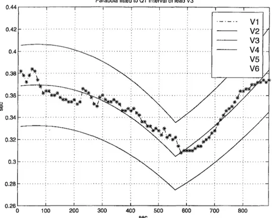
T -e n d V erifica tion Since there is relatively a low number of measurements for the T-end compared to that of the Q point and T-peak we use a different process for eliminating the erroneous T-end measurements. Our algorithm models the variation of the T-end in the exercise and the recovery phases of each lead by using two different second-degree parabolas. If there are more then 8 accepted T-end measurements in
each phase, we fit a parabola to Tendi{n). Otherwise we reject all the
Parabola fitted to QT interval of lead V3
Figure 2.9: T-end elimination.
measurements within that phase. Once we fit a parabola, we reject the T-end measurements which fall out of the ±8 % amplitude range (empir- icall}^ determined) of the parabola. Thus we exclude the outliers. This rejection is also based on the continuity assumption. Figure 2.9 shows
a sample Tendi{n) with the fitted parabolas in exercise and recovery
phases. The ±8 % amplitude ranges are also shown.
2.3.2
QTd Computation
After verifying and eliminating all wrong measures we end up with 12 QT inter
val vectors one vector for each lead. A lead vector Tendi{n) contains the valid
T-end measurements and -Is which correspond to unavailable measurements.
We also obtain a different set of lead vectors Tpki containing the valid T-peak
measurements and -Is for unavailable measurements. We use this second set of calculating Q-Tpeak dispersion trends.
We define QTd vectors as;
QTdij{n) = QTi{n) - QTj{n) if QTi{n) ^ -1 and QTj{n) -1
o.w
QTdij{n) presents the QT interval difference between lead i and lead j for all
available samples. We calculate QTdi/s for all possible pairs of leads. This
results in 64 different QT different QTd^s .
Then we divide the exercise phase into five equal segments. The fifth slice corresponds to the highest stage of exercise. The heart rate is very high at this stage and we expect a high amount of dispersion. However due to the increase in the heart rate, TP fusion at this stage is very high. As a result of this, T-end detection is difficult and not reliable. The first slice corresponds to early stages of exercise and we do not expect to observe effects of ischemia in this part. For this reason we do only consider the second, third and fourth segments of the exercise phase. Let A be the interval corresponding to these segments.
Despite the fact that TP fusion is low on A it is still possible that T-end mea surements are not available due to noise or low T wave magnitude. For more
reliable analyzes we decided to reject QTdi/s with less than 70% of maximum
number of possible measurements in Is ■ We set our 70% threshold for lead rejection after analyzing the effect of different thresholds on the generated QTd trend visually. By setting the threshold to 70% we ended up with more contin uous QTd trends. The rejected leads were checked visually for 5 patients. We have observed a high correlation between the number of beats detected and the reliability of the measures taken within the defined interval.
The mean values of each remaining QTdi/s are calculated as:
QTdij[n] = E
n & I s a n d Q T d i j [ n ] ^ — I
QTd,
■ IJ
number o f valid measurements on IgThen we pick the five QTdifs with highest mean values. Our aim is to
select lead pairs with highest difference in QT intervals. The QTd trend is
defined as the average of these five QTdifs.
We use the same set of leads, which is selected previously for calculating QTd, to calculate Q-Tpk dispersion trends (QTpkd trends). Similar^, we define a set of Q-Tpk difference vectors as follows:
QTpki{n) — QTpkj(n) if QTpki{n) % -1 and QTpkj{n) ^ -1
— 1 o.w
QTpkdij{n)
We then calculate the mean values of these Q-Tpk difference vectors as follows.
________________ e I s a n d Q T p k d i j [ n ] # - 1 .
QTpkdijln] = ---;---m---1-------T number o j valid T — peak measurements on T
We define QTpkd as the average of the five Q-Tpk difference vectors with highest mean values.
The most common definition for QTd dispersion is QTd = QT^ax — QTmn
This definition did not perform well in exercise EGG because this definition
uses different leads throughout the exercise. This resulted in inconsistencj^ in the QTd trends (section 4.3). So we decided to use the same pair of leads throughout all the exercise. Figure 2.10 shows an example of generated QTd
Patient No;175Leads:12 prO= 68% pr1=60% selectcr=70%
Figure 2.10: An example of QTd trend.
trend (top figure solid line). On the same figure the heart rate is plotted in
dashed lines. The second graph shows the five difference vectors {QTdi/s)
used for calculating the QTd trend. The first three QTdi/s show discontinuity
around the exercise peak. This is due to critical TP fusion on leads 6,5 and 7. The numbers under the lowest figure in Figure 2.10 show the percentages of measurements that were possible for each lead. Leads 6,5 and 7 have less num
ber of detected points than leads 9, 10 and 11 that’s why QTd^^g, QTdr^^.yciud
QTdj^Q are discontinuous while QTdg^u and QTdw^u are not. From this ex ample it is clear that using more than one difference vector provides more continuous QTd trends because when ever we fail to have any measurements on a vector it is more likely that we have a valid measurement at least on one of the other four. For this patient it was possible to detect QT end on 68% of the exercise test. An extra 8% was rejected during reliability checks. So the QTd trend was generated using only 60% of the exercise test.
2.4
Decision Making
We use a set of decision rules to differentiate between positive and negative QTd trends. A decision rule depends either on the characteristics of the gen erated QTd trends or on the relation between the QTd trends and the heart
rate. In the following the proceeding terms are used: QTpkd: Trend generated
using the T-peak, QTd: Trend generated using the T-end, (HR): Heart rate
trend.
Correlation : We calculate the cross correlation coefficients (ccc) between
QTpkd , QTd and the heart(Wi?) as follows:
cccl QT d{i)QTpkd{i) a n d Q T p k d ( i ) : ^ ~ l a n d Q T p k d ( i ) ^ —l QTpkd^{i). ^Li,QTd(i)vt-i a n d Q T p k d { i ) · ^ —! QTd?{i) (2.9) ccc2 = QTd{i)HR{i) a n d HR?{i).J2UQT,(i)^-^QTd^{{) a n d H a n d H ccc2 = Q T p k d { i ) ^ - l QTpkd{i)HR{i) a n d H R { i ) : ^ —\ Y ] i = l , Q T p k d ( i ) ^ - l X:Li,QTp*d(o.i-i QTpkd^(i)
(
2.
10)
(
2.
11)
and H R { i ) j i - l a n d H R ( i ) : ^ —1As heart rate increases the depolarization time between healthj^ and injured parts of the heart increases in ischemic patients. It was reported in previous studies [7] [8] that this will lead to an increase in QTd trends and QTd measures for patients with ischemic heart disease. Such changes are expected to lead to higher cross correlation coefficient between the QTd trends and the heart rate. We also calculate the cross correlation between the trends of the T-peak and the T-end dis])ersions in order to investigate the relation between the two measures and its variation for different patients.
Threshold : We can assign a single threshold value, instead of investigating the entire behavior of the QTd trend. We calculate the QTd maximum range for all trends as follows:
QTdT^jidMiN
f^ ^ Q T p k d — Q 'd ^ ^ ’d^pk^A X Q '^ ^ '^ p k M J N
(2.12)
(2.13)
We report a patient positive if either mxQTd or mxQxd exceeds a certain thresh
old value.
Slope : We fit two least square lines Lex and Lj-e respectively to the recovery and the exercise phases of the QTd trends. We then determine the slopes of
the lines Sex and Sres- We define S as the absolute sum of the two slopes. We
expect higher values of S for QTd positive trends.
Chapter 3
E X P E R IM E N T S
3.1
Data Set
We use ECG test data for 54 different patients for this study. The raw exercise ECG data recordings for these patients were obtained at the Eskişehir hospital through the use of the Kardisosis PC based exercise ECG system. For each patient 12 leads ECGs were recorded during a Bruce Protocol based exercise test with a sampling rate of 500 samples/sec and at a 2.9-microvolt resolution. We also use a different data set of 24 patients for decision rule evaluation. Four of these patients had coronary angiography results, which is an invasive diag nostic tool for ischemic heart disease. Only one of these patients was reported non-ischemic. A qualified cardiologist examined ST plots, which represent a non-invasive tool for ischemia diagnosis, for all 24 patients. Twelve of these patients were reported ST positive.
3.2
Analysis
The software Efor from ΤΕΡΑ is used for averaging and filtering. The algorithm for fiducial point detection is developed using Borland C whereas the algorithm for producing QTd trends is implemented using Matlab.
We process the data for the 78 patients using those programs. We obtain 78 different QTd trends. Since the first data set was not clinically verified we have only used QTd trends from the second data set for our analysis (Appendix A). A decision is made on this data and the results are compared with clinical data. Throughout the development and analysis of EGG features the viewer software of Massachusetts Institute of Technology (MIT) and Beth Israel Hospital (BIH) had been used for checking visually the detected fiducial points. We have developed another program using Matlab for checking parabola fitting and T-end detection.
3.3
Results
The generated QTd trends do not show any significant variation with increasing exercise (Appendix A). For some patients, we have noticed some slight increase in QTd trends generated using 0% rejection ratio. On the other hand, the QTd trends generated using 70% rejection ratio for the same patients, do not show any significant changes. We have verified the rejected leads visually. We have noticed that the measurements for these leads are not reliable and are most of the time biased by a high TP fusion.
We have used a specificity-sensitivity test [9] for all proposed decision rules.
The results of the test are shown in Appendix B.
True Positives Sensitivity =
Specificity =
True positives + False negatives True Negatives
True positives + False negatives
We have calculated the area under the Receiver Operator Characteristics (ROC) curve (Table 3.1). The maximum value measured is 76%. It is mea sured using the maximum QTd trend difference. The rest of the decision rules show lower percentage area values.
Decision rule Percentage area under
the ROC curve
cccl{QTpkd and QTd) 51.7% ccc2{HR and QTd) 30 % ccc3{HR and QTpkd) 42% mxQTd 76% mx QTpkd 40% S (slope) 55%
Table 3.1: Results of the Specificity-Sensitivity Test
Chapter 4
D ISCUSSIO N
4.1
Significance of the QTd Trends and the
Decision Rules as a Discriminative Factor
between Healthy and Cardiac Subjects:
Neither the generated QTd trends nor the associated decision rules are dis criminative between cardiac and healthy patients. The low correlation between the QTd and the ST segment analysis propose that both tools are measuring- different characteristics of the exercise EGG. Unlike T-end measurement, de termining the ST level is less problematic, which may explain the reliability of this measure as diagnosis tool. In our study the number of patients with angiography rcisults is limited (only four patients). As a future work it is im portant to inv('stigate the QTd behavior on a larger population with clinically verified data.
4.2
Comparison between the Least Square
Line Fit (LSF) and the Least Square
Parabola Fit (LSPF) Algorithms:
The LSF algorithm introduced earlier was reported to be more reproducible than any other QT-end detection algorithm [1]. For comparing our algorithm (LSPF) with the LSF algorithm and due to the absence of reproducible data we use a linear regression test. For conducting this test we use exercise test data from 15 different patients (11550 QRS complexes). The statistics of the measurements are shown in Table 4.1:
All measurements Rest measurements
Total number of beats 11550 330
Number of beats on
which LSPF failed
3830 138
Number of beats on
which LSI failed
5010 158
Number of beats on
which both algorithms failed
3575 135
Mean Std
LSPF 307.9 (msec) 38.22
LSF 321.4(msec) 39.22
Table 4.1; Statistical comparision between the LSF and LSPF algorithms
An algorithm fails when it does not succeed to determine a T-end for a certain average beat. The criteria of failure depend on both the algorithm performance and the signal quality. According to the statistics the LSF failed on 47% of the rest measurements and on 43.8% of the total number of mea surements. On the other hand, the LSPF algorithm failed on 41% of the rest
measurements and on 33.16% of the total number of measurements. The mea sured T-end’s were verified visually. We have noticed that the T-end values measured using the LSF algorithm are higher (mean=321.4 milliseconds) than the T-end values measured using the LSPF algorithm (mean=307.9 millisec onds). This can be explained by the fact that in the LSF algorithm we fit our line to the maximum derivative point while in the LSPF algorithm our line is fitted to the 80% of absolute parabola maximum level. That is, the slope of the fitted lines and the points to which the lines are fitted are different in both algorithms. A plot of the QT intervals measured by the LSF algorithm versus QT intervals measured using the LSPF algorithm is shown in Figure 4.1.
500
Comparision between the LSF and the LSPF algorithms
250 300 350 400
QT intervals detected with LSPF (msec) 500
Figure 4.1: Comparision between the LSF and the LSPF algorithms.
The shape of the graph proposes that both algorithms are measuring the same characteristics of the average beat. A least square line is fitted to the data. The parameters of the line are slope — 0.94 and x-axis intersect = 31.86. To investigate the difference between the two algorithms we have to consider the outliers. These correspond to average beats where both algorithms detect significantly different T-peaks for the same average beats. After visual verification we have noted that the shape of the falling edge of the T wave varies between successive beats due to several artifacts. The LSPF algorithm is more robust to these variations. This is mainly because the LSPF algorithm uses more sample points than the LSF algorithm. This makes it more stable to noise and regional changes effecting the falling edge of the T wave.
4.3
Comparison of our QTd Definition with
Previous Definitions
In this section we compare our QTd definition with other QTd definitions appearing in literature. We take into consideration the following definitions: Range (QTdc), Standard deviation (SD) and Interquartile range (IQR). We have generated QTd trends for all four definitions (Figure 4.2). The classical definition shows a very noisy QTd trend. This is due to the fact that the classical definition uses different pair of leads for different time instants while our definition calculates QT difference for the same lead pairs throughout all exercise.
The statistics in Table 4.2 show that the QT dispersion values vary signif icantly throughout exercise and recovery phases for all three definitions. This
Figure 4.2: QTd trends generated by different QT dispersion formulae.
suggests that using a single value of QTd dispersion for exercise does not reflect the entire behavior of the heart, moreover if this value is selected at exercise peak [5] it is less reliable due to the effect of TP fusion on QTd end mea surements. In some studies corrected QT dispersion was used *[4]. The QT
QTd definition Range Standard deviation Interquartile range
Rest QTd 42 12.15 16.5 QTd range (exr) 50 12.9 20 Mean(exr) 36.7 10.54 15.15 SD(exr) 13.82 3.43 5.77 QTd range(rec) 46 21 35 Mean(rec) 49.3 13.55 4.24 SD(rec) 15.98 19 6.62
Table 4.2: Comparision between different QTd formulae
correction formulae were derived for canceling the effect of different heart rates when comparing QT intervals from different patients at rest. These formu lae were generated for heart rates less then 120 bpm, hence we think it is an appropriate to use them for exercise ECG.
4.4
Effect of TP Fusion on T-end and QTd
Measurements:
It is known that T-end detection for exercise EGG is very critical. Though presence of noise and baseline wonder can highly effect the reliability of the measures, TP fusion can be considered the most important limitation for gen erating reliable QTd trends. The latter makes it difficult to investigate QTd behavior at most advanced stages of exercise where we expect higher level of ischemia. Due to The presence of TP fusion a number QT intervals is not measured. As heart rate increases the RR interval shortens and the P wave is gradually pushed towards the T-end. This means that leads with maximum QT interval are first effected by TP fusion. As exercise level increases no more measurements are possible on these leads and this is reflected by a gap in the QTd trend round the exercise peak. The omitted leads are expected to carry significant information since they are commonly the leads leading to maxi mum dispersion. As future work it seems necessary to develop more efficient algorithms that can deal with TP fused beats;
Chapter 5
C O N C LU SIO N
QTd analysis is proposed as a non-invasive method for measuring dispersion of ventricular depolarization. At most three values, for rest, exercise, and recovery, of QTd dispersion were used in the previous studies. In our study we proposed a new approach for calculating QTd. Unlike previous studies we generate QTd trends for all exercise and recovery data. We defined these QTd trends on the same set of leads throughout all exercise and recovery phases. The set of leads on wTich we calculated the QTd trends were selected so that they give maximum amount of dispersion. We checked our QTd definition on data and we did improve it in order to eliminate the effect of noisy leads. We compared our QTd definition with classical QT dispersion definitions. We showed that using the complete QTd trend is more reliable than using single sample points for QT dispersion. This is because the classical definitions, which use at most three values of the QTd trend, do not reflect the entire behavior of ventricular depolarization due to the variations in the QTd trends.
For calculating the QTd we used a new T-end detection algorithm (LSPF) which is based on fitting a parabola to the peak of the T wave and determining the T-end using the tangent to this parabola. We compared the performance of the algorithm with previous algorithms. Our algorithm proved more stable than any other T-end detection algorithm. This is mainly because the fitted parabola to the T wave is mathematically well defined and fitting a line to the falling edge of the parabola is less problematic then fitting a line to the falling edge of the T wave. However, our algorithm is not immune to TP fusion and a big set of leads was rejected due to critical TP fusion.
In our study our aim was to investigate QTd for ischemic patients but due to the lack of validated data we evaluated QTd using data collected from patients wnth general cardiac abnormalities. We evaluated our algorithm using 24 exercise EGG recordings, 12 from normal patients and 12 from cardiac patients. A qualified cardiologist evaluated this data u.sing ST diagnosis tests. We conducted discrimination analysis on this data set. The following decision rules were evaluated: Cross correlation (CCC), Threshold, and Slope. We used Specificity-sensitivity measures for evaluating the efficiency of the decision rules. Despite the improvement in T-end detection, thus in QTd measurements, the ROC curve analysis showed that QTd during exercise is not a clinically valuable discriminate of cardiac patients. The threshold decision achieved the maximum ROC curve area and it was 76%.
As future work I suggest that our algorithm be evaluated in a larger and clinically verified data set. The diagnosis of the patients in such a data set must be done by clinical means, like angiography. The results presented in this thesis are mainly a comparison of the clinically validated ST diagnosis tool and
our proposed QTd algorithm. TP fusion should also be further investigated in order to reduce the effect of missing leads on the calculated QT dispersion.
Finallj', we should point out that the importance of cooperating with a hos pital clinic in biomedical engineering research, which requires validated clinical data, could not be overemphasized. This project also required clinically val idated exercise EGG records obtained in clinical environment. We tried to cooperate with many local cardiology clinics, and eventually agreed on a par ticular protocol (this protocol involved classification of patients according to gender, age and other validated test information), with a clinic in Iskisheir. However, it was again not possible to have the clinic implementation of this protocol. This situation is common, probably without any exception, and is well known among researchers in biomedical engineering who need to work on validated clinical data in this country. We believe that the current situation presents limitation for further development of the field of biomedical engineer ing science.
A P P E N D IX A
QTd Trends Plots
These are the QTd trends generated for 24 patients. For each patient two dif
ferent sets of trends are generated. One with a rejection ratio R ej = 70%,
that is only leads with more then 70% detections in Ig were selected (see
section). For the second set all leads were included for generating the QTd trends(i?.ej = 0%). ’A ’ stands for the angiography results and ’ST’ for the ST diagnosis results.
