MOLECULAR KARYOTYPING OF HUMAN HEPATOCELLULAR
CARCINOMA CELL LINES USING SINGLE-NUCLEOTIDE
POLYMORPHISM ARRAYS
A THESIS SUBMITTED TO
THE DEPARTMENT OF MOLECULAR BIOLOGY AND GENETICS AND THE INSTITUTE OF ENGINEERING AND SCIENCE OF
BILKENT UNIVERSITY
IN PARTIAL FULFILLMENT OF THE REQUIREMENTS FOR THE DEGREE OF MASTER OF SCIENCE
BY
KUBİLAY DEMİR AUGUST 2007
I certify that I have read this thesis and that in my opinion it is fully adequate, in scope and in quality, as a thesis for the degree of Master of Science.
________________________ Assist. Prof Cengiz YAKICIER
I certify that I have read this thesis and that in my opinion it is fully adequate, in scope and in quality, as a thesis for the degree of Master of Science.
________________________ Assist. Prof Uygar TAZEBAY
I certify that I have read this thesis and that in my opinion it is fully adequate, in scope and in quality, as a thesis for the degree of Master of Science.
________________________ Assist. Prof Ayşe Elif ERSON
Approved for the Institute of Engineering and Science
________________________ Prof. Dr. Mehmet BARAY Director of Institute of Engineering and Science
ABSTRACT
MOLECULAR KARYOTYPING OF HUMAN HEPATOCELLULAR CARCINOMA CELL LINES USING SINGLE-NUCLEOTIDE POLYMORPHISM ARRAYS
KUBİLAY DEMİR
M.Sc. in Molecular Biology and Genetics Thesis Supervisor: Assist. Prof. Cengiz Yakıcıer
August 2007, 110 Pages
Hepatocellular carcinoma (HCC) etiology is genetically heterogeneous; multiple different mechanisms have been shown to promote hepatocarcinogenesis. However, chromosomal aberrations (CAs) and signaling pathways that they alter are still poorly understood. Changes in chromosome number (aneuploidies) or structural chromosomal aberrations, such as; amplifications, deletions, loss of heterozygosity and recessive mutations are important mechanisms for tumor evolution.
Recently developed single nucleotide polymorphism (SNP) microarrays provide high-throughput quantitative and qualitative screening of genomic DNA with higher resolution compared to conventional methods such as fluorescent in situ hybridization (FISH) and comparative genomic hybridization (CGH). In cancer research, SNP arrays ease the screening of structural changes as well as aneuploidies with exact physical position. In the framework of this study, we aimed to detect DNA copy number alterations in a panel of 14 HCC cell lines. We screened all the autosomal chromosomes and the X-chromosome and found previously undescribed novel regions that harbor homozygous and hemizygous deletions at 13q12 and Xq21; amplifications at 8p23, 8q13, 8q24, 9p22-21, 12p1, 14q12, 15q9p22-21, 16q23, 17p12-p11, 17q11, 22q11 and Xp22. In our knowledge, our results are the first comprehensive high-throughput screen of commonly used HCC
ÖZET
İNSAN HEPATOSELÜLER KARSİNOM HÜCRE HATLARININ TEK NÜKLEOTID POLİMORFİZM YONGALARI KULLANILARAK KARYOTİPLENDİRİLMESİ
KUBİLAY DEMİR
Moleküler Biyoloji ve Genetik Bölümü Yüksek Lisansı Tez Yöneticisi: Yard. Doç. Cengiz Yakıcıer
Ağustos 2007, 110 Sayfa
Hepatoselüler karsinom (HSK) etiyolojisi çeşitli genetik özellikler göstermektedir ve HSK oluşumuna sebebiyet veren birçok değişik işleyiş şekli daha önce gösterilmiştir. Ancak, kromozomsal bozukluklar ve düzensizliğe sebebiyet verdikleri sinyal yolakları halen tamamiyle açıklığa kavuşmamıştır. Koromozom sayısındaki değişimler (aneuploidik) veya yapısal kromozom bozuklukları, örneğin; amplifikasyonlar, delesyonlar, tek kopya kaybı ve resesif mutasyonlar tümör evrimi için önemli mekanizmalardandır.
Yakın bir zaman önce kullanılmaya başlanan tekli nükleotid polimorfizm (SNP) mikroarraylari yüksek çıktılı nitelik ve nicelikte genomik DNA taranmasında kullanılmakta ve geleneksel yöntemlere göre, örneğin florasan in sitü hibridizasyon (FISH) ve karşılaştırmalı genomik hibridizasyon (CGH), daha yüksek çözününürlük sağlamaktadır. Kanser araştırmalarında SNP mikroarraylari yapısal kromozom değişimlerini ve aneuploidileri tam fiziksel genomik pozisyonları ile birlikte vermektedir. Bu çalışma çerçevesinde, 14 HSK hücre hattı panelinde DNA kopya sayısı değişimlerini ortaya çıkarmayı hedefledik. Tüm otozomal kromozomları ve X-kromozomunu taradık ve daha önce tanımlanmamış olan 13q12 ve Xq21 homozigot ve hemizgot kayıplarını ve 8p23, 8q13, 8q24, 9p22-21, 12p1, 14q12, 15q21, 16q23, 17p12-p11, 17q11, 22q11 ve Xp22 amplifikasyonlarını bulduk. Sonuçlarımız bilgilerimiz dahilinde, yaygın HSK hücre hatlarının en kapsamlı, yüksek çıktılı tarama çalışmasıdır.
ACKNOWLEDGEMENTS
First of all, I wish to express my greatest thanks to Assist. Prof. Dr. Cengiz Yakıcıer for his invaluable guidance, endless patience, and true scientific support. This thesis would not be complete without his valuable critics and inspiring comments, support and encouragement for personal development in research.
I am deeply thankful to my thesis jury members Assist. Prof. Dr. Uygar Tazebay and Assist. Prof. Dr. Ayşe Elif Erson for their invaluable comments and discussions.
I am grateful to Prof. Dr. Mehmet Öztürk and all other MBG Faculty Members for their efforts and help during my growth as a young scientist and in providing us an effective and pleasant atmosphere for doing research.
I am also indebted to Prof. Dr. Neşe Atabey, Assist. Prof. Dr. Esra Erdal and Assist. Prof. Dr. Hilal Özdağ for their kindness in providing expression microarray data of cell lines. I would like to thank Tolga Acun for his help in my work and friendship.
I would like to thank Biter Bilen who helped me in improving my computational skills. I would like to thank all MBG family for their help, suggestions and friendships.
I am also indebted to Assoc. Prof. Dr. Mehmet Alikaşifoğlu and Hacettepe Üniversitesi Tıp Fakültesi Çocuk Sağlığı ve Hastalıkları Ana Bilim Dalı Genetik Ünitesi & Çocuk Sağlığı Enstitüsü Temel Bilimler Ana Bilim Dalı Genetik Bilim Dalı Faculty Members for giving me the opportunity to use their microarray experiment equipments.
I am also indebted to Ay-Ka Ltd; namely to Ayşegül Akman & Kayhan Akman, for providing Affymetrix SNP chips and kits used in this work; and people at Ay-Ka Ltd for technical assistance and help in performing microarray experiments.
TABLE OF CONTENTS
SIGNATURE PAGE ii
ABSTRACT iii
ÖZET iv
ACKNOWLEDGEMENTS vi
TABLE OF CONTENTS vii
LIST OF FIGURES ix
LIST OF TABLES x
ABBREVIATIONS xi
1. INTRODUCTION 1
1.1 Epidemiology and Etiology of Hepatocellular Carcinoma 1 1.1.1 Epidemiology of Hepatocellular Carcinoma 1
1.1.2 Etiology of Hepatocellular Carcinoma 2
1.1.2.1 Hepatitis B Virus (HBV) 2
1.1.2.2 Hepatitis C Virus (HCV) 4
1.1.2.3 Aflatoxin B1 5
1.2 Genetic and Epigenetic Changes in Hepatocellular Carcinoma 6
1.2.1 Chromosomal Abnormalities 7 1.2.2 Mutations 12 1.2.3 Epigenetic Alterations 16 2. HYPOTHESIS 17 3. METHODOLOGY 18 3.1 Materials 18
3.1.1 Hepatocellular Carcinoma Cell Lines 18
3.1.2 Reagents 18
3.2 Methods 21
3.2.1.3 Subculturing of Cell Lines 22
3.2.1.4 Preparation of Cell Pellets 23
3.2.5 Genomic DNA Isolation 23
3.2.2 SNP Microarray Assay 23 3.2.3 Microarray Analysis 26 3.2.3.1 Pre-analysis 26 3.2.3.2 Advance Analysis 26 3.2.4 Genomic PCR Analysis 27 3.2.4.1 Oligonucleotide Design 27 3.2.4.2 PCR Purification 28
3.2.4.3 Agarose Gel Electrophoresis 28
3.2.4.4 Sequencing 28
4. RESULTS 29
4.1 Homozygous & Hemizygous Deletions 33
4.2 Amplifications 41
5. DISCUSSION 66
5.1 Homozygous & Hemizygous Deletions 69
5.2 Amplifications 72
LIST OF FIGURES
Figure 1.1: Mechanisms of Hepatocarcinogenesis for different risk factors 6 Figure 1.2: Chromosomal evolution in human solid tumor progression 8 Figure 1.3: Mechanisms by which chromosomal aberrations result in 9 Figure 1.4: Histopathological progression and molecular features of HCC 13 Figure 3.1: Outline of SNP microarray assay 24 Figure 3.2: Preparation of target from genomic DNA 25 Figure 4.1.1: Homozygous deletion at 9p23 in Mahlavu, PLC, SkHep1, 34
Snu182, Snu387 and Snu423
Figure 4.1.2: Homozygous deletion at 9p22.1-p21.2 in SkHep1, Snu387 35 and Snu449
Figure 4.1.3: Homozygous deletion at 13q12.11 in Huh7 an SkHep1 36 Figure 4.1.4: Hemizygous deletion at Xq21.1-21.33 in Huh7 37
Figure 4.2.1: Amplification at 8p23.1 in Hep40 42
Figure 4.2.2: Amplification at 8q13.3-q21.11 in Hep40 43
Figure 4.2.3: Amplification at 8q24.13 in Hep40 44
Figure 4.2.4: Amplification at 9p22.1-p21.2 in Snu398 45 Figure 4.2.5: Amplification at 12p11.21-p11 in Snu475 46 Figure 4.2.6: Amplification at 14q12-q13.1 in Huh7 47
Figure 4.2.7: Amplification at 15q21.3 in Hep40 48
Figure 4.2.8: Amplification at 16q21.3 in Hep40 49
Figure 4.2.9: Amplification at 17p13.1-q11.1 in Snu182 and Snu475 50 Figure 4.2.10: Amplification at 17q21.2 in Snu475 51 Figure 4.2.11: Amplification at 19q13.31-q13.32 in Focus and Mahlavu 52 Figure 4.2.12: Amplification at 22q11.21-q11.22 in Snu182 53 Figure 4.2.13: Amplification at Xp22.11 in Snu182 54
LIST OF TABLES
Table 3.1: Characteristics of the HCC cell lines 18
Table 3.2: Primers used in the PCR assays 27
Table 4.a: Overall disturbances in HCC cell lines; red and green 30 represent amplifications and deletions, respectively.
Table 4.b: Types of gene products mapping to disturbed regions 31 Table 4.c: Localization patterns of proteins in disturbed regions 32 Table 4.1.1: Hom. del. at 9p23 and 9p22.1-p21.2 in Mahlavu, PLC, 38
SkHep1, Snu182, Snu387, Snu423 and Snu449
Table 4.1.2: Homozygous deletion at 13q12.11 in Huh7 and SkHep1 39 Table 4.1.3: Hemizygous deletion at Xq21.1-21.33 in Huh7 40 Table 4.2.1: Amplifications at 8p23.1 and 8q13.3-q21.11 in Hep40 55
Table 4.2.2: Amplification at 8q24.13 in Hep40 56
Table 4.2.3: Amplification at 9p22.1-p21.2 in Snu398 57 Table 4.2.4: Amplifications at 12p11.21-p11 in Snu475, 58
14q12-q13.1 in Huh7, 15q21.3 in Hep40
Table 4.2.5: Amplification at 16q21.3 in Hep40 59
Table 4.2.6: Amplification at 17p13.1-q11.1 in Snu182 and Snu475 60 Table 4.2.7: Amplification at 17p13.1-q11.1 in Snu182 and Snu475 (cont.) 61
Table 4.2.8: Amplification at 17q21.2 in Snu475 62
Table 4.2.9: Amplification at 19q13.31-q13.32 in Focus and Mahlavu 63 Table 4.2.10: Amplification at 19q13.31-q13.32 in Focus and Mahlavu (cont.) 64 Table 4.2.11: Amplification at 22q11.21-q11.22 and Xp22.11 in Snu182 65
ABBREVIATIONS
AFB1 Aflatoxin B1
AML Acute Myeloid Leukemia APC Apolipoprotein C
BMP Bone Morphogenetic Protein
bp Base Pair
BRCA Breast Cancer BRCA2 Breast Cancer 2
BWS Beckwith-Wiedemann Syndrome CA Chromosomal Aberrations Cdk Cyclindependent Kinase cDNA Complementary DNA
CGH Comparative Genomic Hybridization CHEK Chk Checkpoint Homolog (S.Pompe) dChip DNA Chip Analyzer
ddH2O Double Distilled Water
DM Double Minutes
DMEM Dulbecco’s Modified Eagle’s Medium DMSO Dimethylsulfoxide
DNA Deoxyribonucleicacid
dNTP Deoxyribonucleotide Triphosphate
dsDNA Double-Stranded DNA
EDTA Ethylene Diamine Tetra-Acetic Acid EGF Epidermal Growth Factor
eIF2a Translation Initiation Factor 2 Alpha
ERBB2 V-Erb-B2 Erythroblastic Leukemia Viral Oncogene EtBr Ethidium Bromide
g Gram
GAPDH Glyceraldehyde-3-Phosphate Dehydrogenase
GEO Gene Omnibus
GSK-3ß Glycogensynthasekinase-3 Beta HBV Hepatis B Virus
HBX Hepatitis Virus Protein HCC Hepatocellular Carcinoma HCV Hepatis C Virus
HER2 Erbb2
HGF Hepatocyte Growth Factor HMM Hidden Markov Model
HNPCC Hereditary Nonpolyposis Colorectal Cancer HSR Homogenously Staining Regions
IGF Insulin-Likegrowthfactor KCl Potassium Chloride LOH Loss of Heterozygosity
MB Mega base-pairs
mg Miligram
MIN Microsatellite Instability
min Minute ml Mililiter mm Milimeter mM Milimolar MMR Mismatchrepair MPF Mitosispromotingfactor mRNA Messenger RNA
MYC Myelocytomatosis Viral Oncogene Homolog (Avian) NaCl Sodium Chloride
NFKB Nuclear Factor Kappa B
P21/CIP1 Cyclin Dependent Kinase Inhibitor 1A PBS Phosphate Buffered Saline
PCR Polymerase Chain Reaction PI3K Protein3 Kinase
Rb Retinoblastoma
RDA Representational Difference Analysis RFU Relative Fluorescence Unit
RLGS Restriction Landmark Genome Scanning RNA Ribo Nucleic Acid
Rpm Revolutions Per Minute RT PCR Reverse Transcription Pcr RTK Receptor Tyrosine Kinase SCLC Small Cell Lung Cancer
Sec Second
SNP Single Nucleotide Polymorphism TAE Tris Acetate Edta Buffer
TBE Tris Boric Acid Edta
TGFß Transfrominggrowthfactorbeta Tm Melting Temperature TP53 Tumor Protein P53 Tris Tris(Hydroxymethyl)-Methylamine UV Ultraviolet v/v Volume/Volume VC Vinyl Chloride
VEGF Vascular Endothelial Growth Factor
w/v Weight/Volume
WHV Woodchuck Hepatitis Virus
Wnt Wingless
XC Xylenecyanol
µg Microgram µl Microliter
1. INTRODUCTION
1.1 Epidemiology and Etiology of Hepatocellular Carcinoma 1.1.1 Epidemiology of Hepatocellular Carcinoma
Hepatocellular carcinoma (HCC) is the most common primary epithelial malignancy of the
liver and is one of the most common malignancies in the world. It is the fifth most
prevalent carcinoma worldwide and the third cause of mortality among deaths from cancer with an annual number of 600 thousand (Parkin et al. 2001).
It is well described that HCC shows a characteristic geographic distribution. High-incidence areas (defined as those with more than 20 cases per year per 100,000 populations) include Sub-Saharan Africa, Southeast Asia, China, Taiwan, Japan, and Hong Kong. Low-incidence areas (less than 5 cases per year per 100,000 populations) include most of the Western Europe, the United Kingdom, the United States, and Canada. However, the incidence of HCC has substantially increased in the United States and Western Europe over the past 25 years. In the United States, the incidence of HCC increased from 1.4 to 2.4 cases per
100,000 populations between 1976 and 1995 (El-Serag and Mason 1999). The incidence
and mortality rates of HCC are expected to double over the next 10–20 years (El Serag and Mason, 1999; Davila et al. 2004; El Serag, 2004).
Like many other cancers, the incidence of HCC increases progressively with age. This probably reflects the time for accumulation of genetic alterations required for HCC development. Younger age of onset is observed in countries endemic for viral hepatitis, and this may be due to increased risk of generating genome alterations during rapid liver regeneration (Stroffolini et al. 1998). Another interesting future of HCC is that it has a male predominance, regardless of geographical differences (Ng et al. 1995; Chen et al. 1997). In HCC-prevalent regions, such as Africa, China, and Hong Kong, the male: female ratio is even higher. In Hong Kong, the male to female ratio for HCC is about 6 to1 (Ng et al. 1995; Chen et al. 1997).
1.1.2 Etiology of Hepatocellular Carcinoma
HCC is one of the few human cancers with clearly established causal etiologies in most of the cases. The etiology of HCC is multi-factorial and consists of chronic viral hepatitis (caused by hepatitis B and C viruses), cirrhosis, aflatoxin B1 intake, alcohol abuse, and inherited metabolic disorders.
1.1.2.1 Hepatitis B Virus (HBV)
The etiologic association between HBV infection and HCC was first demonstrated by epidemiological studies. The incidence of HCC worldwide parallels the incidence of HBV infection. Variations in HCC incidence within a region generally relate to differences in HBV carrier rates. For chronic hepatitis B (hepatitis B surface antigen [HBsAg] carriers), the life-long risk of developing HCC has been estimated to be up to 40-50% (Beasley 1988). Animal studies have provided additional evidence to support the role of HBV infection in HCC development. Persistent infection of woodchucks with woodchuck hepatitis virus (WHV), which is a HBV-like hepadnavirus, resulted in HCC in almost all animals (Snyder et al. 1982). However, the molecular mechanisms underlying HBV-induced HCC remained obscure.
HBV infection has been shown to promote carcinogenesis by at lest three different mechanisms. First, integration of the viral DNA in the host genome can induce chromosomal instability (Aoki et al. 1996). Persistent HBV infection may provide a cellular environment for hepatocarcinogenesis through non-specific mechanisms such as increase of mutation rate and genome instability associated with rapid cell turnover caused by liver injury and subsequent regeneration. Second, insertional mutations at HBV integration sites may disrupt cellular genes and result in activation of endogenous genes such as retinoic acid β-receptor (Dejean et al. 1986), cyclin A (Wang et al. 1990)
controlling cell proliferation, viability and differentiation suggesting that HBV integration at particular sites are mechanisms frequently involved in HBV hepatocarcinogenesis (Ferber et al. 2003; Horikawa and Barrett. 2003; Paterlini-Brechot et al. 2003). But, unlike WHV-induced HCC, HBV-DNA integration is not specific and is not frequently associated with activation of any cellular proto-oncogenes (Brechot et al.
2000). Third, expression of viral protein HBX has been shown to modulate cell
proliferation and viability (Andrisani and Barnabas, 1999; Diao et al. 2001). HBX binds to p53 which results in abnormal p53-dependent activities such as p53-mediated apoptosis (Feitelson et al. 1993). There are also additional studies suggesting HBX can activate NF-κB signaling pathway, as well as other growth regulatory genes such as c-fos, c-jun, c-myc, and EGF (Feitelson 1999; Brechot et al. 2000; Yeh 2000). In addition, HBV ‘X’ gene transgenic mice frequently develop HCC (Di Bisceglie et al. 1998; Yu et al. 1999). Sequencing of HBV DNA from HCC and adjacent nontumorous liver tissues has shown a high rate of mutations (Di Bisceglie et al. 1998). Recent evidence has shown that mutations in the HBV ‘X’ gene in HCC can abolish both HBX-induced growth arrest and apoptosis. These naturally occurring mutations might therefore render the hepatocytes susceptible to uncontrolled growth and contribute to multi-step hepatocarcinogenesis associated with HBV-infection (Sirma et al. 1999).
There are increasing bodies of evidence supporting that HBV itself may also play a direct oncogenic role in hepatocarcinogenesis. HBV-DNA has been shown to be integrated into the genomes of HCC cell lines and of liver cells of long-term asymptomatic HBsAg carriers. In woodchuck model, WHV genome was found to be frequently integrated into the cellular
N-myc gene (Wei et al. 1992). Insertional activation of this proto-oncogene was believed to be
responsible for the transformation phenotype. However, HBV-mediated HCC does not follow a similar pattern. Unlike WHV-induced HCC, HBV-DNA integration is usually not specific and not associated with activation of any cellular proto-oncogenes (Brechot et al. 2000).
Previous studies have shown that HBX (a viral protein encoded by the ‘X’ gene in HBV genome) physically binds to and inactivates the wild-type p53 tumor suppressor protein
studies suggesting that HBX can activate NF-κB signaling pathway, as well as other growth regulatory genes such as c-fos, c-jun, c-myc, and EGF (Feitelson 1999; Brechot et al. 2000; Yeh 2000). In addition, HBV ‘X’ gene transgenic mice frequently develop HCC (Di Bisceglie et al. 1998; Yu et al. 1999). On the other hand, some reports have indicated that HBX expression can induce G1 cell cycle arrest and apoptosis through a p53-independent pathway (Terradillos et al. 1998; Sirma et al. 1999).
1.1.2.2 Hepatitis C Virus (HCV)
In a series of HCV epidemiology studies, HCV has been detected in 6-75% of patients with HCC, and chronic HCV infection was found to be the major etiological factor for HCC in Japan, Europe, and the United States (Colombo et al. 1989; Chen et al. 1990; Hasan et al. 1990; Saito et al. 1990; Vargas et al. 1990; Yu et al. 1990; Kaklamani et al. 1991; Nishioka et al. 1991). A prospective follow-up study indicated that the incidence of HCC in patients with chronic hepatitis C was 2.7 times higher than patients with chronic hepatitis B (Takano et al. 1995).
The molecular mechanism of HCV-related hepatocarcinogenesis is still obscure. Genome instability and mutations, occurring in regenerating hepatocytes associated with immune-mediated turnover during chronic inflammation and cirrhosis remains a leading hypothesis for HCV-related hepatocarcinogenesis.
Some recent experimental data suggest that HCV may be directly involved in hepatocarcinogenesis. The core protein of HCV is a likely oncogenic candidate. HCV core protein was found to cooperate with Ras in cellular transformation. Primary rat embryo fibroblast cells co-transfected with HCV core gene and H-ras exhibited rapid proliferation, anchor-independent growth, and tumor formation in athymic nude mice (Ray et al. 1996). Other data suggest that amino acid residue 80-122 of HCV core
model. The incidence rate of HCC in transgenic mice harboring HCV core gene was significantly higher than that in non-transgenic mice (Moriya et al. 1998). Interestingly, HCC developed in these transgenic mice followed a stepwise transformation and closely resembled the histopathological characteristics of the early stages of HCC in patients with chronic hepatitis C. The neoplastic lesions first appeared as adenomas, and then HCC developed from the adenomas, presenting a 'nodule-in-nodule' manner (Moriya et al. 1998).
1.1.2.3 Aflatoxin B1
Aflatoxins are mycotoxins produced by the common fungus Aspergillus flavus. Aflatoxins are powerful carcinogens for animals. Field studies have shown a close association between aflatoxin intake and high incidence of HCC in poor countries, where fungal contamination in
food is common. In geographies where AFB1 exposure level is high, such as
Qidong-China and Mozambique, G-T transversion at codon 249 has been reported in more than 50% of the cases (Hsu et al. 1991; Bressac et al. 1991). This mutation at codon 249 of TP53, leading to the amino-acid substitution R249S, is exceptionally found in HCC from geographical regions without AFB1 exposure supporting the hypothesis that this mutagen has a causative role in hepatocarcinogenesis. Molecular mechanisms of AFB1–DNA binding and mutagenesis have been elucidated in human tumors, animal models and in vitro (Smela et al. 2001). These results contrast with p53 mutations reported in other regions of China and Japan where aflatoxin is not the risk factor of HCC (Hayashi et al. 1993; Li et al. 1993a; Fujimoto et al. 1994). Thus, this mutation specificity can be considered as a finger print of aflatoxin B1 exposure.
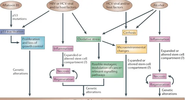
Figure 1.1: Mechanisms of Hepatocarcinogenesis for different risk factors. Commonalities are shown in the same color (Farazi and DePinho, 2006)
1.2 Genetic and Epigenetic Changes in Hepatocellular Carcinoma
Cancer is a DNA disease which emerges through accumulation of genetic alterations in the genes controlling cell cycle, proliferation, differentiation and apoptosis; hepatocellular carcinoma is no exception.
HCC has been extensively studied in terms of genetic alterations in the past ten years which resulted in an increase in our knowledge of altered pathways in hepatocarcinogenesis. Likewise in other solid tumors, a large number of genetic alterations accumulate during the hepatocarcinogenesis process. Genetic and epigenetic alterations have been observed in cirrhotic nodules and half of them have been found to have a monoclonal origin by examining the X-chromosome methylation pattern (Piao et
cell dysplasia (Yeh et al. 2001). Various genetic alterations have been described in primary liver tumors including activating mutations of oncogenes and inactivating mutations of tumor suppressor genes have been only found in HCC and liver adenomas but not in cirrhosis.
1.2.1 Chromosomal Abnormalities
Human cancers are characterized by the presence and accumulation of genetic alterations which target genes or genomic loci. Chromosomal aberrations (CA) are changes in chromosome structure and morphology which are indicators of genetic damage in cancer. CAs are involved in tumor genesis and progression by altering the functions of genes that positively or negatively regulate several aspects of cell proliferation, apoptosis, genome stability, angiogenesis, invasion and metastasis. Their pattern varies between malignancies, ranging from simple balanced rearrangements to complex abnormalities affecting both chromosome structure and euploidy. Subchromosomic abnormalities are often related with genetic alterations, including formation of fusion gene products and swapping of promoter elements which consequently lead to dysregulated gene expression (Aman et al. 1999). The majority of malignant solid tumors, however, exhibit a complex pattern of chromosomal abnormalities, rarely showing any direct association with specific morphological or prognostic subgroups. Many common aggressive epithelial tumors, such as high-grade pancreatic, ovarian, and lung cancer, fall within this category (Pejovic et al. 1992; Johansson et al. 1995; Gorunova et al. 1998), so do many sarcomas, such as osteosarcoma, leiomysorcoma, and malignant peripheral nerve sheath tumor (Mandahl 1996). The molecular genetic alterations corresponding to these complex cytogenetic anomalies are not well characterized, although abnormal activation of oncogenes and losses of tumor suppressor genes are common. These changes are rarely subtype specific. However, the total number of chromosomal aberrations is roughly proportional to the risk of metastasis (Mitelman et al. 1997).
Figure 1.2: Chromosomal evolution in human solid tumor progression: Cells may begin to proliferate excessively owing to loss of tissue architecture, abrogation of checkpoints and other factors. Relatively few aberrations occur before development of in situ cancer and the incidence of genomic aberrations increases during the development of in situ disease (Albertson et al. 2003)
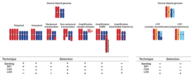
CAs can be studied with an increasing number of large-scale genomic and molecular genetic technologies such as chromosome banding (Mitelman Database of Chromosome Aberrations in Cancer), high-throughput analysis of loss of heterozygosity (LOH) analysis (Hampton et al. 1996), comparative genomic hybridization (CGH) (Pinkel et al. 1998), fluorescence in situ hybridization (FISH) (Schrock et al. 1996), restriction landmark genome scanning (RLGS) (Imoto et al. 1994), representational difference analysis (RDA) (Lisitsyn et al. 1993) and recently introduced SNP microarrays. These techniques differ in which they detect whether balanced or unbalanced aberrations. RLGS, analysis of LOH, RDA and SNP arrays detect allelic imbalances that occur by
or loss of chromosomes and chromosome portions and structural rearrangements. SNP arrays differ from FISH and CGH in detecting unbalanced rearrangements only. Structural changes involve equal exchange of material between two chromosome regions (balanced) or non-reciprocal, such as portions of the genomes are gained or lost. These methods analyze genome-wide DNA content and provide clear information about sporadic and recurrent chromosomal aberrations. The most frequently deleted chromosome arms are 17p, 8p, 16q, 16p, 4q, 9p, 13q, 1p and 6q; and the most frequent gains are observed at 1q, 7q, 8q and 17q (Fujimoto et al. 1994; Boige et al. 1997; Marchio et al. 1997; Nagai et al. 1997; Piao et al. 1998; Guan et al. 2000; Wong et al. 2000; Balsara et al. 2001; Laurent-Puig et al. 2001; Nishimura et al. 2002). Today, Mitelman Database of Chromosome Aberrations in Cancer and University of Helsinki’s Laboratory of Cytomolecular Genetics harbor extensive catalog chromosomal abnormalities in a wide range of tumors.
Figure 1.3: Mechanisms by which chromosomal aberrations result in aneuploidy and common techniques used in detection (Albertson et al. 2003)
Amplification is likely to be initiated by a DNA double-strand break. It can occur only in cells that are able to progress inappropriately through the cell cycle with this damaged DNA whereas normal cells would arrest due to activation of cell-cycle checkpoints. A segment of the chromosomes are copied many times and result in extra copies of genetic material. If extra copies are fused head-to-tail in long tandem arrays within a
chromosomal segment, it is called homogeneously staining regions (HSRs). A segment may also be detached from a chromosome and replicate as an autonomous extrachromosomal entity. Such formations result in subchromosomal fragments termed “double minutes” (DMs). HSRs and DMs increase the copy number of genes they carry and resulting in gene amplifications and are unbalanced. In cancer, amplified regions are likely to carry genes with oncogenic character that bypass cellular control barriers and favor proliferation. Gene amplifications can result in copy number increases from duplications to high level amplifications (700 copies) (Schwab et al. 1999). Today, there is a wide agreement that only less than half of the amplifications result in overexpression of the genes that they harbor. In a recent study with a panel of breast cancer cell lines, 40% of the amplified genes were overexpressed (Hyman et al. 2002).
Interstitial deletions occur when a segment in the middle of a chromosome arm is discarded and the flanking chromosomal regions are rejoined. Interstitial deletions may be rare but they dramatically affect cellular behavior. Such deletions may originate by chromosome breakage and subsequent loss of acentric segment or unequal crossover between misaligned homologous chromosomes or sister chromatids. Chromosome losses are frequent mechanisms of inactivation of one allele of a tumor suppressor gene in solid tumors, recurrent losses at precise loci may point the presence of tumor suppressor genes. In HCC, LOH events have been reported targeting loci in 17p, 13q, 16p, 9p and 6q and inactivating tumor suppressor genes TP53, RB1 (retinoblastoma 1), AXIN1 (axis inhibition protein 1), CDKN2A (cyclin-dependent kinase inhibitor 2A) and IGF2R (insulin-like growth factor 2 receptor), respectively. On the other hand, no tumor suppressor genes have been identified on 1p, 4q, 8p and 16q although high-resolution methods have been used to define consensus boundaries of deletions in these regions (Koyama et al. 1999; Piao et al. 1999; Pineau et al. 1999; Balsara et al. 2001; Yakicier et al. 2001; Bluteau et al. 2002a).
suppressive character whose expression levels are altered by genomic changes. In solid tumors, amplification of ERBB2, MYC and CCND1 have been reported (Slamon et al. 1989; Hinds et al. 1994). Amplification also plays an important role in the development of drug resistance. Cultured cells selected for resistance to N-(phosho-nacetyl)-L-aspartate frequently amplify CAD (Wahl et al. 1979; Schimke et al. 1978) and DHFR is amplified in cultured cells with methotrexate resistance (Banerjee et al. 2002). Similarly, BCR-ABL is amplified in individuals resistant to STI571 (Gorre et al. 2001). Other aberrations include loss of specific regions of the genome. Tumor suppressor genes such as PTEN, CDKN2A have been reported to be lost by homozygous deletions (Li et al. 1997; Orlow et al. 1995). Recessive mutations along with LOH have been shown in the elimination of the functions RB1, BRCA1, BRCA2, PTPRJ and TP53 (Nagai et al. 1994; Cavenee et al. 1983; Baker et al. 1990; Ruivenkamp et al. 2002).
Cytogenetic studies have identified many chromosomal changes in tumors but relatively few of them are recurrent and are involved in tumorigenesis. On the other hand, recurrent abnormalities are frequent transforming events in sarcomas, leukemias and lymphomas (Rowley et al. 1998). Identification of driver genes in the disturbed regions is not easy because these regions often contain multiple genes and more than one gene may important in tumor formation. For example, growth factors FDF19, FGF4, FGF3 and actin-binding oncogene EMS1 are in close proximity to CCND1 and they are amplified together with CCND1 (Bekri et al. 1997). Similarly, growth factor receptor-bound protein GRB7 maps in close proximity to ERBB2 and amplified together. Additionally, cancer genomes may involve many disturbed regions with tens of genes resulting in a complex alteration of different signaling pathways. In such cases, it is harder to establish the driver mechanisms in tumor formation. Finally, the presences of extra copies of individual chromosomes have been reported to be associated with higher cancer risk (Willenbucher et al. 1999).
Genomes of tumor hepatocytes in HCC accumulate a large number of chromosome rearrangements leading to highly abnormal karyotypes, like in other solid tumors. Cytometric analyses have been reported that most HCC cases acquire a global gain of
genetic material (Ezaki et al. 1988; Fujimoto et al. 1991; Chiu et al. 1992). Hyperploidy is also seen in nearly half of the dysplastic lesions observed in cirrhotic disease (Thomas et al. 1992) and its incidence increases in higher grade dysplastic lesions suggesting that chromosome losses followed by endomitosis are early steps in hepatocarcinogenesis. As we already mentioned HCC is genetically heterogeneous and mostly these changes are related to etiological factors. Even though there are several studies addressing chromosomal changes in HCC, new studies with techniques providing higher resolution will probably reveal unknown genetic alterations in HCCs.
1.2.2 Mutations
In human cancers, the most frequently altered gene is the TP53 located at 17p13.1 (Hollstein et al. 1991, Isobe et al. 1986; Miller et al. 1986). Li-Fraumeni syndrome was described as germline mutations of this gene which results in predisposition to cancer in some individuals (Malkin et al. 1990). P53 is a multifunctional transcription factor involved in the control of the cell cycle, apoptosis, senescence, differentiation and development, transcription, DNA replication, DNA repair and maintenance of genome integrity. In HCC, the specific TP53 mutation R249S is found in about 50% of tumors in populations exposed to AFB1 (Bressac et al. 1991; Hsu et al. 1991). In contrast, patients who have not been exposed to this carcinogen have a lower prevalence of TP53 gene mutations (10–30%) and codon 249 is rarely altered. Another frequent mutation in HCC is the hereditary hemochromatosis at codon 220 (Vautier et al. 1999).
Figure 1.4: Histopathological progression and molecular features of HCC: After hepatic injury incurred by any one of several factors (hepatitis B virus (HBV), hepatitis C virus (HCV), alcohol and aflatoxin B1), there is necrosis followed by hepatocyte proliferation. Continuous cycles of this destructive–regenerative process foster a chronic liver disease condition that culminates in liver cirrhosis. Cirrhosis is characterized by abnormal liver nodule formation surrounded by collagen deposition and scarring of the liver. Subsequently, hyperplastic nodules are observed, followed by dysplastic nodules and ultimately hepatocellular carcinoma (HCC), which can be further classified into well differentiated, moderately differentiated and poorly differentiated tumours — the last of which represents the most malignant form of primary HCC. Telomere shortening is a feature of chronic liver disease and cirrhosis. Telomerase reactivation has been associated with hepatocarcinogenesis (its activation in the early versus late stages of disease is still a point of debate, and is discussed in the text). Loss and/or mutation of p53 and genomic instability also characterize hepatocarcinogenesis. p53 loss and/or mutation is shown to occur during progression to HCC, however, there is some evidence that loss and mutation of p53 might also occur in the initial stages of hepatocarcinogenesis (Farazi and DePinho, 2006)
β-catenin is the ortholog of armadillo in Drosophila melanogaster. It is both involved in cell – cell adhesion and Wnt signaling. β-catenin forms complexes with E-cadherin and catenins in adherent junctions. In Wnt signaling, β-catenin may acquire oncogenic character by dominant gain of function mutations in its N-terminus (Morin et al. 1997). These mutations result in the loss of phosphorylation sites in its negative regulation by GSK3β/APC/axin complex. The inhibition of its negative regulation results in higher levels of β-catenin in the cytoplasm and in nuclei leading to abnormal activation of Wnt target genes GLP1 and GRP49. In HCC, β-catenin activating mutations have been
reported in human and mouse models (de La Coste et al. 1998; Miyoshi et al. 1998). In hepatoblastomas and hepatocellular adenomas, β-catenin has also been reported to carry mutations (Koch et al. 1999; Wei et al. 2000; Chen et al. 2002).
AXIN1 maps to 16p13 and this region is frequently (~30%) deleted in HCC (Laurent-Puig et al. 2001). This gene encodes a protein of the GSK3β/APC/axin complex and negatively regulates Wnt pathway. In HCC, LOH events along with mutations and homozygous deletions have been reported in biallelic inactivation of AXIN1 (Satoh et al. 2000; Laurent-Puig et al. 2001). These mutations prevent phosphorylation of β-catenin leading to accumulation of hyperactivation of Wnt target genes.
RB1 locus maps to 13q14 region which is frequently involved in LOH events (Boige et al. 1997; Nagai et al. 1997; Laurent-Puig et al. 2001). RB1 plays major roles in cell division, differentiation and apoptosis. Point mutations and epigenetic regulations along with LOH have been reported in RB1 inactivation (Zhang et al. 1994; Lin et al. 1996). P16INK4 codes for cyclin D-dependent kinase inhibitor 2(CDKN2) and ARF which are involved in p53 mediated apoptosis. These gene products function as tumor suppressors in the RB pathway (Hickman et al. 2002). P16INK4A maps to 9p21 which has been reported to show LOH in 20% of HCC cases (Boige et al. 1997; Nagai et al. 1997; Laurent-Puig et al. 2001). Epigenetic silencing of the p16INK4A promoter has been reported in 30-70% of the tumors (Liew et al. 1999; Matsuda et al. 1999; Jin et al. 2000; Weihrauch et al. 2001). Homozygous deletions of this gene have been reported in HCC, as well (Biden et al. 1997; Jin et al. 2000).
TGF-β pathway is altered in 10-30 % of HCC cases. In TGF-β signaling pathway, inactivating mutations of mannose 6-phosphate/insulin-like growth factor 2 receptor (M6P/IGF2R) have been reported in HCC (Motyka et al. 2000). IGFR also have been shown to carry amino acid substations in two HCC screens (De Souza et al. 1995; Oka et al. 2002). Amino acid substitutions have also been reported in MADH2/Smad2 and
(phosphatidylinositol 3-kinase) in HCC which leads to activation of AKT pathway (Lee et al. 2005).
Vinyl chloride (VC) exposure has been reported be involved in KRAS mutations in hepatocellular carcinoma. VC is a carcinogen associated with the development of liver angiosarcomas and rarely with HCC. Recently, the presence of KRAS2 mutations was observed in 33% of 18 vinyl chloride-associated HCCs and three mutations were found in adjacent non-neoplastic liver tissue (Weihrauch et al. 2001). KRAS mutations are rarely observed in HCCs that are not associated with vinyl chloride exposure which suggest that KRAS2 mutations play an important role in the carcinogenetic pathway linked to vinyl chloride exposure.
Recent reports showed that TCF1 gene (12q24.2) carry biallelic mutation in 60% of a sample of liver cell adenoma cases (Bluteau et al. 2002). TCF1, transcription factor 1, encodes hepatocyte nuclear factor 1α (HNF1α) and function in hepatocyte differentiation and involved in liver specific expression of various genes including β-fibrinogen, albumin and α1-antitrypsin (Frain et al. 1989; Baumhueter et al. 1990; Cereghini et al. 1990; Chouard et al. 1990). In liver cell adenomas, inactivation of both TCF1/HNF1α alleles is usually observed; in 90% of the cases both mutations are of somatic origin. Most of the hepatoblastomas are of sporadic origin. Hepatoblastomas have been reported in Beckwith–Wiedemann syndrome (BWS) and in the familial adenomatous polyposis (FAP). In most hepatoblastomas, β-catenin N-terminal domain harbor interstitial deletions or missense mutations in the GSK3β phosphorylation motif (Koch et al. 1999; Wei et al. 2000; Buendia, 2002). In other hepatoblastoma cases, hyperactivity of the Wnt/β-catenin pathway is related with AXIN2 mutations (Koch et al. 2004)
1.2.3 Epigenetic Alterations
Aberrant DNA methylation patterns have been reported in HCC ( Thorgeirsson et al. 2002; Kanai et al; 1996,1999 & 2000; Yu et al. 2003). Methylation has been reported in the earliest stages of hepatocarcinogenesis and extensively in tumor progression. Molecular analysis of human HCC has shown many epigenetic alterations that result in the deregulation of several oncogenes and tumor suppressor genes including TP53, β – catenin, ErbB receptor family member, MET and its ligand hepatocyte growth factor (HGF), p16 (INK4A), E-cadherin and cyclooxygenase 2 (COX2), apoptosis – associated speck- like – kinase (ASC) and deleted in liver cancer 1 (DLC1) (Feitelson et al. 2002; Wong et al. 1999; Matsuda et al. 1999; Liew et al. 1999; Murata et al. 2004, Kubo et al. 2004; Wong et al. 2003; Maeta et al. 2005). Recently, secreted frizzled-related protein 1 gene (SFRP1) has been reported to be epigenetically silenced in HCC cell lines and primary tumors along with LOH events (Shih et al. 2007). Phosphatase and tensin homologue (PTEN) has been shown to be downregulated by promoter methylation and other epigenetic mechanism in HCC tissues (Wang et al. 2007). Zinc fingers and homeoboxes protein 2 (ZHX2), glutathione S-transferase pi (GSTP1), Ras association domain family 1 (RASSF1), methylation-induced silencing 1 (TMS1), tissue factor pathway inhibitor-2 (TFPI-2), spleen tyrosine kinase (SYK) and LINE-1 type transposase domain containing 1 are other genes that have recently been shown to be downregulated in HCC by methylation (Lv et al. 2006; Wang et al. 2006; Di Gioia et al. 2006; Zhang et al. 2007; Wong et al. 2007; Yuan et al. 2007; Tangkijvanich et al. 2007). Suppressor of cytokine signaling 1 (SOCS1), which is a negative regulator of the JAK/STAT pathway, has been shown to be silenced by methylation in HCC (Yoshikawa et al. 2001).
2. HYPOTHESIS
Amplifications and deletions are common genetic alterations in epithelial cancers. Numerous oncogenes and tumor suppressor genes located in these regions have been identified in cancers. New techniques which provide higher resolution may reveal unknown small chromosomal alterations where important genes for carcinogenesis may be located. DNA copy number changes in HCC still have not been studied with recently available high – throughput molecular methods which provide higher resolution.
In the framework of this study, we aimed to screen HCC cell lines for their DNA copy number changes. We think cell lines are ideal models for this study because their genomic DNAs are available as homogenous and high – quality (intact) which are crucial requirements for SNP microarray analysis. Unlike tissue samples, their genomic DNAs are pure, without any contamination of neighboring normal cells or infiltrating blood DNA which gives better estimates for low – copy number changes. Moreover, screening a panel of commonly used HCC cell lines may provide us independent abnormalities as well as recurrent ones. Thus, our results may reveal new regions of abnormality in HCC genome, in which oncogenes or tumor suppressor genes may reside. Analysis of these new candidates may contribute to our understanding of hepatocarcinogenesis by introducing new mechanisms and related pathways.
3. METHODOLOGY
3.1 Materials
3.1.1 Hepatocellular Carcinoma Cell Lines
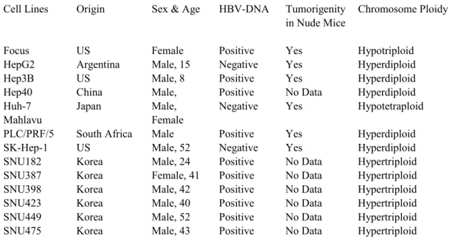
In the framework of this study, 14 Hepatocellular carcinoma (HCC) cell lines were used as shown in Table 3.1
Cell Lines Origin Sex & Age HBV-DNA Tumorigenity Chromosome Ploidy in Nude Mice
Focus US Female Positive Yes Hypotriploid
HepG2 Argentina Male, 15 Negative Yes Hyperdiploid
Hep3B US Male, 8 Positive Yes Hyperdiploid
Hep40 China Male, Positive No Data Hyperdiploid
Huh-7 Japan Male, Negative Yes Hypotetraploid
Mahlavu Female
PLC/PRF/5 South Africa Male Positive Yes Hyperdiploid
SK-Hep-1 US Male, 52 Negative Yes Hyperdiploid
SNU182 Korea Male, 24 Positive No Data Hypertriploid
SNU387 Korea Female, 41 Positive No Data Hypertriploid
SNU398 Korea Male, 42 Positive No Data Hypertriploid
SNU423 Korea Male, 40 Positive No Data Hypertriploid
SNU449 Korea Male, 52 Positive No Data Hypertriploid
SNU475 Korea Male, 43 Positive No Data Hypertriploid
Table 3.1: Characteristics of the HCC cell lines.
3.1.2 Reagents
Ethidium Bromide (EtBr);
10mg/ml in water (stock solution) 30ng/ml (working solution)
10X TBE Buffer Solution; 108g Tris
55g Boric Acid 8.3g EDTA
Dissolved in 1lt of deionized water. 6X Loading Buffer Solution
30% Glycerol 0.04% Bromphenolblue 0.04% Xylene Cyanol ∆dH2O 10x Phosphate-Buffered Saline (PBS) 80 g NaCl 2 g KCl 14.4 g Na2HPO4 2.4 g KH2PO4
Dissolved in 1 lt of water and pH is adjusted to 7.4. 50x TAE Buffer (Tris-Acetate-4EDTA)
242 g Tris Base 57.1 ml Acetic Acid 100ml 0.5M EDTA
ddH2O was added to 1 liter and adjust pH to 8.5 Wash A: Non-Stringent Wash Buffer
(6X SSPE, 0.01% Tween 20) For 1000 mL:
300 mL of 20X SSPE 1.0 mL of 10% Tween-20
Wash B: Stringent Wash Buffer (0.6X SSPE, 0.01% Tween 20) For 1000 mL:
30 mL of 20X SSPE 1.0 mL of 10% Tween-20
969 mL of water, filtered through a 0.2 μm filter 0.5 mg/mL Anti-Streptavdin Antibody
Resuspend 0.5 mg in 1 mL of water 12X MES Stock Buffer
(1.22M MES, 0.89M [Na+]) For 1,000 mL:
70.4g of MES hydrate 193.3g of MES Sodium Salt
800 mL of Molecular Biology Grade water Mix and adjust volume to 1,000 mL. The pH should be between 6.5 and 6.7. Filtered through a 0.2 μm filter
1X Array Holding Buffer
(Final 1X concentration is 100 mM MES, 1M [Na+], 0.01% Tween-20) For 100 mL:
8.3 mL of 12X MES Stock Buffer 18.5 mL of 5M NaCl
0.1 mL of 10% Tween-20 73.1 mL of water
Stain Buffer H2O 666.7 μL SSPE (20X) 300 μL 6X Tween-20 (3%) 3.3 0.01% Denhardt’s (50X) 20 1X Subtotal 990 μL Subtotal / 2 495 μL SAPE Solution Mix Stain Buffer 495 μL 1X
1 mg/mL Streptavidin Phycoerythrin (SAPE) 5.0 μL 10 μg/mL Total 500 μL
Antibody Solution Mix Stain Buffer 495 μL 1X
0.5 mg/mL biotinylated antibody 5 μL 5 μg/mL Total 500 μL
3.2 Methods
3.2.1 Tissue Culture
All cell lines were cultured in 75ml flasks (Greigner-Bio) as monolayers. Cell lines were either grown in RPMI-1640 (Biological Industries) or Dulbecco’s Modified Eagle Medium (DMEM) (Biochrom AG) supplied with 10% FBS (Sigma), 50mg/ml penicillin / streptomycin and non-essential amino acids (Biochrom AG). Cell lines were culture at 37°C incubator with 5% CO2 (Heto-Holten, Surrey, UK). Cells were handled in sterile laminar hoods (Heto-Holten, Surrey, UK). Medias and solutions were kept at 4°C and preheated to 37°C before use.
3.2.1.1 Cyropreservation of Cell Lines
Exponentially growing cells were harvested with trypsin and fresh medium was added to inactivate trypsin through neutralization. The numbers of cells were counted with hemocytometer and precipitated at 250g for 5 minute at room temperature. Following, cells were resuspended with freezing media at a concentration of 5 million / ml in one vial. Freezing medium was prepared as 90% FBS and 10%DMSO (Sigma). Cryotubes were incubated at -20°C for 1 hour, following -80°C overnight and kept in liquid nitrogen tank for long term storage.
3.2.1.2 Culturing of Cell Lines
After removal from liquid nitrogen tanks, cells were thawed at 37°C and 5ml of growth medium was added. Following centrifugation at 1500 rpm for 5 minutes, supernatant was discarded and fresh medium was added. Cells were then grown in 25ml flasks in the incubator.
3.2.1.3 Subculturing of Cell Lines
Cells were grown at a confluency of app 80%. Old medium was removed from the flasks with vacuum and the cells were washed with PBS twice. Trypsin was added to flasks and cells were incubated for 2-3 minutes with trypsin/EDTA solution. After detaching from the flask surface, fresh medium was added to inactivate the trypsin. Collected cells were then transferred to new plates.
3.2.1.4 Preparation of Cell Pellets
Cell pellets were prepared for gDNA and RNA isolation. When the cells reached 80% confluency, medium was removed and cells were washed with PBS twice. Cells were detached with trypsin and fresh medium was added. After centrifugation for 5 minutes at 1500 rpm, supernatant was discarded and the pellet was washed with PBS twice. Pellets were immediately placed -80°C refrigerator.
3.2.1.5 Genomic DNA Isolation
Frozen cell pellets were thawed at room temperature and gDNAs were isolated by using Qiagen DNeasy Tissue Kit according to manufacturer’s recommendation. Isolated genomic DNAs (gDNA) were either dissolved in manufacturer’s buffer or in water. Quality of gDNA was checked on 0.75 % agarose gel and concentration was measured with Nanodrop Spectrophotometer (Nanodrop Technologies). gDNAs were stored at-20°C for long term.
3.2.2 SNP Microarray Assay
Probe preparation for SNP microarray hybridization experiments were done according to manufacturer’s manual (Affymetrix, 10K2.0 Assay). The overall assay is shown in Figure 3.1
Figure 3.1: Outline of SNP microarray assay.
Briefly, genomic DNAs were diluted to 50ng/µl in water. 250ng of each gDNA was subjected to restriction digestion with XbaI enzyme (New England Biolabs) for two hours at 37°C in thermal cycler (Tech e), in replicate. One replicate was run on a 0.75% agarose gel to check if the digestion assay performed well; the other replica was continued with adaptor ligation. T4 DNA ligase (New England Biolabs) was used to attach adaptor Xba (Affymetrix) to XbaI restriction sites. The ligation assay was done at 16°C for 2 hours. Adaptor Xba contains a binding site for Xba Primer (Affymetrix). Later, ligated restriction fragments were diluted to 4 fold with H2O and used in whole genome PCR as template. Hot Star Taq Plus polymerase (Qiagen) was used in the amplification process. The thermo profile was as follows: 94°C for 2 minutes and 30
seconds. PCR amplicons were purified with Qiagen Qiaquick PCR purification kit. 20µg of PCR was fragmented to a range between 35bp – 200bp using DNAseI (Affymetrix). The fragment sizes were checked by electrophoresis on 4% agarose gel. Following, the fragmented PCR products were end labeled with Biotin-labeled reagent (Affymetrix) using Terminal Deoxynucleotidyl Transferase (Affymetrix). Probe DNA was then denatured at 95°C in hybridization buffer containing TMACL (Sigma), DMSO (Sigma), Denhardt’s Solution (Sigma), MES (Sigma), Herring Sperm DNA (Promega), EDTA (Ambion), Tween-20 (Sigma), Human Cot-1 DNA (Invitrogen) and Oligonucletide control (Affymetrix). Following, denatured probe was injected to the array (Affymetrix 10K2.0). The hybridization was done at 48°C, 60 rpm for 16 hours in the hybridization oven (Affymetrix). After hybridization, the probe mix was discarded and the array was washed with Wash A and B buffers (6X SSPE, 0.01% Tween 20, 0.6X SSPE, 0.01% Tween 20, respectively) in fluidics station (Affymetrix). Then, the chips were stained with buffers containing Biotinylated Anti-Streptividin antibody (Vector), SAPE (Streptavidin, R-phycoerythrin conjugate), Acetylated Bovine Serum Albumin, 20X SSPE, Denhardt’s Solution and Tween-20. The stained chip was scanned at the scanner (Affymetrix) and pre-analyzed with GeneChip Operating Software (GCOS) and GeneChip DNA Analysis Software (GDAS) software bundle (Affymetrix). All these steps above are briefly shown in figure 3.2.
3.2.3 Microarray Analysis 3.2.3.1 Pre-Analysis
Pre-analysis of the SNP microarrays were performed with GCOS and GDAS software bundle. The bundle operates the scanner and builds the raw data captured by the sensor. It uses the specific library files containing the information about the probesets on the array using pre-defined settings and algorithms (Affymetrix 10K2.0 Manual). The bundle then extracted probeset information from raw data and generates CEL files containing the signal intensity of each probeset along with a chip report file. The chip report file provided information about the performance of the hybridization, such as; average signal intensity of probesets, background and oligonucleotide controls along with a pseudo-image of the chip. Later, using the CEL files, genotype calls of the each probeset was calculated with their intensities.
3.2.3.2 Advance-Analysis
Advance analysis was performed with DNA-Chip Analyzer (dChip) (Harvard University) Software freely available for academic users at www.dchip.org. dChip is a Windows software package for probe-level and high-level analysis of gene expression microarrays and SNP microarrays (Li and Wong 2001, Lin et al. 2004). At the probe level, dChip can display and normalize the CEL files, and the model-based approach allows pooling information across multiple arrays and automatic probe selection to handle cross-hybridization and image contamination. High-level analysis in dChip includes comparing samples, hierarchical clustering, view expression and SNP data along chromosome, LOH and copy number analysis of SNP arrays, and linkage analysis. In these functions the gene information and sample information are correlated with the analysis results. In the analysis, model-based expression was selected with perfect
method. Hidden Markov Model and median smoothing were used in inferred copy number analysis.
3.2.4 Genomic DNA PCR
All PCR reactions were performed using Techne-512 equipment (Techne Inc). Primers were first checked for their optimal conditions by altering magnesium levels in a thermo-gradient PCR. ). A reaction mixture of 2.5µl 10X reaction buffer, 2.5µl MgCl2 (25mM), 1µl dNTP (10μM), 1µl of each primer (10pmol), and 0.5µl Taq DNA polymerase (5u/μL) was prepared per 250ng of gDNA. The thermo profile was 94°C for 2 minutes and 30 seconds; denaturation at 94°C for 30 seconds, annealing temperature (differs) for 45 seconds, extension at 72°C for 30 seconds, and final extension for 5 minutes and 30 seconds.
3.2.4.1 Oligonucleotide Design
All oligonucleotide primers were designed by using Primer3 algorithm available at http://frodo.wi.mit.edu/. Oligonucletides were purchased from Iontek (Iontek) as lyophilized. Primer sequences are listed in Table 3.2
TPTE2_1F ATGGACACATTTAGTTCGACTTC TPTE2_1R CAGCCTTCTCATCAGCTTTT HSA_MIR_31_F ATACACAGCAATACACGAAGGACT HSA_MIR_31_R GGTGAAAGGAAAAATTTTGGAA GAPDH_070228_cDNA_F GGCTGAGAACGGGAAGCTTGTCAT GAPDH_070228_cDNA_R CAGCCTTCTCCATGGTGGTGAAGA Mir124a1-F GTCGGTCGCTCCTTCCTT Mir124a1-R TCTACCCACCCCTCTTCCTT SATL1_F GGGGACAATCCCCTTTTCTAC
SATL1_R AAAGTACCTTGCCAGTCCATGA
NUBPL_gDNA_F AGTTCCGATTTTGTTTCTTTCCA
NUBPL_R ACAATTGGCTGGCCTGTATCT
Table 3.2: Primers used in the PCR assays.
3.2.4.2 PCR Purification
All PCR products were purified by using the Qiagen Qiaquick PCR purification system according to manufacturer’s recommendation except a few modifications. After washing the membrane containing the PCR products with ethanol containing was buffer, an additional step of centrifugation was performed at 20,000g for 5 minutes with caps open. This allowed complete evaporation of PCR products and then H20 was used for reconstitution of the PCR products.
3.2.8.3 Agarose Gel Electrophoresis
2µl of 6X DNA loading dye was added to 10µl of each PCR product. PCR products were then loaded in 30ng/µl ethidium bromide containing 1% (w/v) agarose gels and were run in horizontal gel electrophoresis equipment in 1X TAE buffer under 90V for 30 minutes. Gene Ruler DNA ladder (Fermentas) was used as DNA size marker. Transilluminator equipment (Bio – Rad) was used for visualization at 340nm wavelength UV along with MultiAnalyst software (Bio – Rad).
3.2.4.4 Sequencing
Selected amplified PCR products were purified by using the Qiagen Qiaquick PCR purification kit and quantified with Nanodrop spectrophotometer. Required amounts of
4. RESULTS
In the framework of this study, we have detected two deletions and 12 amplifications which are novel in hepatocellular carcinoma. These disturbed regions harbor more approximately 570 transcripts. Some these genes are well described in cell cycle and tumorigenesis, other’s role are still poorly understood. Among the described ones, a high percentage of these genes code for enzymes, transcription regulators, cytokines, transporters and kinases. Concordantly, most of the gene products of these trascipts are found in cytoplasm, nucleus and extracellular spaces. Below are the figures that show overall results along with protein functions and cellular distribution.
4.1 Homozygous and Hemizygous Deletions
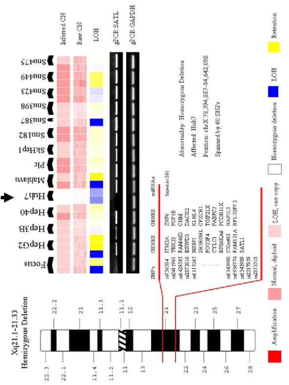
We have observed three homozygous and one hemizygous deletions. Homozygous deletions are located at 9p23 in Mahlavu, PLC, SkHep1, Snu182, Snu387 and Snu423; 9p22.1-p21.2 in SkHep1, Snu387 and Snu449; 13q12.11 in Huh7 and SkHep1; hemizygous deletion maps to Xq21.1-21.33 in Huh7 (male origin). All the deletions are in concordance with the microarray expression data (not shown) and they have also been confirmed by PCR.
4.2 Amplifications
We have observed 11 amplifications at 8p23.1 in Hep40; 8q13.3-q21.11 in Hep40; 8q24.13 in Hep40; 9p22.1-p21.2 in Snu398; 12p11.21-p.11 in Snu475; 14q12-q13.1 in Huh7, 15q21.3 in Hep40; 16q21.3 in Hep40; 17p13.1-q11.1 in Snu182 and Snu475; 17q21.2 in Snu475; 19q13.31-q13.32 in Focus and Mahlavu; 22q11.21-q11.22 in Snu182 and Xp22.11 in Huh7. Expression analysis results mostly did not reflect any signs of amplifications in this amplicons. We confirmed selected regions by PCR methods.
5. DISCUSSION
In the framework of this study, we searched for DNA copy number changes in the genomic DNAs of 14 HCC cell lines. We used commercially available SNP microarrays consist of approximately 10 thousand SNP markers representing the whole genome with a mean physical inter-marker distance of 210KB and 0.32 cM of genetic distance. These SNP markers spanned all the autosomal chromosomes and the X-chromosome. We performed two biological replicates for each cell line except Focus (three) and Snu387 (one).
SNP markers, in principle, provide two types of information which can be classified as qualitative and quantitative. Qualitative information refers to genotyping of the DNA to be investigated. Each SNP marker is chosen from a pool of highly heterozygous SNPs (0.37 on average) representing Caucasian, Asian and Afro-American populations. High heterozygosity values of these bi-allelic markers enable genotyping of genomic DNA. Briefly, each allele of the SNP markers are spotted as different probesets on the array and genotyping is performed based on the hybridization efficiencies of each allele’s probesets. Genotyping calls can be used in two ways; first, “no calls” which theoretically refer to non or mis-hybridization, may point homozygous deletions; second, homozygous calls of a number of consecutive SNPs may suggest loss of heterozygosity regions. The former can be used as a deletion marker if they include at least three consecutive SNPs. In such deleted regions, inter-SNP marker distances should also be checked. In the latter, the unlikely probability of homozygous calls for consecutive SNPs is calculated as the possibility of LOH events. Moreover, in the analysis of SNP array data, the source of specimen to be investigated (such as cell lines or peripheral blood DNA etc.) and copy number neutral events should also be concerned while drawing conclusions.
Quantitative information is described as the percentage of saturation of each probeset by the interrogated DNA during hybridization. Briefly, amplified regions saturate probesets
and mismatch probesets. Similar to the genotype values, in the quantitative analysis of SNP data, a number of consecutive SNP markers are expected to behave similarly to conclude as copy number gains or losses. Finally, qualitative and quantitative values for each SNP are expected to be in accordance to obtain significant results.
In the present study, we benefited from both genotype and copy number values of the probesets to achieve significant results with minimal regions and maximum confidentiality. For genotype calls, we expected to have at least three consecutive SNP markers to be present as no calls in order to represent homozygous deletions; therefore our resolution is expected to be around 600-KB. We also considered the possibility of failure in the restriction enzyme digestion and subsequent whole genomic DNA PCR amplification. This may cause under-representation of target and result in false-positive deletions. This type of false-positive errors can be batch specific, observed as common no calls at particular SNP markers in most of the samples and they usually behave unlike adjacent SNPs. Some SNPs with no call values may have normal copy number values in contrast to deletions, therefore we checked raw copy number values each no call SNPs and excluded the ones with values higher than 0.5.
For quantitative measurement, we used dChip Software to analyze saturation values (Li and Wong 2001, Lin et al. 2004). We first tried peripheral blood genomic DNA results of four healthy individuals as reference controls to obtain copy number values since we had no chance to use match-controls for our cell lines. We noticed that these individuals have characteristic copy number polymorphisms in their genome and behaved differently than the nature of the cell lines’ gDNA. Therefore, we excluded these controls in the analysis and performed no-reference analysis by introducing all the cell lines as normal to the software. This approach significantly reduced the noise and disturbance. Moreover, we also considered the concordance of genotype data with copy number data whether they are in accordance with homozygous deletion and LOH regions. Noteworthy, we also checked inter-marker distances and saturation signatures at the raw copy number values for an additional level of evaluation of the significance and to set the physical margins. We mapped the physical positions of disturbances using UCSC Genome
Browser Build March 2006. In some chromosomal regions, SNP markers can be very few and the distance between SNP markers in an imbalanced region and the neighboring normal region SNP marker can be as large as a few MB. On such occasions, the disturbed regions might exceed the imbalance region defined by the borderline SNP markers. To be on the confident side, we preferred to use the last SNPs as the margins of imbalance and neglect if there are any genes neighboring, but we checked if these regions contain interesting genes.
In this study, we preferred to report only homozygous deletions and amplifications with copy number values equal to or greater than four. We excluded LOH profiles based on genotype calls due to cell line’s nature. The cell lines used in this study are hyperploid; therefore using genotyping calls as a qualitative marker would be erroneous. Although qualitative use of genotype calls from SNP chips are invaluable information in linkage and association studies in which the target DNA is usually from blood or tissue (Ozturk et al. 2006). When we analyzed our cell lines for their LOH profile, we observed LOH in less than half of the whole genomes of the cell lines; therefore we preferred not to report them. On the other hand, we also did not include copy number changes smaller than 4 in our report although we observed quite few hemizygous duplications which can be as large as whole chromosomes.
Furthermore, we compared our copy number data with the available microarray expression data of the cell lines and primary tumors. We accessed the raw expression data (Affymetrix U133 Plus 2.0 Platform) of primary tumors (GSE6764, Wurmbach et al. 2007), HepG2 (GSE6368, Wang et al. 2006) through Gene Omnibus (GEO). Huh7, SkHep1 and Hep40 cell lines’ expression data were obtained through personal communications. Although the aims of these experiments were quite different than ours, we used them only with purpose of comparing the expression signatures in regions of our interest. One advantage of these expression data is that it has higher resolution compared to our SNP array. The expression arrays we analyzed have more probesets and cover all
Basically, we expected no expression of consecutively mapped genes in the deleted regions. For copy number gains, we did not expect abnormally high expression of consecutive genes in all of amplified regions, because not all the amplifications result in overexpression of the genes they contain. Therefore, the expression data of the available cell lines and the primary tumors allowed us to check and confirm our findings in gDNA at transcript level and gave us a chance to correlate it with primary tumors.
5.1 Homozygous Deletions
Our results showed three homozygous deletions on chromosomes 9, 13 and X. In 9p23, Mahlavu, Plc, Skhep1, Snu182, Snu387 and Snu423 contain a homozygous deletion site within Mahlavu and Snu182 the largest. This region spans 1-MB and maps to a part of protein tyrosine phosphatase, receptor type, D gene (PTPRD) which is a large gene and spans a region of 2.3-MB. This gene has partially been shown to be deleted in other cancers and no data is available for HCC (Sato et al. 2005). At present, the pathogenic significance of PTPRD deletion is unclear, but, frequent deletions at this locus indicates that the inactivation of this gene may have a major role in tumorigenesis. Expression array results also support our findings that this gene is downregulated in cell lines, cirrhotic and HCC tumor tissue compared to normal liver.
Another homozygous deletion maps to 9p21.3-p21.2 region in SkHep1, Snu387 and Snu449 and it spans 6-MB. Genomic DNA PCR targeting hsa-mir-31 region in this deletion confirmed our SNP array results. This region harbors important tumor suppressor locus of cyclin-dependent kinase inhibitors 2 (CDKN2A/p14ARF/CDKN2B) which encode negative regulators of cell growth. The region has been shown to be frequently inactivated by homozygous deletions in HCC, lung and other cancers (Liew et al. 1999; Liggett and Sidransky, 1998). In addition to deletions, this locus is also inactivated by epigenetic regulation, LOH and mutations (Lukas et al. 1995). Among the three cell lines, SkHep1 has the narrowest deletion targeting this locus with a span of 1.4-MB, while the span of deleted regions in Snu387 and Snu449 are 3.5 and 5-1.4-MB,


