FLUORESCENCE DETECTION OF BIOLOGICAL THIOLS
AND AXIALLY CHIRAL BODIPY DERIVATIVES &
ALTERNATIVE METHODOLOGIES FOR SINGLET OXYGEN
GENERATION FOR PHOTODYNAMIC ACTION
A DISSERTATION SUBMITTED TO
MATERIALS SCIENCE AND NANOTECHNOLOGY PROGRAM OF THE GRADUATE SCHOOL OF ENGINEERING AND SCIENCE
OF BILKENT UNIVERSITY
IN PARTIAL FULFILLMENT OF THE REQUIREMENTS FOR THE DEGREE OF
DOCTOR OF PHILOSOPHY
By
SAFACAN KÖLEMEN September, 2014
I certify that I have read this thesis and that in my opinion it is fully adequate, in scope and in quality, as a thesis of the degree of Doctor of Philosophy.
………. Prof. Dr. Engin U. Akkaya (Advisor)
I certify that I have read this thesis and that in my opinion it is fully adequate, in scope and in quality, as a thesis of the degree of Doctor of Philosophy.
………. Prof. Dr. Metin Zora
I certify that I have read this thesis and that in my opinion it is fully adequate, in scope and in quality, as a thesis of the degree of Doctor of Philosophy.
………. Assoc. Prof. Dr. Tamer Uyar
I certify that I have read this thesis and that in my opinion it is fully adequate, in scope and in quality, as a thesis of the degree of Doctor of Philosophy.
………. Assoc. Prof. Dr. Özgür Altan Bozdemir
I certify that I have read this thesis and that in my opinion it is fully adequate, in scope and in quality, as a thesis of the degree of Doctor of Philosophy.
………. Assist. Prof. Dr. Bilge Baytekin
Approved for the Graduate School of Engineering and Science:
………. Prof. Dr. Levent Onural
ABSTRACT
FLUORESCENCE DETECTION OF BIOLOGICAL THIOLS AND AXIALLY CHIRAL BODIPY DERIVATIVES &
ALTERNATIVE METHODOLOGIES FOR SINGLET OXYGEN GENERATION FOR PHOTODYNAMIC ACTION
SAFACAN KÖLEMEN
PHD IN MATERIALS SCIENCE AND NANOTECHNOLOGY SUPERVISOR: PROF. DR. ENGİN UMUT AKKAYA
SEPTEMBER, 2014
Calorimetric and luminescent detection of biological thiols namely cysteine (Cys), homocysteine (Hcy) and glutathione (GSH) have attracted great interest due to the their biological significance. There are many reported fluorescent probes for Cys and Hcy, however selective probe designs for GSH remained elusive. We represented in thesis (Chapter 3) a BODIPY based selective fluorescent probe for the in vitro detection of GSH in cancer cell lines. Photodynamic therapy (PDT) is one of the promising and developing treatment modality for certain indications. Therapeutic action is achieved by the generation of cytotoxic singlet oxygen (SO). Most critical compartment of SO production pathway is the sensitizer molecule. In order to get effective inter-system crossing, which is highly needed for singlet oxygen generation, common strategy is to incorporate heavy atoms on sensitizers. However, presence of heavy atoms increases the dark toxicity that is not desired in clinical applications. In Chapter 4, we are introducing a new concept for activatable heavy atom free sensitization of PDT by designing novel orthogonal BODIPY derivatives and detailed computational analysis of this new concept. While dealing with orthogonal BODIPYs, we synthesized for the first time two axially chiral BODIPY derivatives and characterized the enantiopure products, which holds great promise for enantioselective sensing applications (Chapter 5). PDT has two major problems, which are light penetration depth of the incident light and the hypoxia. These two restrictions are addressed in chapter 6, by combining gold nanorods and aromatic endoperoxides.
Keywords: GSH probe, axial chirality, photodynamic therapy, singlet oxygen, gold
ÖZET
BİYOLOJİK TİYOLLERİN FLORESAN TAYİNİ VE AKSİYAL KİRAL BODIPY TÜREVLERİ & FOTODİNAMİK ETKİ İÇİN ALTERNATİF YOLLAR İLE
SİNGLET OKSİJEN ÜRETİMİ
SAFACAN KÖLEMEN
MALZEME BİLİMİ VE NANOTEKNOLOJİ, DOKTORA TEZ YÖNETİCİSİ: PROF. DR. ENGİN UMUT AKKAYA
EYLÜL, 2014
Biyolojik tiyoller sistein (Cys), homosistein (Hcy) ve glütatyonun (GSH) floresan moleküler ajanlar ile tayin edilmesi, bu tiyollerin taşıdıkları biyolojik rollerden ötürü çok büyük önem arz etmektedir. Literatürde yer alan pek çok çalışmada Cys ve Hcy tayini için dizayn edilmiş ajanlar bulunabilmektedir. Ancak GSH için seçici floresan moleküler görüntüleme ajanları çok az sayıdadır. Bu tez kapsamında GSH’ın kanser hücre kültürleri içerisindeki tayini seçici olarak BODIPY tabanlı floresan boya yardımı ile yapılmıştır (Bölüm 3). Fotodinamik terapi (PDT) fazlaca umut vaat eden ve gelişmekte olan bir kanser tedavi yöntemidir. PDT kapsamındaki terapatik etki zararlı singlet oksijen (SO) üretimi ile gerçekleşmektedir. SO üretimi sırasında yer alan parçalardan en önemlisi duyarlaştırıcı moleküldür. SO elde edilmesi için gerekli olan sistemler arası geçişin sağlanabilmesi için sıklıkla uygulanan dizayn prensibi duyarlaştırıcı molekülü ağır atomlar ile modifiye etmektir. Ancak ağır atomların varlığı duyarlaştırıcının uyarıcı ışık olmaksızın toksik etki göstermesine neden olmaktadır. Bu doğrultuda bölüm 4’te yer alan çalışmalarda ağır atom içermeyen ve sadece kanserli hücrede aktifleşebilen yeni duyarlaştırıcı dizaynları ve ortaya çıkarılan yeni konseptin detaylı teorik çalışması sunulmuştur. BODIPY kimyası üzerinde yürüttüğümüz çalışmalar sonunda ilk kez aksiyal kiral özelliğe sahip BODIPY türevleri tasarlamış, sentezlenmiş ve karakterize edilmiştir. PDT mekanizmasının iki önemli problemi mevcuttur. Bunlar uyarma ışığının dokulardan kısıtlı bir miktarda geçiş yapması ve hipoksiyadır. Bölüm 6’da altın nanoçubuklar ve aromatik endoperoksitler yardımı ile bu sorunlara çözüm sunulmaktadır.
Anahtar Kelimeler: GSH ajanı, aksiyal kiral, fotodinamik terapi, singlet oksijen, altın
ACKNOWLEDGEMENTS
I would like to start acknowledgement part with my PhD supervisor Prof. Engin U. Akkaya, whom I will definitely miss after finishing my studies at Bilkent University. He is the smartest, foreseeing, enthusiastic, kind and the funniest person that I have ever met. I feel proud and lucky for being a member of his research group. With immense respect, I would like to express my gratitude to him for his guidance, support, patience and for teaching us how to become a good scientist and more importantly a good person. I would also like to thank him for always giving us the opportunity to try new things and pursue our own ideas. I appreciate all his contributions, discussions and valuable thoughts to make my PhD experience productive and sensational. I will never forget him and his support throughout my life.
I am sincerely grateful to my PhD thesis progress committee members, Prof. Metin Zora and Assoc. Prof. Dr. Tamer Uyar for their valuable advices and fruitful discussions.
I owe a special thank to Assoc. Prof. Dr. Özgür Altan Bozdemir for him endless help and support, guidance to improve my skills in the field of supramolecular and organic chemistry as well as his unlimited knowledge and experience that I have benefited from greatly. I would also like to express my graduate for him for his valuable friendship.
I would like to express my special thanks to Dr. Yusuf Çakmak, Dr. Ruslan Guliyev, Dr. Murat Işık, Yiğit Altay, Tuğçe Karataş, Ziya Köstereli and Cpt. Bilal Kılıç for their partnership in my PhD studies. Their support patience and contributions are so valuable and unforgettable.
I am sincerely grateful to my close friend Tuğba Özdemir Kütük. She made my PhD period joyful, tolerable and easy with her precious friendship, endless support, everlasting help and understanding. I am sure everything would have been much difficult without her. I will thoroughly miss working with her.
I want to thank our present and past group members Dr. Fazlı Sözmen, Dr. Sündüs Erbaş Çakmak, Dr. Esra Tanrıverdi, Dr. Onur Büyükçakır, Dr. Serdar Atılgan, Dr. Ali Çoşkun, Taha Bilal Uyar, Ahmet Atılgan, Tuba Yaşar, Tuğçe Durgut, Nisa Yeşilgül, Sencer&Hande Selçuk, Hale Atılgan, Jose Bila, Ceren Çamur, Darika Okeava, Cansu Kaya, Melek Baydar, Veli Polat, Ahmet Bekdemir, Elif Ertem, Şeyma Öztürk Guliyeva, Dr. Dilek Taşgın, Işın Sakallıoğlu, Dr. Özlem Seven, Seylan Ayan, Özge Yılmaz, İlke Şimşek Turan, Dr. Seda Demirel, Deniz Yıldız and rest of the SCL (Supramolecular Chemistry Laboratory) members for great friendships, wonderful collaborations, and perfect ambiance in the laboratory. They are very precious for me. It was wonderful to work with them.
I also would like to thank Dr. Yavuz Dede, Muhammed Buyuktemiz and Selin Duman for breakthrough computational studies, helpful discussions and collaborations.
I am grateful to Dr. Lale Doğan, Dr. Dicle Güç, Dr. Turgay Tekinay, Dr. Juyoung Yoon and Berna Şentürk for their impressive efforts during the cell culture studies. My thanks also go out to Bora Bilgiç for fantastic and creative graphical designs. I also wish to express my warm and sincere thanks to my friends İlker Kütük, Oya Ertunç Bilgiç, Dr. Görkem Günbaş, Dr. Asuman Günbaş, and Nurhan&Reşat Çiftçi for being with me all the time.
Special thanks are extending to my beloved mother Filiz Kölemen and father Fikret Kölemen for their infinite support, huge love and understanding. It is really reassuring to known that there are always some people waiting there for you.
During the last four years, a lot of things have changed in my life but the most beautiful change happened when I met with Birsu Teoman (soon to be Kölemen). Finally and most importantly, with great love, I would like to thank my intended wife for her understanding, love, patience, tolerance and support. It would not have been possible to write this thesis without her apprehension and help.
LIST OF ABBREVIATIONS
BODIPY : 4,4-difluoro-4-bora-3a,4a-diaza-s-indacene
CASSCF : Complete active-space self consistent field methodology CHCl3 : Chloroform
DCM : Dichloromethane
DS-TR : Doubly substituted-tetra radicalic DFT : Density functional theory
ISC : Intersystem crossing
FRET : Förster resonance energy transfer HOMO : Highest occupied molecular orbital LUMO : Lowest unoccupied molecular orbital MALDI : Matrix-assisted laser desorption/ionization MS : Mass spectrometry
NMR : Nuclear magnetic resonance
NOON : Natural orbitals and occupation numbers PDT : Photodynamic therapy
PS : Photosensitizer SS : Singly substituted
SOMO : Singly occupied molecular orbital TLC : Thin layer chromatography
TABLE OF CONTENTS
1. INTRODUCTION ... 1
2. BACKGROUND ... 6
2.1. Fluorescent Molecular Sensors ... 6
2.1.1. General Information ... 6
2.1.2. Overview of Fluorescence ... 8
2.1.3. Fluorescent Probes ... 9
2.2. BODIPY Dyes ... 12
2.3. Photophysics of Sensing Mechanism ... 16
2.3.1. Photo-induced Electron Transfer (PeT) ... 16
2.3.2. Photo-induced Charge Transfer (PCT) ... 18
2.3.3. Fluorescence Resonance Energy Transfer (FRET) ... 20
2.3.4. Excimer Formation ... 21
2.3.5. Aggregation Induced Emission (AIE) ... 21
2.4. Detection of Biological Thiols ... 22
2.4.1. Michael Addition ... 23
2.4.2. Cyclization with Aldehydes ... 27
2.4.3. Conjugate Addition & Cyclization ... 28
2.4.4. Cleavage of Sulfonamide and Sulfonate Esters ... 28
2.4.5. Thiol-Halogen Nucleophilic Substitution ... 29
2.4.6. Disulfide Exchange Reaction ... 30
2.4.7. Metal Complexation ... 30
2.5. Photodynamic Therapy ... 31
2.5.1. General Information ... 31
2.5.3. Mechanism of PDT Action ... 33
2.5.4. Basic Components of PDT ... 36
2.5.5. PDT Effects on Tumors ... 37
2.6. Triplet Photosensitization for PDT: Design Principles ... 39
2.6.1. Heavy Atom Effect ... 40
2.6.2. Heavy Atom Free Photosensitization ... 41
2.6.3. Chromophores with Low-Lying n-π* Transitions ... 42
2.6.4. Exciton Coupling ... 43
2.6.5. Spin Convertors ... 44
2.6.6. Glutathione Mediated Activation of PDT Sensitizers and Low pH of Cancer Cells ... 45
2.7. Axial Chirality ... 48
2.7.1. Atropisomerism ... 50
2.7.2. Chiral BODIPY Dyes ... 52
2.8. Polycyclic Aromatic Endoperoxides ... 54
2.8.1. Preparation of Endoperoxides ... 55
2.8.2. Thermolysis of Endoperoxides ... 56
2.9. Gold Nanorods ... 58
2.9.1. Preparation of Gold Nanorods ... 60
2.9.2. Functionalization of Gold Nanorods ... 62
2.9.3. Plasmonic Properties and Photothermal Conversion ... 63
3. Selective Fluorescence Sensing of Glutathione Using a Bifunctional Probe ... 67
3.1. Objective ... 68
3.2. Introduction ... 68
3.3. Results and Discussion ... 70
3.5. Conclusion ... 83
4. Activatable Heavy Atom Free Sensitizers for Photodynamic Therapy Application ... 85
4.1. Singlet Oxygen Generation by Using Heavy Atom Free Orthogonal BODIPY Derivatives ... 86
4.1.1. Objective ... 86
4.1.2. Introduction ... 86
4.1.3. Results and Discussion ... 88
4.1.4. Experimental Details ... 105
4.1.5. Conclusion ... 116
4.2. Activatable Heavy Atom Free Sensitizers: Selective Enhancement of Photocytotoxicity ... 117
4.2.1. Objective ... 117
4.2.2. Introduction ... 117
4.2.3. Results and Discussion ... 118
4.2.4. Experimental Details ... 131
4.2.5. Conclusion ... 141
5. Axially Chiral BODIPY Derivatives ... 142
5.1. Objective ... 143
5.2. Introduction ... 143
5.3. Results and Discussion ... 144
5.4. Experimental Details ... 148
5.5. Conclusion ... 152
6. Controlled Singlet Oxygen Generation on Gold Nanorods: Advancing Photodynamic Therapy ... 153
6.1. Objective ... 154
6.3. Results and Discussion ... 157 6.4. Experimental Details ... 164 6.5. Conclusion ... 168 7. CONCLUSION ... 169 BIBLIOGRAPHY ... 173 APPENDIX ... 187
LIST OF FIGURES
Figure 1. Tsien’s Ca2+ probe.4 ... 7
Figure 2. The Jablonski diagram. ... 8
Figure 3. Stokes shift. ... 9
Figure 4. Molecular structures of common fluorescent probes. ... 11
Figure 5. Molecular structures of BODIPY and its precursors. ... 12
Figure 6. Synthetic pathways for meso-functionalized symmetric BODIPYs. ... 13
Figure 7. Synthetic pathways for meso-unsubstituted symmetric BODIPYs. ... 14
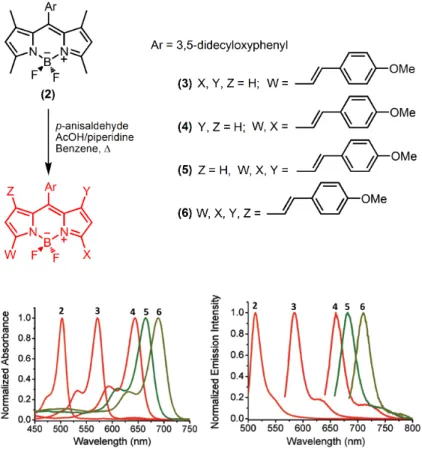
Figure 8. Functionalization of 3,5 and 1,7 positions by Knoevenagel condensation reaction. Copyright © 2009, American Chemical Society. Reprinted with permission from ref (41).41 ... 16
Figure 9. Schematic representation of PeT. Copyright © 2009, Royal Society of Chemistry. Reprinted with permission from ref (62).62 ... 17
Figure 10. Schematic representation of reverse-PeT. ... 18
Figure 11. Some PeT based fluorescent probes. ... 18
Figure 12. Schematic representation of PCT. Copyright © 2007, American Chemical Society. Reprinted with permission from ref (2). ... 19
Figure 13. Some PCT based probes. ... 20
Figure 14. 1-methyl-1,2,3,4,5-pentaphenylsilole (13) as an AIE probe. ... 21
Figure 15. Molecular structures of bio-thiols. ... 22
Figure 16. Maleimide substituted probes for bio-thiol detection. ... 23
Figure 17. Coumarin and fluorescein based Michael addition mechanism for selective detection of bio-thiols. ... 24
Figure 18. Intracellular detection of Cys via Michael addition. ... 25
Figure 20. A FRET based chemodosimeter. ... 26
Figure 21. Coumarin based detection of bio-thiols via cyclization mechanism. ... 27
Figure 22. Conjugate addition and cyclization approach for Cys detection. ... 28
Figure 23. Cleavage of sulfonate ester bonds with bio-thiols. ... 29
Figure 24. BODIPY based thiol-halogen substitution. ... 29
Figure 25. Disulfide exchange reaction mechanism on a naphthalimide derivative. . 30
Figure 26. Metal complexation approach for the detection of bio-thiols. ... 31
Figure 27. Molecular structures of Photofrin and Foscan. ... 33
Figure 28. Modified Jablonski diagram. ... 36
Figure 29. Heavy atom mediated ISC. ... 40
Figure 30. Near-IR absorbing, water-soluble BODIPY sensitizer. ... 41
Figure 31. Charge transfer mediated ISC. ... 43
Figure 32. BODIPY based exciton-coupling behavior. ... 43
Figure 33. BODIPY-C60 conjugate. Copyright © 2012, American Chemical Society. Reprinted with permission from ref (127).127 ... 44
Figure 34. GSH activated phthalocyanine sensitizer. ... 46
Figure 35. GSH activated graphene oxide-chlorine 6 conjugate for PDT. Copyright © 2012, Royal Society of Chemistry. Reprinted with permission from ref (136). ... 47
Figure 36. Some common molecules with axial chirality. ... 48
Figure 37. Schematic representation of axially chiral plane. ... 48
Figure 38. Schematic representation of atropisomerism. ... 50
Figure 39. Nomenclature of atropisomeric compounds. ... 51
Figure 40. Enantioselective detection of α-Hydroxycarboxylic acids. ... 52 Figure 41. Boron asymmetry on a BODIPY dye. Copyright © 2010, American
Chemical Society. Reprinted with permission from ref (148).148 ... 53
Figure 42. Molecular structure of binaphthalene-BODIPY conjugate. ... 53
Figure 43. Axially chiral BODIPY derivatives. ... 54
Figure 44. Synthesis of aromatic endoperoxides. ... 55
Figure 45. Thermolysis pathways for aromatic endoperoxides. ... 56
Figure 46. Activation parameters for the thermolysis of various aromatic endoperoxides and singlet oxygen generation yields. ... 57
Figure 47. Formation and decomposition of naphthalene based endoperoxides. ... 58
Figure 48. Temperature dependence of anthracene (51) thermolysis. Copyright © 2008, Elsevier. Reprinted with permission from ref (160). 160 ... 58
Figure 49. Comparative LSPR peaks of nano-sphere, shell and rod. Copyright © 2010, John Wiley and Sons. Reprinted with permission from ref (166).166 ... 60
Figure 50. Preparation of gold NRs according to El-Sayed’s seed mediated synthesis. Copyright © 2009, John Wiley and Sons. Reprinted with permission from ref (164)164 ... 61
Figure 51. Control of plasmonic properties by adjusting the aspect ratio of NRs. Copyright © 2013, Royal Society of Chemistry. Reprinted with permission from ref (165) 165 ... 64
Figure 52. Selective photothermal therapy of cancer cells with anti-EGFR/Au. At 80 mW cancer cells (HSC and HOC) are injured while there is no effect on healthy cells (HaCat) Copyright © 2006, American Chemical Society. Reprinted with permission from ref (183). ... 65
Figure 53. Schematic illustration of gold nanorods-PDT complex for NIR imaging and PDT. Copyright © 2011, American Chemical Society. Reprinted with permission from ref (184). ... 66
Figure 54. The structure and the signal modulation sites of the target probe. The distance between the terminal amine and the thiol suggests a much better match for GSH and the probe, than the other two biological thiols. Copyright © 2014, American Chemical Society. Reprinted with permission from ref (185). ... 69 Figure 55. Synthetic route for the synthesis of GSH probe (Dye 1). Copyright ©
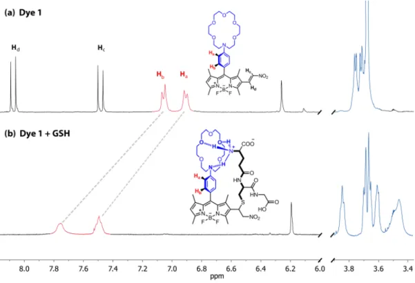
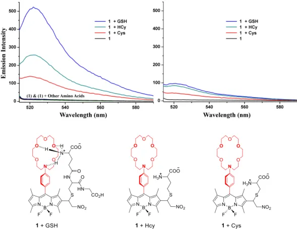
2014, American Chemical Society. Reprinted with permission from ref (185). 70 Figure 56. Absorbance spectra of (Dye 1) (8.0×10⁻⁶ M) and (Dye 1) + Thiols (200 equivalents) at pH 6.0 in 60% MES Buffer (30 mM) / 40% MeCN and at pH 7.4 in 60% MOPS Buffer (30 mM) / 40% MeCN. Copyright © 2014, American Chemical Society. Reprinted with permission from ref (185). ... 71 Figure 57. Partial 1H-NMR (in CD3OD, 298 K) spectra depicting the changes on GSH conjugate addition to the (Dye 1): The adduct was isolated by preparative HPLC following a room temperature reaction of the probe and GSH in aqueous acetonitrile. Trans-coupled protons disappear in the product, and the azacrown peaks show a more spread out cluster of peaks, suggesting an emergence of non-covalent, non-symmetric interaction. Copyright © 2014, American Chemical Society. Reprinted with permission from ref (185). ... 72 Figure 58. Emission response to biological thiols at two different pH values, 7.4 (left, 60:40, 30 mM MES buffer/acetonitrile) and 6.0 (right, 60:40, 30 mM MOPS buffer/acetonitrile). Small change in pH causes more than 5-fold increases in emission intensity for the GSH – (Dye 1) adduct. For Cys and Hcy, the change is approximately 2.5 fold. The concentrations of the thiols and other amino acids (Gly, His, Ile, Leu, Met, Phe, Pro, Ser, Thr, Tyr, Val) were 1.6 mM and the (Dye 1) concentration was 8.0 µM. Excitation was at 500 nm, with 5 nm slit-widths. Copyright © 2014, American Chemical Society. Reprinted with permission from ref (185). ... 73 Figure 59. Selective emission response of the (Dye 1). Blue bars correspond to the emission enhancement ratios (Io being the emission intensity of the probe) when biological thiols are introduced at their respective intracellular concentrations79,196,197 in aqueous medium (60:40, pH 6.0, 30 mM MOPS buffer/acetonitrile) with 8.0 µM (Dye 1) concentration. Red bars show the emission enhancement when all analytes are introduced at 1.6 mM, in the same solvent system and probe concentration. Excitation was at 500 nm, emission data at 522 nm (I and I0) were used in calculations. Copyright © 2014, American Chemical Society. Reprinted with permission from ref (185). ... 74 Figure 60. Electronic absorption and emission spectra of (Dye 1) (8.0 µM) in 30 mM MES:CH3CN (60:40, v/v, pH=6.0, 25 °C) in increasing GSH concentrations (0 to 500 eq.). Excitation wavelength is 500 nm. Copyright © 2014, American Chemical Society. Reprinted with permission from ref (185). ... 75 Figure 61. Following synthetic route was pursued to get (Dye 3). Copyright © 2014, American Chemical Society. Reprinted with permission from ref (185). ... 75
Figure 62. The structures of control BODIPY (Dye 2) and (Dye 3). (Dye 2) has a binding site for ammonium moiety, but no reactive group for thiols. (left) (Dye 3) is thiol reactive, but it does not have a site for ammonium recognition. (right) Dye concentrations were 8.0 µM, and the biological thiols were introduced at 1.6 mM. Aqueous buffer solutions were used as solvents, for pH 7.4 (60:40, 30 mM MES buffer/acetonitrile) and for pH 6.0 (60:40, 30 mM MOPS buffer/acetonitrile). Excitation was at 500 nm, with 5 nm slit-widths. Copyright © 2014, American Chemical Society. Reprinted with permission from ref (185). ... 76 Figure 63. Time-lapse confocal microscopy pictures of Human breast adenocarcinoma cells (MCF-7) cells incubated with (Dye 1) at 0.5 µM (a: 0 min, b: 1 min, c: 120 min). d, e, 120 min, fluorescence, optical images and f: merged image. Human umbilical vein endothelial cells (HUVEC) cells pre-treated with 5 mM BSO (g), DIC image (h), merge (i). The selective inhibition of GSH synthesis reduces fluorescence emission from the cells to a very low level, attesting the selectivity of the designed probe. Copyright © 2014, American Chemical Society. Reprinted with permission from ref (185). ... 77 Figure 64. Frontier molecular orbitals. HOMO (left) LUMO (right), of the parent BODIPY core at B3LYP/6-31G(d,p) level of theory. Surfaces are plotted at 0.2 a.u. Copyright © 2011, John Wiley and Sons. Reprinted with permission from ref (121). ... 88 Figure 65. Orthogonal BODIPY designs. Copyright © 2011, John Wiley and Sons. Reprinted with permission from ref (121). ... 89 Figure 66. Structures of the dimeric Bodipys. a) X-ray diffraction structure of the orthogonal 8,2’ dimer (3). b) Optimized geometry of orthogonal 8,8’ dimer (6) at CAS(6e in 6o)/cc-pVDZ level. Dihedral angle between the two Bodipy units is very close (± 0.5o) to 90o in both dimers. Copyright © 2011, John Wiley and Sons. Reprinted with permission from ref (121). ... 89 Figure 67. Building up bis-BODIPY S1 wave function. MO energies are ... 90 Figure 68. Left: (Top) BODIPY HOMO and LUMO. (Bottom) Frontier orbitals and natural orbital occupation numbers of S1 states of dimers (3) and (6). Right: Leading configurations of S0, S1, and T1 CASSCF wave functions for (3) and (6). Copyright © 2012, American Chemical Society. Reprinted with permission from ref (227). ... 91 Figure 69. Synthesis of the target photosensitizers. a) POCl3, DMF, ClCH2CH2Cl,
50oC; b) 2,4-dimethylpyrrole, trifluoroacetic acid (TFA), CH2Cl2, p-chloranil, Et3N, BF3·OEt2; c) TFA, CH2Cl2, p-chloranil, Et3N, BF3·OEt2. Copyright © 2011, John Wiley and Sons. Reprinted with permission from ref (121). ... 92 Figure 70. Singlet oxygen phosphorescence with sensitization from BODIPY derivatives: (3) (blue), (6) (black) and (7) (red), (1) (green) in CHCl3 at equal absorbances (0.2) at the peak wavelength of their respective absorbances. Copyright © 2011, John Wiley and Sons. Reprinted with permission from ref (121). ... 93 Figure 71. Comparative singlet oxygen generation experiment. Absorbance decrease of DPBF at 414 nm with time in dichloromethane in the presence of BODIPY photosensitizers: (3) (black), (6) (blue), (7) (red) and reference photosensitizer methylene blue (green). Copyright © 2011, John Wiley and Sons. Reprinted with permission from ref (121). ... 94 Figure 72. Singlet oxygen generation experiment in aqueous solution. Decrease in Absorbance spectrum of trap molecule (anthracene derivative) in the presence of 1.69 µM compound (3) in phosphate buffer saline (PBS). Details are given in singlet oxygen measurements part. Copyright © 2011, John Wiley and Sons. Reprinted with permission from ref (121). ... 95 Figure 73. Photocytotoxicity of the sensitizer (3) as demonstrated by MTT assay. Cell suspensions (K562, human erythroleukemia cells) were seeded in 96-well flat-bottom plates and varying concentrations of the sensitizers were added into each well. Cells were kept either in dark, or under illumination with a green (520 nm) LED array at 2.5 mW/cm2 fluence rate for a period of 4 h at 370C in a humidified incubator containing 5% CO2. Copyright © 2011, John Wiley and Sons. Reprinted with permission from ref (121). ... 96 Figure 74. Photocytotoxic activity of the dimeric BODIPY (3) visualized via confocal microscopy. Cell suspensions (K562, human erythroleukemia cells) were seeded in 24-well plates. a, Cells in Control 1 (upper plates) wells were incubated in dark for 24 hours; b, Cells in Control 2 wells (second row plates) were incubated with 500 µl/well Cremophor-EL solubilized (3) (at a final concentration of 164 nM) and kept in dark for 24 hours in the same incubator, c, Cells in Control 3 well were illuminated for 4 h without the sensitizer and incubated for a further 20 h in dark at the incubator. d, Cells in Control 4 (bottom row of plates) well were illuminated for 4 h after addition of 164 nM (3) and incubated for a further 20 h in dark at the incubator. Cells in wells were collected after 24 h and imaged at 40x magnification. Live cells are preferentially stained with acridine orange (AO, green) (a), whereas dead cells
are preferentially stained with the dye propidium iodide (PI, red) due to increased cellular permeability (b). Copyright © 2011, John Wiley and Sons. Reprinted with permission from ref (121). ... 97 Figure 75. Effect of inter-plane angle (θ) variation on S0→S1 excitation characteristics (DS-TR/SS). Copyright © 2012, American Chemical Society. Reprinted with permission from ref (227). ... 98 Figure 76. Synthesis of (bis-10a’). Copyright © 2012, American Chemical Society. Reprinted with permission from ref (227). ... 99 Figure 77. Electronic absorption spectrum of (bis-10a’). Copyright © 2012, American Chemical Society. Reprinted with permission from ref (227). ... 99 Figure 78. The set of monomers and dimers studied. Copyright © 2012, American Chemical Society. Reprinted with permission from ref (227). ... 100 Figure 79. Selected natural orbitals and occupation numbers for (bis-9), (bis-10) and (bis-10a). Calculations were done at CAS(6,6)/CEP-31G//(U)B3LYP/CEP-31G level. Copyright © 2012, American Chemical Society. Reprinted with permission from ref (227). ... 103 Figure 80. Relation of DS-TR character of S1 to the extent of π conjugation in butadienyl- and styryl-substituted BODIPY dimers (bis-9′) and (bis-10′). Copyright © 2012, American Chemical Society. Reprinted with permission from ref (227). ... 104 Figure 81. Size distribution of compound micellar compound (3). Cremophor EL was used for embedding the dye. Copyright © 2011, John Wiley and Sons. Reprinted with permission from ref (121). ... 109 Figure 82. Reaction of singlet oxygen with 1,3-Diphenylisobenzofuran. ... 110 Figure 83. Reaction of singlet oxygen with
2,2'-(Anthracene-9,10-diylbis(methylene))dimalonic acid. ... 110 Figure 84. Decrease in absorbance of DPBF in dichloromethane in the presence of compound (3) in medium. Details are given in singlet oxygen measurements part. Copyright © 2011, John Wiley and Sons. Reprinted with permission from ref (121). ... 111 Figure 85. Decrease in absorbance of DPBF in dichloromethane in the presence of compound (6) in medium. Details are given in singlet oxygen measurements
part. Copyright © 2011, John Wiley and Sons. Reprinted with permission from ref (121). ... 111 Figure 86. Decrease in absorbance of DPBF in dichloromethane in the presence of compound (7) in medium. Details are given in singlet oxygen measurements part. Copyright © 2011, John Wiley and Sons. Reprinted with permission from ref (121). ... 112 Figure 87. Decrease in absorbance of DPBF in dichloromethane in the presence of MB in medium. Details are given in singlet oxygen measurements part. Copyright © 2011, John Wiley and Sons. Reprinted with permission from ref (121). ... 112 Figure 88. Packing diagrams of compound (3). Dashed lines indicate inter H-bond between C2 and F4 atoms. Copyright © 2011, John Wiley and Sons. Reprinted with permission from ref (121). ... 113 Figure 89. Design principles (top) and Orbital energies (eV) and plots of (PS) and (actPS) relevant to the CT excitation from BODIPY to methylpyridinium (bottom). ... 119 Figure 90. Synthesis of activatable sensitizer (PS). ... 121 Figure 91. Reduction of compound (PS) in the presence of mercaptoethanol. 1H NMR spectra of (PS) and (actPS). HR-MS spectrum of (actPS) (Δ (ppm) = 4.9 ppm). ... 122 Figure 92. Normalized electronic absorption spectra of (dimBDY), (pydimBDY), (PS) and (actPS) in DCM. ... 123 Figure 93. Relative 1O2 generation efficiency of top: (dimBDY), (PS) and (actPS) in DCM and bottom: (PS) and (actPS) in 90:10 (Hepes buffer, pH 7.2, 20mM : MeCN) detected by the absorbance decrease of DPBF at 414 nm and 2'-(Anthracene-9,10 diylbis(methylene))dimalonic acid (ADMDA) at 378 nm respectively with time. During first 60 seconds (right) and 15 minutes (left), the samples were kept in the dark. ... 124 Figure 94. Left: The T1-Tn transient absorption spectra of (actPS) in argon saturated THF and MeCN with laser excitation at 355 nm (the absorbance at 355 nm is 0.402). Right: The decay of triplet state T1 of (actPS) at 425 nm in argon saturated THF and MeCN with laser excitation at 355 nm (the absorbance at 355 nm is 0.402). The triplet lifetimes are 8.7 µs (MeCN) and 27.6 µs (THF). ... 125
Figure 95. Comparison of electronic absorption spectrum of left: (PS) (30µM) and (PS) (30µM)+ GSH (5mM), right: (PS) (25µM) and (PS) (25µM) + Cys (25µM) & Hcy (25µM) in 90:10 (Hepes buffer, pH 7.2, 20mM : MeCN). Reaction time is 12 hours in all cases. ... 127 Figure 96. Photocytotoxicity of the sensitizer (PS) (blue) and (actPS) (red) as demonstrated by MTT assay. Cells were kept either in the dark (bottom), or under illumination with a green (520 nm) LED array (top). ... 128 Figure 97. Cytotoxic effects of sample (PS) (blue), (actPS) (red). Cells were incubated with (PS), (actPS) for 4 hr and irradiated with green LED. Cytotoxic effects were examined by MTT assay. Normal cell lines : NIH 3T3, WI38 VA13, cancer cell lines : HeLa, SK Hep 1. ... 128 Figure 98. The HeLa cells were incubated with (PS), (actPS) for 4 hrs. Cells were irradiated with green LED for 30 min and incubated another 3 hrs or kept under dark and stained with Annexin V-AF594 (apoptosis marker). Fluorescence images were acquired by confocal microscopy. For nucleus stain, 1 µg/ml DAPI costained for 30 min. (a) DAPI (ex. 405 nm, em. 430-455 nm), (b) (PS), (actPS) (ex. 473 nm, em. 490-590 nm), (c) Annexin V-AF594 (ex. 559 nm, em. 575-675 nm), (d) DIC, (e) Merge. Scale bar: 10 µm ... 129 Figure 99. Michael addition of mercaptoethanol to compound (PS) in the absence of base in aqueous solution (90:10 (HEPES buffer, pH 7.2, 20mM : MeCN)). HR-MS spectrum of (Michael adduct) (Δ (ppm) = 31.8 ppm). ... 130 Figure 100. The normalized fluorescence emission spectra of (dimBDY), (pydimBDY), and (PS) in DCM. ... 132 Figure 101. The normalized fluorescence emission spectra of (actPS) in different solvents with excitation at 418 nm (absorbance 0.090). ... 132 Figure 102. Fluorescence decay of (actPS) in EtOH with excitation at 509 nm diode laser (150 ps), the emission was monitored at 520 nm, the concentration of dyes is ca. 3.0 µM. ... 133 Figure 103. Decrease in absorbance of DPBF in DCM in the presence of (dimBDY) (1X10⁻⁶ M). Excitation @508 nm. ... 135 Figure 104. Decrease in absorbance of DPBF in DCM in the presence of (PS) (1X10
-6 M). Excitation @508 nm. ... 135 Figure 105. Decrease in absorbance of DPBF in DCM in the presence of (PS) (1X10
-6 M). Excitation @607 nm. ... 135 Figure 106. Decrease in absorbance of ADMDA in 90:10 (20 mM Hepes buffer:MeCN) in the presence of (PS) (1X10⁻⁶ M). Excitation @513 nm. ... 136 Figure 107. Decrease in absorbance of ADMDA in 90:10 (20 mM Hepes buffer:MeCN) in the presence of (PS) (1X10⁻⁶ M). Excitation @590 nm. ... 136 Figure 108. Decrease in absorbance of DPBF in DCM in the presence of (actPS) (1X10⁻⁶ M). Excitation @511 nm. ... 136 Figure 109. Decrease in absorbance of ADMDA in 90:10 (20 mM Hepes buffer:MeCN) in the presence of (5r) (1X10⁻⁶ M). Excitation @508 nm. ... 137 Figure 110. Synthesis of the atropisomeric BODIPY derivative (4) and (5). As expected, (4) and (5) are produced as racemates. For both (4) and (5) (R) enantiomers are depicted. Copyright © 2014, American Chemical Society. Reprinted with permission from ref (150). ... 144 Figure 111. HPLC separation of compound (4) to (4a) (20.3 min) and (4b) (33.4 min), (95% / 5 %, heptane / IPA, detection @270 nm) (top), and compound (5) to (5a) (18.5 min) and (5b) (20.6 min), (98% / 2%, heptane / ethanol, detection @510 nm) (bottom). Copyright © 2014, American Chemical Society. Reprinted with permission from ref (150). ... 145 Figure 112. Electronic absorption (red) and emission (blue) spectra of (4) (left) and (5) (right). Ext @ 500 nm for (4) and 510 nm for (5). Copyright © 2014, American Chemical Society. Reprinted with permission from ref (150). ... 146 Figure 113. Electronic absorption (red) and emission (blue) spectra of (4a)(left) and (4b) (right). Ext @ 500 nm. Copyright © 2014, American Chemical Society. Reprinted with permission from ref (150). ... 146 Figure 114. Electronic absorption (red) and emission (blue) spectra of (5a) (left) and (5b) (right). Ext @ 510 nm. Copyright © 2014, American Chemical Society. Reprinted with permission from ref (150). ... 146 Figure 115. Circular dichroism spectra of racemic mixture (4) and the two enantiomers following resolution using Chiralcel-OD columns via preparative HPLC. The structures shown on the figure are the energy-minimized structures (employing the MM method as implemented in Spartan 08) and the assignment is arbitrary. Copyright © 2014, American Chemical Society. Reprinted with permission from ref (150). ... 147
Figure 116. Circular dichroism spectra of racemic mixture (5) and the two enantiomers following resolution using Chiralcel-OD columns via preparative HPLC. The structures shown on the figure are the energy-minimized (employing the MM method as implemented in Spartan 08) structures and the assignment is arbitrary. Copyright © 2014, American Chemical Society. Reprinted with permission from ref (150). ... 147 Figure 117. Electronic absorption (solid line) and emission (dashed line) spectra of compounds (4) and (5). Copyright © 2014, American Chemical Society. Reprinted with permission from ref (150). ... 148 Figure 118. Therapeutic design. ... 156 Figure 119. TEM images of gold nanorods (~40 nm) and electronic absorption spectrum of GNRs in HEPES buffer (pH 7.2, 20 mM). ... 158 Figure 120. Reaction scheme for the target compound. ... 158 Figure 121. Top: Absorption spectra that is taken during the endoperoxide formation reaction. Bottom: Thermolysis of endoperoxide (8) to parent compound (7). 159 Figure 122. Left: Thermolysis of 5x10⁻⁵ M (9) in DMSO at different temperature. Right: Absorbance vs. temperature graph of the thermolysis. The time of heating was 30 min for all selected temperatures. ... 160 Figure 123. Thermolysis of 5x10⁻⁵ M (9) in DMSO at 95oC for 6 hours. Inset: Absorbance of 5x10⁻⁵ M (9) in DMSO at 37oC. Sample was heated for 6 hours. ... 161 Figure 124. Thermolysis of 1x10⁻⁵ M (9) at 100oC for 1 hr in HEPES buffer (pH 7.2, 20 mM). ... 161 Figure 125. Decrease in absorbance of DPBF in DMSO in the presence of 1x10⁻⁵ M upon heating at 70oC (9) and the reappearance of parent anthracene absorbance bands. ... 162 Figure 126. Left: TEM image of GNR-(9) conjugate. Right: Normalized electronic absorption spectra of CTAB-GNR, (9)-GNR(in HEPES (pH 7.2, 20 mM) and (9)-GNR in DMSO. ... 162 Figure 127. Decrease in absorbance of DPBF at 414 nm in DMSO in the presence of (9)-GNR upon heating in oil bath. ... 163 Figure 128. Decrease in absorbance of DPBF at 414 nm in DMSO in the presence of
(9)-GNR. Excitation @830 nm. ... 163 Figure 129. Absorbance of DPBF in DMSO in the presence of GNRs only (0.125 nM). Excitation @830 nm. ... 164 Figure 130. Relative 1O2 generation efficiency of (9)-GNR and GNR only in DMSO detected by the absorbance decrease of DPBF @414 nm with time. During first 4 min the samples were kept in the dark. ... 164
LIST OF TABLES
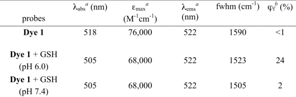
Table 1. FDA approved PDT drugs.93,107 ... 34 Table 2. Selected photophysical parameters for the (Dye 1) and its conjugate adduct.
... 71 Table 3. Comparative spectroscopic properties of BODIPY compounds. ... 93 Table 4. Photophysical Properties of the Synthesized BODIPY Compounds. ... 100 Table 5. Selected Parameters for Modified BODIPY Cores and Associated Orthogonal Bis-BODIPYs. ... 102 Table 6. Relative Energies (eV) of S2 and S3 States for Selected Species at CAS(6,6)/CEP-31G level of theory. ... 105 Table 7. Photophysical characterization of sensitizers. ... 123 Table 8. The quantum yields for T1 state formation of (actPS) in different solvents
... 126 Table 9. Fluorescence quantum yield (Φf) and lifetime (τf) of (actPS) in different
solvents. ... 133 Table 10. Photophysical characterization of compound (4) and (5). ... 148
CHAPTER 1
1.
INTRODUCTION
Cancer appears to be one of the most challenging health problems faced by large amount of people all around the world. Number of patients is expected to reach more extended life spans in the coming years. State-of-the-art diagnostic tools and treatments must emerge in order to have more effective results, decreased invasiveness, fewer side effects and more patient compliance with respect to the conventional methods (chemotherapy and radiotherapy).
Conventional imaging methods for diagnosis applications mostly rely on contrast agents. These agents emit signal continuously regardless of target cell (“always on”). As a natural consequence of this, sensitivity cannot be achieved sufficiently due to the low target to background signal ratios. At that point, molecular optical imaging by means of fluorescent probes arises as a good alternative. These probes can be designed in such a way that they become active only under certain conditions (“turned on”). Thus, signal from well-designed probe can only be detected after selective interaction between target cells and probe that increases the specificity and selectivity. This improves target to background ratio and provides opportunity for detection of even very small tumours. Furthermore, molecular fluorescence imaging offers simple and cheap instrumentation as well as real time analysis.
Design, synthesis and characterization of molecular fluorescent probes, which are selective to various diseases marker is the key of targeted fluorescent imaging. At that point, BODIPY (4,4-difluoro-4-bora-3a,4a-diaza-s-indacene) dyes with their high fluorescence quantum yields/absorption coefficients, tunable absorption/fluorescence peaks, ease of synthetic methods and various sides for functionalization were most widely employed in fluorescence imaging studies during
the last decade. Therefore, in chapter 3, we introduced a BODIPY based glutathione selective turn-on fluorescent probe. Levels of biological thiols such as cystein (Cys), homocystein (Hcy) and glutathione (GSH) in living systems are known to be important parameters in health and sickness. Since their presences are vital for the maintenance of cellular redox status and alterations in their levels is linked to a number of debilitating diseases, probes that respond to these thiols by color change, emission wavelength change, or both are highly valued. Additionally, level of GSH in cancer cells is 2-50 fold higher than health cells, which makes GSH a promising target for cancer diagnosis. A vast amount of examples in which Cys and/or HCY are selectively detected and monitored appeared in the literature, however there are only few examples for GSH probes. This is the result of limited design principles for bio-thiols detection, which are mainly based on Michael addition, cyclization and cleavage of the disulfide bond. Structural differences between GSH and other two bio-thiols make GSH monitoring a challenging task. In chapter 3, for the purpose of selective GSH sensing, we thought that two binding sites on a probe are essential. We modified the BODIPY core with a crown ether moiety at meso position. Crown moiety is the modulation site for photo-induced electron transfer (PET) and it is a very well known binding site for the ammonium end of the GSH. In addition to that we incorporated a nitro styryl group on the BODIPY core in order to create a thiol-binding site. In this work, we have shown that selectivity for reaction-based probes can be improved by applying additional photophysical manipulation sites.
Photodynamic therapy (PDT) is a promising candidate for treatment of certain malignant (skin, head and neck, gastrointestinal, gynecological cancers), premalignant (actinic keratosis), and nonmalignant (psoriasis, AMD-age related macular degeneration) indications since it offers non-invasive and activatable therapy alternatives. PDT utilizes light, sensitizer and molecular oxygen to cause cancer cell death via apoptosis or necrosis. The therapeutic action is taking place upon the generation of cytotoxic singlet oxygen through excitation of a particular chromophore (sensitizer) followed by an energy transfer to the dissolved oxygen in tumor tissues.
One of the most substantial parts of the PDT designs is the choice of sensitizer. An effective sensitizer should hold some unique characteristics such as low toxicity in the absence of light, high extinction coefficients, photostability, amphiphilicity and biocompatibility. More importantly, a sensitizer should have high triplet quantum yield such that transition of an excited electron from singlet excited state to triplet excited (inter-system crossing (ISC)) takes place smoothly. Common strategy for achieving efficient ISC is to decorate sensitizers with heavy atoms such as iodine and bromine. However, presence of heavy atoms increases the dark toxicity of sensitizers. We have been interested in finding out some possible different ways of achieving increased intersystem crossing (ISC) without incorporating heavy atoms in order to minimize dark toxicity for photodynamic therapy applications, turning our attention to the excited state properties of the sensitizers. In chapter 4, we are introducing a new excited state concept, which we named it as doubly substituted tetra radical state (DS-TR) by realizing orthogonal arrangement of BODIPY monomers. DS-TR character of S1 was shown to have a strong correlation with S1→T1 ISC yielding 1O2.
Detailed study of DS-TR excited state was also provided by π-extended BODIPY derivatives in Chapter 4. Near-IR absorption, desired for potential photodynamic therapy applications, was not pursuable for bis-chromophores by the standard strategy of π -extension, as DS singlet states are destabilized. Decreased exchange coupling in π -extended cases appears to be responsible for this destabilization. On the other hand, by using the accumulated knowledge of excited state configurations and triplet photosensitization, we further improved our new concept through activatable heavy-atom free PDT sensitization. There is no doubt that targeted sensitizers increase the therapy efficacy and decrease the side effects. To do so, we extended the π-conjugation on the orthogonal dimer core, which removes the DS-TR picture and makes our sensitizer inactive. In the presence of GSH only, a cancer marker, double bond is reduced and the conjugation is broken that yields effective charge transfer mediated ISC. The detailed results of this project have been given in chapter 4.
While we were dealing with orthogonal BODIPY dimers for PDT applications, we recognized the potential of our dimers for atropisomerism. Synthesis, separation and detection of chiral molecules are at the heart of drug discovery studies. If a chiral molecule do have fluorescence property than its application areas can be expanded. For instance, fluorescence enantioselective sensing is one of the most up-to-date and improvable topics among the optical sensing studies due to its potential as a simple detection tool in chiral assays. This detection is quite important for drug discovery, biological labeling, catalysts, and understanding the mechanism of molecular recognition in biological systems. There are number of fluorescent sensors for recognition of chiral amines, carboxylic acids, alcohols and amino acids. Especially biologically important hydroxy acids and amino acids have been attracting much interest.
Rich BODIPY chemistry allows us to design, synthesize and characterize axially chiral BODIPY derivatives. In chapter 5, we presented the first example of atropisomeric BODIPY derivatives with persistent chirality due to multiple methyl group clashes. Circular dichroism studies clearly demonstrate successful separation of the two enantiomers. The orthogonally linked BODIPY compounds have been demonstrated to have interesting photophysical properties but also we show that they can be isolated as atropisomeric chiral compounds.
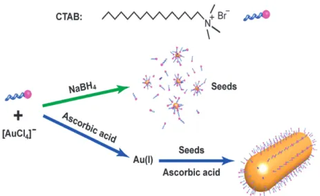
In chapter 6, I switched my attention back to PDT concept. On the way of fight against cancer, nanotechnology that combines life science, medicine, electronics and biomaterials is a good choice of weapon. As a result of increasing nanotechnology based biomedical applications and out coming new properties, there is a growing interest to use inorganic nanoparticles in cancer treatments. Nanomaterials can be used in various therapies (Photothermal, photodynamic therapies), ultrasensitive detection of cancer markers, imaging techniques as contrast agents, carriers for drug delivery, control release, and regenerative medicine. Among possible nanomaterials, gold nanoparticles have many advantages and unique properties that make them suitable for therapy, diagnosis and theranostic (therapy + diagnosis) applications. Biocompatibility, bioinertness, presence of tunable surface plasmon resonance peak,
which covers almost all of the electromagnetic spectrum, well establish surface chemistry for easy functionalization (targeting), compact size and insignificant toxicity are some of the well known properties of gold nanoparticles. It is important to note that; localized surface plasmon resonance (LSPR) wavelength of a gold nanoparticle can be tuned by simply adjusting the size and the shape of the nanoparticle during the chemical synthesis. For instance gold nanorods have red shifted LSPR peaks that span through near-IR region, which make them highly suitable for biomedical studies. Gold nanorods like other types of gold nanoparticles can be heated by external factors such as light (near IR). In fact, since these nanoparticles can be targeted to the tumor tissue, this heating alone is being considered as a viable treatment modality (photothermal therapy).
Applicability of PDT is severely limited by two major factors, penetration depth of the excitation light and hypoxia (low oxygen concentration) in tumor tissues. In chapter 6, we tried to address these restrictions by combining aromatic endoperoxides and gold nanorods. Our primary goal is to deliver the principal cytotoxic agent of photodynamic therapy, singlet oxygen, directly by thermal decomposition of otherwise stable endoperoxides, which are located on gold nanorods and observe cell death due to the thermal and singlet oxygen effects. Near-IR excitation mediated temperature rise on the surface of gold nanorods triggers the thermal decomposition and release of trapped singlet oxygen from endoperoxides. Thus, this design is not using heat as an end product as in the case of classical photothermal therapy. This project, which is a radical rewriting of the photodynamic therapy concept, is poised to revamp photodynamic therapy, offering potential solutions for major problems hitherto blocking broader clinical applications.
CHAPTER 2
2.
BACKGROUND
2.1. Fluorescent Molecular Sensors
2.1.1. General InformationA sensor is a device, which yields a measurable output (signal) upon interacting with matter or energy.1 Sensors are considered only as macroscopic devices (such as pH electrode or thermometer) until the beginning of the nineties.1 Followed by the increased recognition of nanotechnology in sensing applications, properly designed molecules are also appeared to be suitable candidates. Today we have an accepted and well-known term molecular sensor, which is a molecule that signals the presence of an analyte by some physical output. Signaling mechanism of molecular sensors basically involves selective interaction of a molecule with an analyte of interest, which forms measurable form of energy that can be detectable with various simple spectroscopic techniques such as optical tools (e.g., UV, visible, fluorescence), or electrochemical methods (e.g., cyclic voltammetry). Optical sensors utilize light-based detection of analytes and are considered to be useful in practical applications because of their high sensitivity, low cost, and simplicity of instrumentation.1 One type of the optical sensing is the calorimetric sensor, which involves the color change (shift in absorbance spectrum) upon interaction with an analyte. Second and more promising method is to sense analyte concentrations with fluorescent signal transduction and these types of molecular sensors can be referred as fluorescent
sensors. In these type of sensors, photophysical mechanisms that control the
response of a molecular probe mainly include photo-induced electron transfer (PeT), photo-induced charge transfer (PCT), excimer/exciplex formation, Förster resonance energy transfer (FRET), and aggregation-induced emission (AIE).2 These
mechanisms will be discussed briefly in the section 2.3.
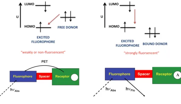
Fluorescent molecular sensor design strategies mostly involve three main approaches:3 (i) binding site-signaling subunit, (ii) displacement approach, and (iii) chemodosimeter designs. In the case of first principle, a molecular sensor contains covalently bonded binding site(s) that is selective to a certain analyte and an optical signaling sub-unit in such a way that the interaction of an analyte with the binding site triggers electronic modulations in the optical signaling unit in the form of fluorescence (emission) change. Second modality (the displacement approach) suggests non-covalent interaction of the binding and signaling sites. Molecular ensemble between these two units is destroyed upon coordination of an analyte to the binding site causes displacement of the binding moiety that yields detectable optical changes. Chemodosimeter designs are realized by the chemical reactions on a molecular probe with specific analytes (a cation, an anion or a molecule) resulting in fluorescence alterations. It involves irreversible bond breaking or formation. Recent studies in the field reveal novel approaches that involve the use of nanoparticles in combination with molecular probes.
Figure 1. Tsien’s Ca2+ probe.4
Fluorescent molecular sensor development has evolved into an attractive field of study after Tsien’s pioneering study4 on fluorescent Ca2+ detection in 1980 (figure 1), followed by large number of promising examples emerging at a steady pace with worldwide participation in this endeavor.5 Major application areas of fluorescent molecular sensors are real time imaging of biological systems/processes and medical diagnosis.6 It is known that living organisms and their environment are composed of elements, ions and molecules, which are constantly forming network of chemical
reactions including acid-base chemistry, electron transfer, metal-ligand interactions and catalytic transformations.6 A molecular level understanding of these processes and detection of rich array of analytes (e.g., ions, molecules) that living organisms provide are real challenges, but also provides great opportunity for researchers to investigate biological systems in their own medium and create new tools especially for medical diagnosis. Towards addressing these challenges, molecular imaging (sensors) offers powerful modality for real time monitoring of living samples with high resolution, quick response and selectivity.
2.1.2. Overview of Fluorescence
Figure 2. The Jablonski diagram.
It is important to review fluorescence concept before getting in to detail with fluorescent molecular probes. The absorption of light by a molecule results in excitation of one electron from ground electronic state (S0) to one of the higher vibrational levels of a singlet-excited state. In a very short time, excited electron relaxes back to lowest vibrational level of the excited state (S1). There are several pathways for an electron to release its stored energy after reaching to S1 state. It can turn back to its ground state by releasing heat only (non-radiative path) or by emitting light, which is known as fluorescence. In addition to these, if the molecule contains suitable modifications and has sufficiently long-lived excited state lifetime, an electron in the singlet excited state can pass to lower energy triplet excited state (inter-system crossing). Relaxation from excited triplet electronic level to ground
state by emitting photons is named as phosphorescence. Time for fluorescence to occur is around nano or microseconds (10-9 - 10-6 s), on the other hand phosphorescence is more time consuming process (10-3 - 102 s) since it involves several transitions.7 Possible deactivation pathways for an excited electron are summarized in the Jablonski diagram (figure 2). One should note that emission wavelength of a molecule is always located at longer wavelengths than absorption wavelength due to the rapid decay of an excited electron to lowest vibrational level S1 (figure 3). This phenomenon is called as Stokes’ shift7 and it is counted as one of the positive characteristics of fluorescent molecular sensors.
Figure 3. Stokes shift.
2.1.3. Fluorescent Probes
Fluorescence is well-accepted optimum signaling mechanism for optical molecular sensors due to some superior properties as mentioned before. Most important characteristic of fluorescent probes (fluorophore) is their extreme sensitivity due to Stokes’ shift that provides different wavelengths for excitation and emission. Thus, analyte detection can be realized by low or almost zero background signals. Fluorescence signal can detect analytes even at picomolar concentrations, whereas with chromogenic probes can detect concentrations as low as micromolar levels (one million times higher).8 It is also important to note that, presence of Stokes’ shift by itself is not sufficient to diminish background signal at some cases. For instance, biological samples have their own autofluorescence signals up to 600 nm. Probes absorbing/emitting at longer wavelengths of electromagnetic spectrum mostly overcome these interferences. So appropriate fluorescent sensors for biological studies should have optimized wavelength range between 600-900 nm so called
therapeutic window.9 Upper limit of this scale is arranged according to water absorption. Thus it is possible to monitor and analyze morphological details in tissues with subcellular resolution both in vitro and in vivo by using non-invasive fluorescence sensing tools. Biochemistry and molecular biology applications such as medical diagnosis, DNA sequencing and bioassays regularly take advantage of fluorescent molecular sensors in daily life practices. Another useful property of this optical method is the widespread availability of instrumentation that requires minimal maintenance. An additional advantage of fluorescence sensing is that detection of analytes can be performed via time resolved measurements.
Basic requirements for a fluorescent molecular probe can be summarized as follows;1 (i) the binding site must have high selectivity for the analyte of interest (ii) there must be signal transduction mechanism between probe and analyte (iii) sensor-analyte conjugate should be bio-compatible and stable in physiological medium (iv) out-coming signal should not be affected by environmental factors (e.g., pH and fluorescent quenchers) (v) irradiation and detection instrumentation have to be user friendly and cheap. It must be emphasized that it is really hard task to combine all of these properties in one probe and requirements may differ according to end-use of a molecular senor.
There are many fluorophores, which are employed in fluorescent detection of certain analytes. Endogenous molecules are an important class. For instance, aromatic amino acids including phenylalanine and tyrosine are weakly emissive compounds.10 Tryptophan (Trp) shows the highest fluorescence among amino acids at 353 nm under UV excitation with fluorescence quantum yield of 0.13.11 Trp can be used to monitor protein folding and ligand binding processes as well as it can be included in FRET12 and PeT13 applications. Other and maybe the most popular naturally occurring fluorescent dye is porphyrin, which has tunable absorption and emission peaks through visible and red region.
Synthetic small molecules such as cyanine, fluorescein, perylene, coumarin, BODIPY and rhodamine are well known and extensively studied fluorescent probes (figure 4), which are applied in many applications including anion, cation and
biologically relevant molecules detection, food analysis, environmental monitoring, medical diagnosis and many other disciplines.14
Figure 4. Molecular structures of common fluorescent probes.
For all of these probes, it is possible to tune absorption and emission wavelengths, however coumarin dyes mostly known as UV probe whereas fluorescein and rhodamine derivatives are well recognized by their strong emission at near-IR region. Suitable chemical modifications that can be done on these dyes favor the incorporation of analyte binding side according to particular purposes. Different classes of dyes show unique properties. For instance coumarin dyes have very large Stokes’ shift, which makes them suitable for biological applications. Most popular coumarin dyes are heteroatom substituted ones. Fluorescein dyes after their first synthesis in 187115 have widely been used in chemosensor designs. It has very high fluorescence quantum yield, water solubility and it also possesses versatile modification sides for many different applications. However, this class of dyes have very high rate of photo-bleaching and pH sensitivity. On the other hand, rhodamine dyes, another well-studied fluorescent probes offer very low pH sensitivity and tunable absorption/emission peaks.16 Cyanine dyes are constructed with two polymethine chains between nitrogens. They are intensively used as DNA stains and
membrane sensors.17 Intramolecular chain twisting makes cyanine dyes flexible and alters their fluorescence properties. Fluorescent probes based on perylene dyes have strong absorption in visible range and high fluorescent quantum yield but difficult derivatization restricts their wide application.18 BODIPY dyes will be discussed in the following section separately since they will show up extensively throughout this dissertation.
2.2. BODIPY Dyes
BODIPY (4,4-difluoro-4-bora-3a,4a-diaza-s-indacene) dye was synthesized for the first time accidently in 1968 while Treibs and Kreuzer were trying to react 2,4-dimethylpyrrole with acetic anhydride in the presence of BF3.OEt2.19 After 17 years, in 1985 Hauglang and Kang reported the fluorescent characteristics of BODIPY dyes.20 Since then BODIPY dyes have been well recognized as fluorescent molecular sensors and employed intensively for bio-labeling applications.21-23
Figure 5. Molecular structures of BODIPY and its precursors.
BODIPY core can be obtained after two consecutive steps.24 In general, dipyrromethene group is obtained by combining two pyrrole moieties with a methine bridge. After that, dipyrromethene unit is coordinated with a disubstituted (mostly a BF2 group) boron atom, which completes the formation of rigid and constrained core structure. Simple BODIPY core structure, its IUPAC numbering and structures for BODIPY precursors are given in figure 5.
There are three different synthetic pathways for the synthesis of unsubstituted BODIPY dyes. According to Bruce et al. procedure,25 after the formation of dipyrromethane core, it is oxidized by DDQ in order to obtain dipyrromethene
structure followed by the addition of tertiary amine base and BF3.OEt2. Second pathway involves the acid-catalyzed condensation of unsubstituted pyrrole and pyrrole-2-carbaldehyde just before the addition of base and BF3.OEt2 complexation.26 In the final procedure, Pene-Cabrera and co-workers introduced the palladium-catalyzed reaction of 8-thiomethyl BODIPY and triethylsilane in the presence of copper(I) thienyl-2-carboxylate (CuTc) with very high yield.27
Figure 6. Synthetic pathways for meso-functionalized symmetric BODIPYs.
Symmetric BODIPY dyes with meso (8-position) functionalization are synthesized by acid-catalyzed condensation of excess pyrroles with aldehydes, acid chlorides or anhydrides in the presence of amine base and BF3.OEt2 (figure 6).28,29 When aldehydes are employed in condensation, initially dipyrromethane intermediate is formed which is then oxidized to dipyrromethene by using quinone derivatives. Substituted BODIPY dyes at meso position are widely used due to their advantageous characteristics with respect to unsubstituted analogues. They provide high stability, enhanced solubility in organic solvents and free functional groups for further modifications depending on the end-use.
It is also possible to synthesize meso-unsubstituted symmetric BODIPY derivatives by the condensation of excess pyrrole derivatives (~2.2 eq) with orthoester.30 Initially dipyrromethene intermediate is formed and afterwards base and
borontrifloride are added to get BODIPY skeleton. Burgess and co-workers propose another possible mechanism in which pyrrole-2-carbaldehyde is used together with POCl3 without any need for excess pyrrole.31 Synthetic pathways for both strategies are given in figure 7. On the other hand, in the case of asymmetric BODIPY synthesis, condensation of pyrrole-2-carbaldehyde with other pyrrole derivatives has to be carried out.32
Figure 7. Synthetic pathways for meso-unsubstituted symmetric BODIPYs.
The parent BODIPY unit has a major absorption peak (S0-S1 transition) near 500-520 nm with moderate to high absorption coefficients (40.000 – 100.000 M-1 cm-1). One should also note that in the absorption spectrum of a BODIPY dye, it is very common to observe a weak peak at high-energy region around 370 ± 10 nm that corresponds to S0-S2 transition.33 A sharp and strong emission peak due to the relaxation of an excited electron from S1 state is mostly observed between 530-550 nm suggesting approximately 30 nm Stokes’ shift. BODIPY dyes have very high fluorescence quantum yield (φf > 0.50) and long fluorescence lifetimes (in the order of nanoseconds). Phosphorescence is very rare since this class of dye normally have very low triplet quantum yields.34 They have high thermal and photostability as well as physiological medium compatibility.24
Simple BODIPY core has some drawbacks such as solubility in aqueous solution, absence of functional groups for further modifications and low absorbance/emission wavelengths for biomedical applications. These challenges can be addressed by suitable derivatization of the core at meso, 3-5, 1-7, 2-6 and boron center.24 Meso (8) functionalization is straightforward since it can be realized by just choosing the