POLYMORPHISMS IN P21 (CODON 31) AND P53 (CODON 72): ASSOCIATION WITH BREAST CANCER SUSCEPTIBILITY
IN THE TURKISH AND GREEK POPULATIONS
A THESIS SUBMITTED TO
THE DEPARTMENT OF MOLECULAR BIOLOGY AND GENETICS AND
THE INSTITUTE OF ENGINEERING AND SCIENCE OF BILKENT UNIVERSITY
IN PARTIAL FULFILLMENT OF THE REQUIREMENTS FOR THE DEGREE OF MASTER OF SCIENCE
BY
GÜLSEN ÇOLAKOĞLU AUGUST, 2003
I certify that I have read the thesis, and that in my opinion it is fully adequate, in scope and in quality, as a thesis for the Master of Science.
Assoc. Prof. Dr. Uğur ÖZBEK
I certify that I have read the thesis, and that in my opinion it is fully adequate, in scope and in quality, as a thesis for the Master of Science.
Assoc. Prof. Dr. Tayfun ÖZÇELİK
I certify that I have read the thesis, and that in my opinion it is fully adequate, in scope and in quality, as a thesis for the Master of Science.
Asst. Prof. Dr. Işık G. YULUĞ
Approved for the Institute of Engineering and Science
Prof. Dr. Mehmet BARAY Director of the Institute of Engineering and Science
ABSTRACT
POLYMORPHISMS IN P21 (CODON 31) AND P53 (CODON 72): ASSOCIATION WITH BREAST CANCER SUSCEPTIBILITY
IN THE TURKISH AND GREEK POPULATIONS
Gülsen ÇOLAKOĞLU
Ms. in Molecular Biology and Genetics Supervisor: Asst. Prof. Dr. Isık G. YULUĞ
August 2003, 121 pages
The aim of this study was to investigate the potential association of p53 codon 72 and/or p21 codon 31 polymorphisms with increased susceptibility for breast cancer either independently or combined in the Turkish and Greek populations. A case-control study was conducted for both populations and the genotypes of the subjects were determined by PCR-RFLP (Turkish; p53 genotypes for 274 cases and 221 controls, p21 genotypes for 322 cases and 246 controls, Greek; p53 genotypes for 138 cases and 138 controls, p21 genotypes for 156 cases and 136 controls were obtained). Binary logistic regression was used to analyze the data. Although the Greek study population alone did not give statistically significant results, the p53 codon 72 Arg/Arg inheritance was found to be significantly associated with breast cancer susceptibility in the Turkish study population (OR=2.16; 95% CI=1.08-4.31) as well as in the combined population of Turkish and Greek subjects (OR=2.35; 95% CI=1.25-4.41). This association was further increased with increased BMI (OR=3.86; 95% CI=1.12-13.26) in the Turkish population but the result should be treated with caution because of the wide confidence interval. The inheritance of the combined p21 codon 31 Arg/Arg or Ser/Arg genotypes increased breast cancer susceptibility in the Turkish study population (OR=1.15; 95% CI=0.75-1.76) although the result is not statistically significant. The most prominent result of this study is that there is an interaction between the p53 Arg72Arg and p21 Arg31Arg or Ser31Arg genotypes for breast cancer susceptibility (OR=2.66; 95% CI=1.06-6.66). These results let us to conclude that there is a strong association between the p53 Arg72Arg genotype and breast cancer risk in the Turkish population and that the combination of high-risk allelic variants of both p53 and its downstream effector protein p21 may have a role as a risk factor for breast cancer development.
ÖZET
P21 (CODON 31) VE P53 (CODON 72) POLİMORFİZMLERİ: TÜRK VE YUNAN POPULASYONLARINDA MEME KANSERİ İLE
İLİŞKİSİ Gülsen ÇOLAKOĞLU
Moleküler Biyoloji ve Genetik Yüksek Lisansı Tez Yöneticisi: Yrd. Doç. Dr. Işık G. YULUĞ
Ağustos 2003, 121 sayfa
Bu çalışmanın amacı, Türk ve Yunan populasyonlarında, p53 kodon 72 ve/veya p21 kodon 31 polimorfizmleri ile meme kanserine yatkınlık arasındaki olası ilişkiyi incelemekti. Her iki populasyon için hasta-kontrol çalışması yapıldı ve tüm örneklerin genotipleri PCR-RFLP yöntemi ile belirlendi (Türklerde; p53 genotipi için 274 hasta ve 221 kontrol, p21 genotipi için 322 hasta ve 246 kontrol, Yunanlılarda; p53 genotipi için 138 hasta ve 138 kontrol, p21 genotipi için 156 hasta ve136 kontrol elde edildi). Verilerin değerlendirilmesi için ikili lojistik regresyon analizi yöntemi kullanıldı. Yunanistan populasyonu tek başına incelendiğinde, istatistiksel açıdan anlamlı bir sonuç bulunamamasına rağmen, Türk ve Türk-Yunan populasyonları birleştirildiğinde anlamlı sonuçlar elde edildi. p53 geninin 72. kodonunun Arg/Arg olması durumu meme kanseri riski ile önemli derecede ilişkiliydi (Türk: olasılıklar oranı OR=2.16; %95 güven aralığı (GA)= 1.08-4.31, Türk-Yunan: OR=2.35; 95% GA=1.25-4.41). Bu ilişki, vücut kütle indeksi yüksek Türk kadınları arasında incelenince, olasılıklar oranı önemli derecede artış gösterdi (OR=3.86; 95% GA=1.12-13.26). Ancak, bu sonuç değerledirilirken güven aralığının geniş olduğu dikkate alınmalıdır. p21 genini incelediğimizde ise, 31. kodonun Arg/Arg ya da Ser/Arg olmasının meme kanserine yakalanma olasılığını arttırdığı görülmüştür, ancak sonuç istatistiksel açıdan anlamlı değildir (OR=1.15; 95% GA=0.75-1.76). Bu çalışmanın belki de en çarpıcı bulgusu, p53 kodon 72 Arg/Arg genotipini ve p21 kodon 31 Arg/Arg ya da Ser/Arg genotiplerinden birini aynı anda taşıyan bireylerin meme kanseri riskinin artmasıdır (OR=2.66; 95% GA=1.06-6.66). Sonuç olarak, Türk populasyonunda meme kanserine yakalanma riski ile p53 Arg72Arg genotipi arasında önemli bir ilişki olduğu ve de ayrıca her iki genin birlikte, belirtilen kodon polimorfizmlerinde yüksek risk genotiplerini taşımasının meme kanserine yakalanma riskini daha da arttırdığı söylenebilir.
ACKNOWLEDGEMENT
I am very grateful to my advisor Asst. Prof. Dr. Isik G. Yulug for her supervision, motivating comments, guidance, and continuous support.
I also would like to thank Prof. Dr. Mehmet Özturk and Assoc. Prof. Dr. Tayfun Özçelik for their valuable advices.
Many thanks to Ebru Demir, Dr. Arzu Atalay, and Bala Gür for supporting me with their knowledge and experience.
I also thank Dr. Betül Bozkurt and Ömer Cengiz from Ankara Numune Research and Teaching Hospital, Prof. Dr. Ali Esat Karakaya, Prof. Dr. Semra Sardaş and Dr. Neslihan Aygün Kocabaş from Gazi University, Faculty of Pharmacy, Prof. Dr. Güven Lüleci from Akdeniz University, Medical School, and Dr. Drakoulis Yannoukakos from National Center for Scientific Research in Greece for providing the samples and clinical data for my work.
I thank all members of MBG department especially İlter Sever, Akın Sevinç and Hilal Çelikkaya for their support and friendship.
Special thanks to my family and Barış Efe for their unconditional support, encouragement and love.
TABLE OF CONTENTS
SIGNATURE PAGE……….i ABSTRACT……….ii ÖZET………...iii ACKNOWLEDGEMENTS……….iv TABLE OF CONTENTS………..v LIST OF TABLES………viii LIST OF FIGURES………..x ABBREVIATIONS……….xi 1. Introduction………...11.1. Cancer: A Loss of Normal Growth Regulation……….1
1.1.1. Neoplastic Transformation and Tumor Progression……….1
1.2. Molecular Genetics of Cancer………...2
1.2.1. Genomic Integrity………..2
1.2.2. Cancer-Critical Genes………...3
1.2.2.1. Oncogenes……….4
1.2.2.2. Tumor Suppressor Genes………..4
1.3. Cell Cycle Regulation and Cancer………5
1.4. Breast Cancer………8
1.4.1. Clinical Information………..8
1.4.1.1. Incidence and Mortality………8
1.4.1.2. Histopathology………..8
1.4.1.3. Risk Factors………..9
1.4.2. Genetics of Breast Cancer………...11
1.4.3. Single Nucleotide Polymorphism Analysis and Importance…………...15
1.4.4. Genetic Polymorphism and Breast Cancer………..17
1.4.4.1. p53 and p21 polymorphisms………...23 1.4.4.1.1. p53 Structure-Function Relationship and Polymorphism……….24 1.4.4.1.2. p21 Structure-Function Relationship and Polymorphism……….28 1.5. Aim………..38
2. Materials and Methods………39 2.1. Materials………..39 2.1.1. Subjects………...39 2.1.1.1. Turkish Population………..39 2.1.1.2. Greek Population………40 2.1.2. Oligonucleotides………..43
2.1.3. Chemicals and Reagents………..44
2.1.4. PCR Materials……….45
2.1.5. Restriction Endonucleases………...45
2.1.6. Purification Kit...45
2.1.7. Sequencing Kit...45
2.1.8. Solutions……….46
2.1.9. Standard DNA Size Marker………47
2.2. Methods………...48
2.2.1. DNA Isolation From Human Blood………48
2.2.2. Polymerase Chain Reaction (PCR)……….49
2.2.3. Restriction Endonuclease Digestion………49
2.2.4. Agarose Gel Electrophoresis………...49
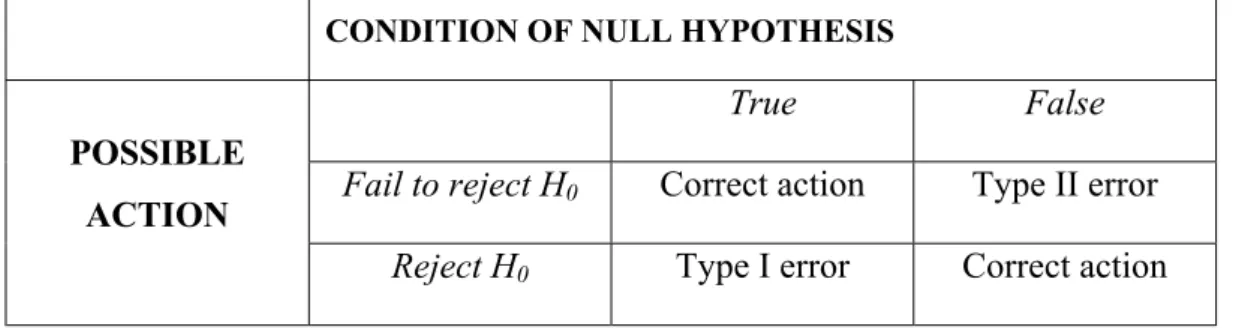
2.2.5. Genotyping of Subjects………...50 2.2.5.1. p21 Codon 31 Genotyping………..50 2.2.5.2. p53 Codon 72 Genotyping………..51 2.2.6. Statistical Analyses………..54 2.2.6.1. Hypothesis Testing………...54 2.2.6.1.1. Types of Errors………...55 2.2.6.1.2. P-Values……….55 2.2.6.1.3. Confidence Interval………56
2.2.6.2. The Chi-Square Distribution Test………...56
2.2.6.3. Relative Risk and Odds Ratio Calculation………..58
3. Results……….62
3.1. Genotyping………..62
3.1.1. Genotyping of p53 codon 72………...62
3.1.2. Genotyping of p21 codon 31………...67
3.2. Statistical Analysis………..71
3.2.1. Characteristics of the Subjects in the Turkish Population………..71
3.2.2. Genotype Distributions in the Turkish Population………..74
3.2.2.1. Distribution of the p53 Codon 72 Genotype………...74
3.2.2.2. Distribution of the p53 Codon 72 Genotype According to BMI………75
3.2.2.3. Distribution of the p21 Codon 31 Genotype………...78
3.2.2.4. Distribution of the p21 Codon 31 Genotype According to BMI………78
3.2.2.5. Combined Analysis of p21 and p53 for Breast Cancer Risk……….82
3.2.3. Characteristics of the Subjects in the Greek Study Population………...84
3.2.4. Genotype distributions………84
3.2.4.1. Distribution of the p53 Codon 72 Genotype………...84
3.2.4.2. Distribution of the p21 Codon 31 Genotype………...85
3.2.5. Genotype Distributions in the Turkish and Greek Populations………...89
3.2.5.1. Distribution of the p53 Codon 72 Genotype……….89
3.2.5.2. Distribution of the p21 Codon 31 Genotype……….89
3.2.6. P-value Calculation………92
4. Discussion………...93
5. Conclusions and Future Perspectives………101
6. References……….103
LIST OF TABLES
Table 1.1: Major Genetic Defects in Breast Cancer………..12
Table 1.2: Somatic Alterations in Breast Cancer………...14
Table 1.3: Genetic Polymorphisms in Relation to Breast Cancer Risk……….21
Table 1.4: p21 Codon 31 Polymorphism and Cancer Association………32
Table 1.5: p53 Codon 72 Polymorphism and Cancer Association………34
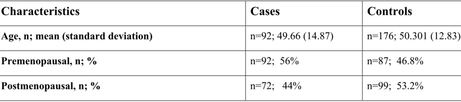
Table 2.1: Selected Characteristics of Breast Cancer Patients and Age-matched Control Subjects in the Turkish Population………...41
Table 2.2: Selected Characteristics of Breast Cancer Patients and Age-matched Control Subjects in the Greek Population……….42
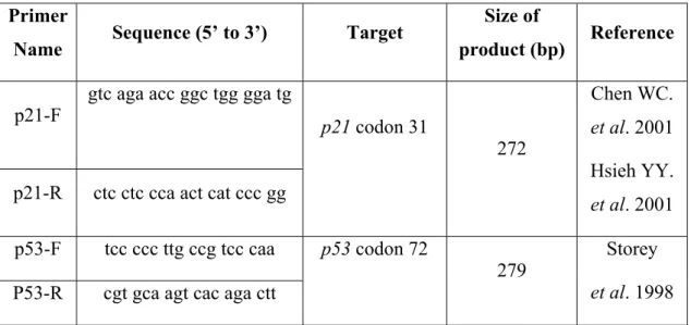
Table 2.3: List of Primers Used for PCR Experiments………..43
Table 2.4: Conditions under which Type I and II Errors may be Committed………55
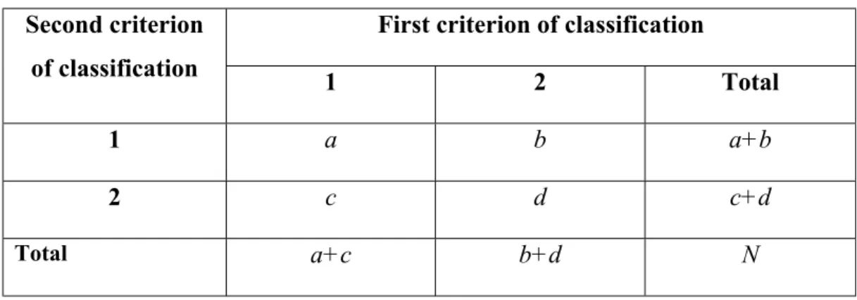
Table 2.5: A 2 X 2 Contingency Table………..58
Table 2.6: Subjects of a Retrospective Study Classified According to Status Relative to Risk Factor and Whether They Are Cases or Controls……….60
Table 3.1: General Characteristics of the Subjects in the Turkish Study Population………..73
Table 3.2: Distribution of the p53 Codon 72 Genotype Stratified According to Menopausal Status in the Age-matched Control and Breast Cancer Patients………76
Table 3.3: Distribution of the p53 Codon 72 Genotype Stratified According to BMI in Cases and Controls………77
Table 3.4: Distribution of the p21 Codon 31 Genotype Stratified According to Menopausal Status in the Age-matched
Control and Breast Cancer Patients……….80 Table 3.5: Distribution of the p21 Codon 31 Genotype Stratified
According to BMI in Cases and Controls………81 Table 3.6: Combination of the p21 Codon 31 Genotype with the
p53 Codon 72 Genotype for Breast Cancer Risk……….83
Table 3.7: Characteristics of Subjects from the Greek Population………86 Table 3.8: Distribution of the p53 Codon 72 Genotype Stratified
According to Menopausal Status in the Age-matched
Control and Breast Cancer Patients in the Greek Population…………..87 Table 3.9. Distribution of the p21 Codon 31 Genotype Stratified
According to Menopausal Status in the Age-matched
Control and Breast Cancer Patients in the Greek Population ………….88 Table 3.10. Distribution of the p53 Codon 72 Genotypes Startified
According to Menopausal status in the Age-matched Control and Breast Cancer Patients in the Turkish and
Greek Populations………90 Table 3.11. Distribution of the p21 Codon 31 Genotypes Startified
According to Menopausal status in the Age-matched Control and Breast Cancer Patients in the Turkish and
Greek Populations………91 Table 3.12. P-values for the p53 Codon 72 and p21 Codon 31 in the
LIST OF FIGURES
Figure 1.1: Pathways in Cancer………...3
Figure 1.2: Schematic Representation of Cell Cycle Regulators……….7
Figure 1.3: Structural Organization of p53 Protein………...24
Figure 1.4: Representation of p21 Protein……….29
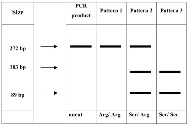
Figure 2.1: Schematic Representation of Genotyping of p21 Codon 31 Polymorphism……….52
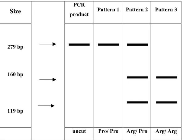
Figure 2.2: Schematic Representation of Genotyping of p53 Codon 72 Polymorphism……….53
Figure 3.1: Agarose Gel Photograph of PCR Products for p53 Region …………...64
Figure 3.2: Genotyping of the p53 Gene at Codon 72………...65
Figure 3.3: Restriction Enzyme Digestion and Sequencing Reaction Results for p53 Samples ………..66
Figure 3.4: Agarose Gel Photograph of PCR Products for p21 Region …………...68
Figure 3.5: Genotyping of the p21 Gene at Codon 31………...69
Figure 3.6: Restriction Enzyme Digestion and Sequencing Reaction Results for p21 Samples………...70
ABBREVIATIONS
AIP1 Apoptosis Induced Protein 1
Arg Arginine
BMI Body Mass Index
Bp Base pairs
BRCA1 Breast Cancer Susceptibility Gene 1 BRCA2 Breast Cancer Susceptibility Gene 2
Cdk Cyclin-dependent kinase
CI Confidence Interval
Cip-1 Cdk-interacting protein 1
cm centimeter
COMT catechol-O-methyltransferase
CYP1A1 Cytochrome P450 1A1
CYP2D6 Cytochrome P450 2D6
CYP2E1 Cytochrome P450 2E1
CYP17 Cytochrome P450, subfamily XVII
CYP19 Cytochrome P450, subfamily XIX
˚C Degree celsius
DNA Deoxyribonucleic Acid
dNTP Deoxynucleotide triphosphate
EGF Epithelial Growth Factor
F Forward
GST Gluthathione S-Transferase
GSTT1 Gluthathione S-Transferase theta 1
GTP Guanosine Triphosphate
HPV Human Papilloma Virus
hrs hour(s) KIP1 Kinase Inhibitory Protein 1
KIP2 Kinase Inhibitory Protein 2
kb kilobase(s)
kDa kilo Dalton
LOH Loss of Heterozygosity
M Molar mg milligram min minute(s) ml milliliter mM millimolar MSH2 Mut S Homolog 2 µl microliter µg microgram n number of subjects
NAT1 N-acetyl transferase Type 1
NAT2 N-acetyl transferase Type 2
ng nanogram
OR Odds Ratio
PCR Polymerase Chain Reaction
pmol picomol Pro Proline R Reverse
rpm revolutions per minute
s second(s) Ser Serine
SNP Single Nucleotide Polymorphism
TP53 Tumor Protein p53
u unit(s)
UV Ultra Violet
V Volt WAF-1 Wild type p53 activated protein-1
w/v weight per volume
X2 Chi-square
1. INTRODUCTION
1.1 . Cancer: A Loss of Normal Growth Regulation
Various disease states can arise when the normal stability of the organization of tissues and organs is disturbed. A tumor or neoplasm (literally, “new growth”) is an example of such tissue in which the control of growth becomes defective. Neoplasms can be defined as benign or malignant based on their likelihood of spreading. Encapsulated nodules of neoplastic tissue that do not spread are called benign tumors. On the other hand, malignant tumors often invade neighboring tissues and even other parts of the body, and thus may become lethal. Cancer is the common term for a malignant tumor. The word is from the Latin term for “crab” because early physicians noticed certain cancers had a crablike appearance (Becker et al. 1996).
1.1.1. Neoplastic Transformation and Tumor Progression
In most cases, malignant tumors develop from a single progenitor cell. The progenitor cell has undergone a series of irreversible (permanent and heritable) and cumulative changes in a process called neoplastic transformation. There are two general characteristics of the transformed cells: they undergo uncontrolled growth and tend to spread. The spread of cancer cells to neighboring tissues is called invasion; the spread to distant organs is called metastasis. The term metastases is used to refer to the tumor nodules that implant at sites distant from the parent tumor (Becker et al. 1996).
Tumor progression is the incremental development of increasingly malignant characteristics by a tumor. Typically, tumors are relatively benign, slowly growing, weakly invasive or noninvasive in the early stages of development. With time, however, they can enter a phase of increasingly rapid growth and become highly invasive and metastatic.
1.2. Molecular Genetics of Cancer
Cancer is a genetic disease resulting from mutations in somatic cells (Alberts et al. 2002). Several lines of evidence indicate that tumorigenesis in humans is a multistep process and that these steps reflect genetic alterations that drive the progressive transformation of normal human cells into highly malignant derivatives (Hanahan et
al. 2000).
1.2.1. Genomic Integrity
Genomic instability, which results in an elevated mutation rate, is a fundamental prerequisite of tumorigenesis (Schmute et al. 1999). Some cancer cells are defective in the ability to repair local DNA damage or to correct replication errors that affect individual nucleotides. These cells tend to accumulate more point mutations than do normal cells. Other cancer cells cannot maintain the integrity of their chromosomes properly and thus display gross abnormalities in their karyotype (Alberts et al. 2002). Cells must protect the integrity of their genome to avoid both the inheritance of deleterious mutations by daughter cells and the accumulation of mutations in genes that control cell proliferation. Although there are many safeguards in cells to protect the genomic integrity, cellular DNA is constantly bombarded by mutagens from endogenous and exogenous sources. DNA replication, gene transcription, DNA repair and cell cycle checkpoints must all interlink to promote cell survival following DNA damage and protect the integrity of chromosomes. A highly coordinated response to DNA damage is the activation of appropriate repair pathways and reversible arrest at cell cycle checkpoints. The cell cycle arrest gives time for repair to be completed (Levitt et al. 2002).
The p53 protein, also known as the “guardian of the genome”, responds in several ways to DNA damage in the cell. p53 acts as a transcription factor, stimulating synthesis of a 21-kDa protein that inhibits cyclin-dependent kinase (Cdk)-cyclin complexes. This block stops the cell cycle when DNA damage has occurred, giving the cell time to repair the damage so that genetic errors are not passed on to daughter cells. If the repair fails, p53 can trigger the damaged cells to undergo apoptosis, or programmed cell death, before their genetic abnormalities are inherited. Recent
directly and indirectly through other proteins. Mutations in the p53 gene not only cause a lack of these protective effects but also stimulate abnormal cell growth (Becker et al. 1996).
1.2.2. Cancer-Critical Genes
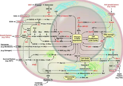
All genes whose mutation may lead to cancer are cancer-critical genes (Alberts et al. 2002). Figure 1.1 summarizes the pathways and genes involved in cancer (Hanahan
et al. 2000). These genes are classified into two groups according to whether the
dangerous mutations they contain are those that cause loss of function or those that lead to gain of function: oncogenes and tumor suppressor genes.
1.2.2.1. Oncogenes
Gain-of-function mutations of proto-oncogenes stimulate cells to increase their ability to proliferate, disseminate, and divide when they should not. These mutations have a dominant effect, and the mutant genes are known as oncogenes (Alberts et al. 2002). There are different types of genetic alterations that can activate proto-oncogenes to become proto-oncogenes. The gene may be altered by a small change in sequence such as a point mutation, by a large scale change such as partial deletion, or by a chromosomal translocation that involves the breakage and rejoining of the DNA helix. These changes can occur in the protein-coding region yielding a hyperactive product, or they can occur in adjacent control regions so that the gene is simply expressed at concentrations that are much higher than normal. Alternatively, the cancer-critical-gene may be over-expressed because extra copies are present due to gene amplification events caused by errors in DNA replication.
Oncogenes influence (directly or indirectly) functions connected with cell growth (Lewin B. et al. 2000). They may function as growth factors (i.e. wnt1- related to wingless), growth factor receptors (i.e. c-erbB- EGF receptor kinase), G protein/signal transduction (i.e. c-ras- GTP-binding protein), intracellular tyrosine kinases (i.e. c-abl- cytosolic), serine/threonine kinases (i.e. c-raf- cytosolic), signaling proteins (i.e. vav- SH2 regulator), and as transcription factors (i.e. myc,
c-fos, c-jun). The common feature is that each type of protein is able to direct general
changes in cell phenotypes, either by initiating or responding to changes associated with cell growth, or by changing gene expression directly.
1.2.2.2. Tumor Suppressor Genes
Tumor suppressor genes protect cells from dysregulated growth and division. Both of their alleles must be inactivated to observe a phenotypic effect. There are two types of tumor suppressor gene: ‘gatekeepers’ and ‘caretakers’ (Levitt et al. 2002). Gatekeeper genes act directly to regulate cell proliferation and are rate limiting for tumorigenesis. The retinoblastoma (Rb) and p53 genes are examples of gatekeeper tumor suppressors. Caretaker genes do not directly regulate proliferation but when mutated lead to accelerated transformation of a normal cell to a neoplastic cell.
characterized in hereditary non-polyposis colon cancer. There are many so-called chromosomal instability disorders (i.e. ataxia telangiectasia) in which germ-line mutations in a caretaker gene lead to both genome instability and a predisposition to cancer. This shows the importance of these genes in suppressing neoplastic transformation.
Tumor suppressor genes act mostly in a recessive manner (Oesterreich et al. 1999). The classical inactivation of tumor suppressor genes is caused by chromosomal loss of one allele and mutation of the other remaining allele. Functional inactivation of tumor suppressor genes can also be caused by hypermethylation, increased degradation, or mislocalization.
The most important tumor suppressor is p53, the cellular gatekeeper for growth and division. More than half of all human cancers have either lost p53 protein or have cells with p53 mutations (Lewin et al. 2000). The p53 mutations fall into the category of dominant negative mutations, and the mutants function by overwhelming the wild-type protein and preventing it from functioning. The most common form of a dominant negative mutant is one that forms heteromeric protein containing both mutant and wild-type subunits, in which the wild-type subunits are unable to function. p53 exists as a tetramer. When mutant and wild-type subunits of p53 associate, the tetramer takes up the mutant conformation. The stability of p53 is another important parameter as it usually has a short half-life. The response to DNA damage stabilizes the protein and transactivates it. The cellular oncoprotein Mdm2 inhibits p53 activity. p53 induces transcription of Mdm2, so the interaction between p53 and Mdm2 forms a negative feedback loop in which the two components limit each other’s activities (Lewin et al. 2000).
1.3. Cell Cycle Regulation and Cancer
An important component in the maintenance of the genome is the coordination and control of DNA replication, repair, and the distribution of DNA to daughter cells during each division cycle. Regulation at two stages of the cell cycle is critical in response to DNA damage: G1-S and G2-M boundaries. Cells delay cell cycle progression in order to facilitate the repair of DNA damage and to ensure that previous steps in the cell cycle are complete before proceeding (Kastan et al. 1997).
The p16-cyclin D1-cdk4-Rb pathway is central to the regulation of the G1-to-S phase transition and to the understanding of human cancers (Figure 2) (Levine et al. 1997). One of these four genes is altered or mutated in nearly every cancer examined. p16 is a negative regulator of cyclin D1-Cdk4, and the gene is heavily methylated in some cancer cells and mutated in other cancers. Cyclin D1 is amplified and over-expressed in a number of cancers (about 16% of breast cancers), and cdk4 mutations (no longer sensitive to p16) and gene amplifications have been reported in selected tumors. The retinoblastoma protein (Rb) is the major target of cyclin D1-Cdk4 for cell cycle regulation and is also present in a mutant form in a number of cancers (such as small-cell lung cancer and osteosarcomas). The Rb protein regulates E2F-DP transcription factor complexes (E2F-1, -2, and –3, and DP-1, -2, and -3), which in turn regulate a number of genes (including those encoding cyclin E, cyclin A, and proliferating cell nuclear antigen) required to initiate or propagate the S phase of the cell cycle. Phosphorylation of Rb by cyclin D1-Cdk4 releases E2F-DP proteins from the Rb complex, relieving repression of these genes or activating their transcription. The Rb protein regulates the restriction point or start, as a “go- no go” signal for cell cycle progression that is sensitive to the impact of various growth factors (via the regulation of cyclin D1-Cdk4 and possibly p16).
In response to some forms of DNA damage, p53 is activated and turns on the transcription of one of its downstream genes, p21 (WAF1, Cip-1), for G1 arrest of the cell cycle. p21 binds to a number of cyclin and Cdk complexes: cyclin D1-Cdk4, cyclin E-Cdk2, cyclin A-Cdk2, and cyclin A-cdc2. One molecule of p21 per complex permits Cdk activity (and may even act as an assembly factor), while two molecules of p21 per complex inhibit kinase activity and block cell cycle progression. p21 also binds to PCNA (Proliferating Cell Nuclear Antigen) (at its C-terminal domain). The available evidence suggests that p21-PCNA complexes block the activity of PCNA in DNA replication, but not its activity in DNA repair. Thus, p21 can act on cyclin-Cdk complexes and PCNA to stop DNA replication (Levine et al. 1997).
1.4. Breast Cancer
1.4.1. Clinical Information
1.4.1.1. Incidence and Mortality
Breast cancer is the most common malignancy in women and constitutes 18% of all cancers in women (Haimov-Kochman R. et al. 2002). Approximately 183,000 women are diagnosed with invasive breast cancer each year and nearly 41,000 women die of the disease (DeVita et al. 2001). In women aged 40 to 55, breast cancer is the leading cause of all mortality.
1.4.1.2. Histopathology
Breast carcinoma arises from the epithelium of the mammary gland, which includes the milk-producing lobules and the ducts that carry milk to the nipple. Malignant transformation of the stromal, vascular, or fatty components of the breast is not included in this definition and is extremely rare. There is increasing evidence that the breast epithelium undergoes a transformation from normal to hyperplasic, followed by the appearance of atypia in association with hyperplasia, ultimately becoming malignant. Malignant cells continue to evolve from noninvasive carcinoma, typified by ductal carcinoma in situ, to invasive carcinoma, and ultimately, to cells with metastatic potential (Vogelstein et al. 2002).
The treatment and prognosis of a woman with breast cancer are strongly influenced by the stage at the time of diagnosis. Multiple staging systems have been proposed, but the most commonly used system is the one adopted by both the American Joint Committee and the International Union against Cancer. This staging system is a detailed TNM (tumor, nodes, metastasis) system but can be summarized as; Stage 0 (carcinoma in situ), Stage I (tumor ≤ 2 cm, negative axillary nodes), stage II (tumor size 2-5 cm and/or mobile positive axillary nodes), Stage III (tumor size > 5 cm and/or fixed axillary nodes; inflammatory breast cancer), Stage IV (distant metastases beyond ipsilateral axillary nodes) (Vogelstein et al. 2002).
1.4.1.3. Risk
Factors
Multiple factors are associated with an increased risk of developing breast cancer, including increasing age, family history, exposure to female reproductive hormones (both endogenous and exogenous), dietary factors, benign breast disease, and environmental factors (DeVita et al. 2001).
Family history of breast cancer: The best studied and most significant risk factor is family history of breast cancer. Shared exposure to another risk factor cannot be excluded, but this most commonly represents heritable factors that increase the likelihood of developing breast cancer. The breast cancer susceptibility genes BRCA1 and BRCA2 represent the most dramatic examples, but since they account for only 15 to 20 percent of the breast cancer cases that cluster in families, other less penetrant but more common heritable factors are also considered (Vogelstein et al. 2002). The risk of developing breast cancer is increased 1.5- to 3.0- fold if a women has a mother or sister with breast cancer. Family history, however, is a heterogeneous risk factor and depends on the number of relatives with breast cancer, the exact relationship, the age of diagnosis, and the number of unaffected relatives. Exposure to female reproductive hormones: The development of breast cancer in many women appears to be related to female reproductive hormones. Epidemiological studies have consistently identified a number of weaker breast cancer risk factors, each of which is associated with increased exposure to endogenous estrogens. Early age at menarche, nulliparity or late age at first full term
pregnancy, and late age at menopause increase the risk of developing breast cancer
(DeVita et al. 2001).
Age at menopause: In postmenopausal women, obesity and hormone therapy, both of which are positively correlated with plasma estrogen and estradiol levels, are associated with increased breast cancer risk. The age specific incidence of breast cancer increases steeply with age until menopause. After menopause, although the incidence continues to increase, the rate of increase decreases to approximately one-sixth of that seen in the premenopausal period. This dramatic slowing of the rate of increase in the age specific incidence curve suggests that ovarian activity plays a major role in the etiology of breast cancer (DeVita et al. 2001). The relative risk of
developing breast cancer for a women with natural menopause before age 45 is one-half that of a woman whose menopause occurs after age 55.
Age at menarche and the establishment of regular ovulatory cycles are strongly linked to breast cancer risk. Earlier age at menarche is associated with an increased risk of breast cancer; there appears to be a 20% decrease in breast cancer risk for each year that menarche is delayed.
Regarding menarche and menopause, it seems that the total duration of exposure to endogenous estrogen is an important parameter in breast cancer risk.
Pregnancy: The relationship between pregnancy and breast cancer risk appears more complicated. Age at first full term pregnancy clearly influences breast cancer risk. Based on epidemiological studies, women whose first full term pregnancy occurs after age 30 have a two- to fivefold increase in breast cancer risk in comparison with women who have their first full term pregnancy before approximately age 18. Additionally, terminal differentiation of breast epithelial cells does not occur until the onset of lactation after the completion of a full term pregnancy. This final stage of differentiation may confer increased resistance to carcinogens (Vogelstein et al. 2002).
Body mass index (BMI; kg/m2): Many studies have examined breast cancer in relation to body weight, height, and overall body size (BMI) (Wrensch et al. 2003). Most case-control and cohort studies of increased height, a variable highly correlated with age at menarche, and risk of breast cancer suggest a positive relationship (DeVita et al. 2001, Brinton et al. 1992). Although being overweight (high BMI) during early adult life has been associated with a lower incidence of premenopausal breast cancer (Wrensch et al. 2003, Brinton et al. 1992, Franceschi et al. 1996), weight gain after age 18 is associated with a significantly increased risk of postmenopausal breast cancer. The protection conferred by increased weight early in life is thought to be secondary to increased irregularity of menstrual cycles in these women, suggesting their exposure to endogenous estrogens is decreased. The increased risk with weight gain in later adult life has been explained by increased estrogen levels in these women secondary to increased production in adipose tissues (DeVita et al. 2001).
1.4.2. Genetics of Breast Cancer
Breast cancer is a complex and heterogeneous disease caused by the interaction of both genetic and non-genetic factors. BRCA1 and BRCA2 are the two high penetrance breast cancer genes. Breast cancer in families with germ-line mutations in these genes appears as an autosomal dominant trait. In addition, mutations in several other genes such as TP53, MSH2, and PTEN have been identified as rare causes of hereditary breast cancer. It is very likely that other lower-penetrance genes, whose susceptibility inheritance pattern does not fit the classic model of Mendelian inheritance, are also responsible for inherited susceptibility to breast cancer. The presence of breast cancer susceptibility genes is directly responsible for 5 to 10 percent of all breast cancers (Vogelstein et al. 2002).
Breast cancer due to inherited susceptibility has several distinctive clinical features: age at diagnosis is considerably lower than in sporadic cases, the prevalence of bilateral breast cancer is higher, and the presence of associated tumors in affected individuals is noted in some families. Associated tumors may include ovarian, colon, prostate, and endometrial cancers and sarcomas. However, breast cancer due to inherited susceptibility does not appear to be distinguished by histologic type, metastatic pattern, or survival characteristics (Vogelstein et al. 2002).
Table 1.1 summarizes the inherited defects in somatic genes responsible for hereditary and familial breast cancers (DeVita et al. 2001).
Table 1.1. Major Genetic Defects in Breast Cancer
ESTABLISHED FAMILIAL BREAST GENES (ALL TUMOR SUPPRESSORS)
Gene Chromosomal location Disease
TP53 (p53) 17p13 (mutated, LOH) Li-Fraumeni syndrome of multiple hereditary cancers
PTEN 10q23 (mutated, LOH) Cowden’s syndrome of multiple hereditary cancers
BRCA-1 17q21 (mutated, LOH) Familial female breast and ovarian cancers
BRCA-2 13q14 (mutated, LOH) Familial female and male breast cancers
ESTABLISHED BREAST CANCER PROGRESSION GENES
Gene Chromosomal location Class Function
C-ERBB2 17q12 (amplified) Oncogene Growth factor receptor subunit
C-MYC 8q24 (amplified) Oncogene Cell-cycle/cell death regulator
CCND1 (Cyclin D1) 11q13 (amplified) Oncogene Cell-cycle G1 regulator
CDKN2 (p16) 9p21 (methylated, LOH) Suppressor gene Cell-cycle G1 regulator
RB-1 13q14 Suppressor gene (mutated, LOH) Cell-cycle G1 and G1/S regulator
TP53 (p53) 17p13 Suppressor gene (mutated, LOH) Cell-cycle/cell death/DNA repair regulator
Another approach to understanding the pathogenesis of breast cancer is the study of non-inherited (sporadic) breast cancers. This is an important complementary approach to the study of germ-line alterations for several reasons. First, the large majority of breast cancers do not arise as a result of inherited mutations in a single breast cancer susceptibility gene, and sporadic tumors may have fundamental molecular genetic differences. Second, genes that are frequently dysregulated or mutated in sporadic breast cancer are candidate genes for susceptibility loci. Third, the study of genetic alterations, such as mutations, deletions, and amplifications, provides clues to the mechanisms that result in the genomic instability in cancer cells. A summary of the genes altered in sporadic breast cancers is given in Table 1.2.
Table 1.2. Somatic Alterations in Breast Cancer
Gene/Region Modification Frequency Growth factors and receptors
EGFR Overexpression 20-40%
HER-2/neu Overexpression 20-40%
FGF1/FGF4 Overexpression 20-30%
TGFα Overexpression Not reported
Intracellular signaling molecules
Ha-ras Mutation 5-10%
c-src Overexpression 50-70%
Regulators of cell cycle
TP53 Mutation/inactivation 30-40%
RB1 Inactivation 20%
Cyclin D Overexpression 35-45%
TGFβ Dysregulation Not reported
Adhesion molecules and proteases
E-cadherin Reduced/absent 60-70% P-cadherin Reduced/absent 30% Cathepsin D Overexpression 20-24% MMPs Increased expression 20-80% Other genes bcl-2 Overexpression 30-45% c-myc Amplification 5-20%
1.4.3. Single Nucleotide Polymorphism Analysis and Importance
Complex diseases do not follow a simple Mendelian mode of inheritance and frequently have an environmental component of causation. Many genes seem to be involved with comparatively low individual impact but, nevertheless, have considerable overall contribution. To understand the genetic contribution to the etiology of complex diseases, model calculations for detection of genes or alleles with modest effect use the approach called “association study” by geneticists or “case-control study” by epidemiologists (Becker et al. 2003).The original Mendelian view of the genome classified alleles as either wild-type or mutant. Subsequently multiple alleles, each with a different effect on the phenotype were recognized. In some cases it may not even be appropriate to define any one allele as “wild-type”. The coexistance of multiple alleles at a locus is called genetic polymorphism (Lewin et al. 2000). An allele is usually defined as polymorphic if multiple alleles exist as stable components and if it is present at a frequency of >1% in the population. An SNP (single nucleotide polymorphism) marker is just a single base change in a DNA sequence, with an alternative of two possible nucleotides at a given position (Vignal et al. 2002). Although in principle any of the four possible nucleotide bases can be present at each position of a sequence stretch, SNPs are usually biallelic in practice. In every 1000 bases along the human chromosomes, on average approximately one nucleotide position is estimated to differ between any two copies of that chromosome (Landegren et al. 1998).
There are different reasons why SNPs are currently utilized in epidemiological studies. One is their use in genome-wide scans as markers for disease genes. Another reason is the interest in allele-specific variation on the population level introduced by functionally relevant SNPs or by susceptibility loci in close linkage with them (Becker et al. 2003). In this instance, it is usual to start with candidate genes whose functional relevance for a disease is known or strongly assumed; and to consider several genes along well-established functional pathways, since most likely more than a single gene is associated with the disease.
There recently has been a shift away from monogenic disorders toward the analysis of complex multifactorial diseases such as osteoporosis, diabetes, cardiovascular and inflammatory diseases, psychiatric disorders and most cancers, which occur at a
much higher frequency than single gene disorders. There is also increasing interest in the genetics of drug response (pharmacogenetics), an understanding of which may allow the ‘tailoring’ of therapies on an individual basis (Gray et al. 2000). The broadly familial nature of complex diseases clearly indicates a significant genetic component. However, in contrast to monogenic conditions, this genetic element is comprised of multiple gene variants each contributing a small effect. The extent of this problem is likely to be so great that the frequency of any polymorphism contributing to a disease phenotype will be only slightly elevated in a disease group compared with unaffected controls. Association studies with a large sample size, where cases of disease are compared with matched controls from the same population, are likely to provide a greater opportunity to detect small effects. Single nucleotide polymorphisms (SNPs) are the most abundant and stable types of DNA sequence variation in the human genome due to low mutation rates. Many SNPs also have functional consequences if they occur in the coding or regulatory regions of a gene (Gray et al. 2000). The SNP markers have gained more and more popularity for their quick, accurate, and inexpensive properties for the genetic analyses of various diseases. The SNP markers provide a new method for identification of complex gene-associated diseases such as breast cancer (Hsieh et al. 2001).
A handful of molecular strategies are in use for SNP analysis. All current methods involve target sequence amplification, and this is followed by distinction of DNA sequence variants by short hybridization probes or by restriction endonucleases, discrimination of mismatched DNA substrates by polymerases or ligases, or by observing the template-dependent choice of nucleotide incorporated by a polymerase (Landegren et al. 1998).
1.4.4. Genetic Polymorphism and Breast Cancer
Breast cancer is a clinically heterogenous disease, as evidenced by the widely variable morphological appearance and distinctive gene expression profiles. Because of possible effects on protein function or expression, it is reasonable to suspect that polymorphisms in genes involved in carcinogen metabolism, estrogen production, DNA repair and cell-cycle control could predispose individuals to the development of breast cancer, as well as influencing the clinical phenotype of the tumor. Genetic variants associated with an amino acid change can obviously have consequences for protein function, while those that occur in promoter or intronic regions could alter the level of gene expression. Alternatively, the genetic variant may have no direct functional implications but could be linked to other polymorphisms that have altered functions relative to the wild-type sequence (Powell et al. 2002).
Although 10-15% of breast cancer cases have some family history of the disease, only 5% can be explained by rare, highly penetrant mutations in genes such as
BRCA1 and BRCA2. First degree relatives of breast cancer patients have a two-fold
increase in risk over the general population, most of which cannot be accounted for by BRCA1 or BRCA2 (Dunning et al. 1999). Apart from shared environmental factors, the remaining familial risk may be due to common, low-penetrance genetic variants that are also referred to as modifier genes. Modifier genes have subtle sequence variants or polymorphisms that are associated with a small to moderate increased relative risk for breast cancer. Such variants are relatively common in the population and may be associated with a much greater proportion of breast cancer risk as a whole than the rare high penetrance genes (Weber et al. 2000).
There are different ways of presenting gene polymorphism data in relation to breast cancer risk, depending on the nature of the polymorphism. In the case of simple biallelic polymorphisms, allele frequencies in cases and controls can be compared using the X2 test to ascertain statistical significance. However, this method does not produce an easily interpretable measure of the magnitude of breast cancer risk and also lacks statistical power compared with some alternatives (Dunning et al. 1999). A more appropriate method is to compare genotype frequencies of three possible genotypes among cases and controls. The relative risk of breast cancer for each genotype is then estimated by the odds ratio (OR). The baseline group is usually the
Depending on the allele frequencies, the number of rare allele homozygotes may be very small, particularly in small studies, and the associated OR will have a wide confidence interval. Under these circumstances, it is common to combine the heterozygotes and rare-allele homozygotes and calculate the rare-allele carrier OR. However, this risk estimate is valid only if the genetic model is dominant, an assumption that should not be made without appropriate evidence (Dunning et al. 1999).
Candidate low penetrance gene products have been chosen on the basis of biological plausibility, in that alterations in the protein would affect a pathway involved in carcinogenesis. Low penetrance candidates are found in a wide variety of pathways, ranging from the detoxification of environmental carcinogens to steroid hormone metabolism and DNA damage repair (Weber et al. 2000). Candidate modifier genes can be divided into three main groups: genes for proteins with roles in steroid hormone metabolism; genes coding for carcinogen metabolism enzymes; and common alleles of genes that have been identified through family studies such as
TP53 and BRCA1. The candidate gene polymorphisms and their possible functional
effects are listed in Table 3 (Dunning et al. 1999). The existence of low-penetrance genetic polymorphisms may explain why some women are more sensitive than others to environmental carcinogens such as replacement estrogens (Coughlin et al. 1999). Steroid hormone metabolism genes: Several factors, such as age at menarche, age at first pregnancy, number of pregnancies, and age at menopause alter exposure to endogenous hormones and many of these alter breast cancer risk. Hence, genes involved in the metabolism of sex hormones are strong candidates for breast cancer susceptibility genes. Those which take part in the sex hormone biosynthesis pathway may affect production of, and thus exposure to estradiol, the most active estrogen. Genes in this pathway include CYP17, CYP19, and the gene for 17β-hydroxysteroid dehydrogenase type 2.
The bioavailability of hormones is partially controlled by catabolism, and catechol estrogens (2 hydroxy-estrogens) are the major breakdown products of estrogens. COMT is a phase II enzyme that methylates catechol-estrogens during their conjugation and inactivation. It has two forms: one membrane-bound and the other cytosolic; both are expressed in breast tissue and share a polymorphism associated
The sex hormones control the activation of responsive genes by first binding to specific receptors and forming complexes that can in turn bind to sequences in the promoters of downstream, hormone-responsive genes, such as Estrogen Receptor, Progesterone Receptor, and Androgen Receptor, which are candidates for breast cancer susceptibility genes (Dunning et al. 1999). The biological role of estrogens, including the growth and differentiation of normal mammary tissue, is mediated through the nuclear receptor protein (ER) that has an estrogen and DNA binding domains (Coughlin et al. 1999).
Carcinogen metabolism genes: Several enzymes function in the detoxification of xenobiotic compounds, and their gene expression is induced in response to the presence of the compound (e.g. polycyclic aromatic hydrocarbons found in tobacco smoke). The actions of phase I and phase II enzymes make susceptible compounds more soluble and more readily excreted and consequently reduce cancer risk. However, the more soluble products of some compounds are even more potent carcinogens than the less soluble form. Hence, a genetic change that increases the expression of the gene or the activity of the protein produced may increase the amount of reactive carcinogen formed and, thus, increase the risk of cancer (Dunning
et al. 1999).
Two phase I enzymes, CYP1A1 and CYP2D6, are induced by, and act on, carcinogens found in tobacco smoke. Both have polymorphic differences in either inducibility or activity. CYP2E1, an enzyme that metabolizes ethanol, is also a candidate because epidemiological studies suggest that breast cancer risk is increased with alcohol consumption.
The GST family are phase II enzymes that are potentially important in regulating susceptibility to cancer because of their ability to metabolize reactive electrophilic intermediates to usually less reactive and more water soluble glutathione conjugates (Mitrunen et al. 2001). For both GSTM1 and GSTT1, a high percentage of the Caucasian populations are homozygous for null alleles (up to 60 and 20%, respectively) and have no detoxifying GST activity. Levels of DNA adducts, sister-chromatid-exchange, and somatic genetic mutations may be increased in carriers of
The N-acetyl transferases, NAT1 and NAT2, are also phase II enzymes, and metabolise aromatic amines, which are present in cigarette smoke and heterocyclic amines in cooked meats (Weber et al. 2000). However, the action of NATs on these carcinogens can produce electrophilic ions that may induce point mutations in DNA (Dunning et al. 1999). Aryl aromatic amines are mammary carcinogens whose rate of metabolic activation is determined by polymorphisms in NAT genes (Weber et al. 2000). Polymorphism in both genes results in two phenotypes: slow acetylators who are homozygous for low-activity alleles, and fast acetylators who carry one or more high-activity alleles (Dunning et al. 1999).
Common alleles of high-penetrance genes: Mutations in the TP53 and BRCA1 genes are associated with a high risk of breast and other cancers. Mutation in the TP53 gene results in decreased p53 activity, which may lead to failure of cells with DNA damage to arrest and thus to continue to replicate with damaged DNA (Dunning et
al. 1999). Polymorphisms in the p21 downstream component of p53 pathway are
also described. In the case of BRCA1, where the protein function is still uncertain, the majority of confirmed mutations generate truncated proteins that are likely to have severely reduced activity. It has been hypothesized that amino acid substitutions outside the major functional domains may confer more moderate breast cancer risks. The majority of these substitutions are rare, and putative functional effects remain unconfirmed (Dunning et al. 1999).
According to the recent review of published case-control studies, polymorphisms in
CYP19, GSTM1, GSTP1 and TP53 appear to be stronger candidates for
low-penetrance breast cancer susceptibility genes, although they too need to be confirmed in larger studies (Dunning et al. 1999).
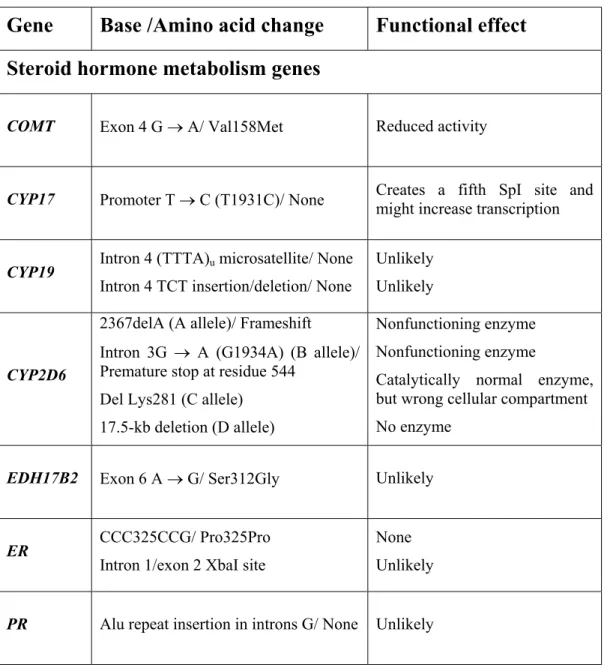
Table 1.3. Genetic Polymorphisms in Relation to Breast Cancer Risk
Gene
Base /Amino acid change
Functional effect
Steroid hormone metabolism genes
COMT Exon 4 G → A/ Val158Met Reduced activity
CYP17 Promoter T → C (T1931C)/ None Creates a fifth SpI site and might increase transcription
CYP19 Intron 4 (TTTA)u microsatellite/ None Intron 4 TCT insertion/deletion/ None
Unlikely Unlikely
CYP2D6
2367delA (A allele)/ Frameshift
Intron 3G → A (G1934A) (B allele)/ Premature stop at residue 544
Del Lys281 (C allele) 17.5-kb deletion (D allele)
Nonfunctioning enzyme Nonfunctioning enzyme
Catalytically normal enzyme, but wrong cellular compartment No enzyme
EDH17B2 Exon 6 A → G/ Ser312Gly Unlikely
ER CCC325CCG/ Pro325Pro Intron 1/exon 2 XbaI site
None Unlikely
Table 1.3. Continued
Gene
Base /Amino acid change
Functional effect
Carcinogen metabolism genes
CYP1A1
Exon 7 A → G (A4889G)/ Ile462Val 3’ UTR T → C (T6235C)/ None Exon 7 C → A (C4887A)/ Thr461Asp 3’ UTR T → C (T5639C)/ None
Uncertain, possible increase in enzyme activity
None Unknown None CYP2E1 Intron 6 unspecified/ None Unlikely
GSTM1 Gene deletion Null individuals have no enzyme GSTP1 A313G/ Ile105Val Reduced enzyme activity
GSTT1 Gene deletion Null individuals have no enzyme activity NAT1 A1088T/ None Possible increase in enzyme activity
NAT2
G191A C481T G590A G857A
Low activity allele Low activity allele Low activity allele Low activity allele
Other genes
BRCA1 C2731T/ Pro871Leu Unknown
BRCA1 G1186A/ Gln356Arg Unknown
HSP70-2 1267/ Silent Unknown
HSP70-hom 2437/ Met493Thr Unknown
TNF-α -308 G → A/ None Increased constitutive and inducible levels of TNF-α
TP53
Exon 3 G → C/ Arg72Pro
16-bp insertion in introns 3/ None Intron 6 G → A/ None
Unknown Unlikely Unlikely
1.4.4.1. p53 and p21 polymorphisms
Polymorphisms in TP53 are considered candidate risk factors because of the very important role played by this gene in the maintenance of genomic integrity following genotoxic insult (Powell et al. 2002). Highly penetrant germline mutations in TP53 are very rare, but polymorphisms are quite common and at least 14 polymorphisms have been described (Keshava et al. 2002). Five of these are in exons (codons 21, 36, 47, 72, and 213), and 9 are in introns (intron numbers 1-3, 6, 7, and 9) (Keshava et
al. 2002). Those investigated for association with breast cancer include a 16 bp
insertion in intron 3, an Arg72Pro polymorphism in exon 4 and a single nucleotide polymorphism in intron 6. Only the codon 72 polymorphism appears to be significantly associated with the risk of breast cancer (Dunning et al. 1999).
A major downstream component of the TP53 tumor suppressor pathway is the p21 cyclin dependent kinase inhibitor, also known as WAF1 or CIP1 (Powell et al. 2002). It was initially thought that somatic mutations in this gene might be involved in tumor formation, particularly for cases having wild type TP53; however, p21 mutations proved to be extremely rare in a variety of cancer types investigated (Powell et al. 2002). Polymorphisms in p21 have been described, with the two most common being Ser31Arg in exon 2 and a single nucleotide polymorphism in the 3’ untranslated region of exon 3, 20 bp downstream from the stop codon (Powell et al. 2002). Another polymorphism, Asp149Gly, has also been reported in an Indian population (Powell et al. 2002). Interestingly, both codon 31 and 149 polymorphisms appear to occur more frequently in patients whose tumors contain wild type TP53 (Powell et al. 2002). Another suspected p21 polymorphism occurs in the 5’ region of intron 2 but this remains to be confirmed (Powell et al. 2002).
Su et al. (2003) reported that different p53 and p21 genotypes or their combinations are associated with an altered human gene expression of p21. The genotype combination involving both the p53 codon 72 Pro allele and the p21 codon 31 Arg allele is associated with a particularly low expression of p21.
1.4.4.1.1. p53 Structure-Function Relationship and Polymorphism
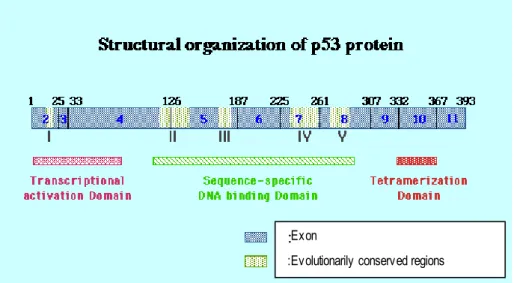
The p53 tumor suppressor gene is located at 17p13.1 and encodes a 53-kDa nuclear phosphoprotein whose primary role is to maintain genomic integrity through cell cycle arrest, DNA repair, and apoptosis. The protein consists of 393 amino acids that can be functionally divided into three domains (Figure 3, Ko et al. 1996). The NH2 terminus (amino acids 1-95) controls the transactivational activity of the protein, the central region (amino acids 102-292) controls the DNA binding activity, and the COOH terminus (amino acids 300-393) is responsible for oligomerization, nonspecific DNA binding, and DNA damage recognition (Powell et al. 2000).Figure 1.3. Structural Organization of p53 Protein
:Exon
:Evolutionarily conserved regions
Polymorphisms in TP53 are considered candidate risk factors because of the very important role played by this gene in the maintenance of genomic integrity following genotoxic insult (Powell et al. 2002). In human populations, the p53 gene has a common polymorphism at codon 72. The alleles of the polymorphism at codon 72 (exon 4) are ‘G’ or ‘C’ at the nucleotide residue 347. When ‘G’ is present it encodes an arginine amino acid (CGC; Arg72) with a positive-charged basic side chain, when ‘C’ is present it encodes a proline residue (CCC; Pro72) with a nonpolar-aliphatic side chain (Langerod et al. 2002). Matlashewski et al. (1987) concluded that this is a
nonconservative amino acid change, and results in a structural change in the protein, since the p53pro variant migrates more slowly than the p53Arg variant in sodium dodecyl sulfate polyacrylamide gel electrophoresis (SDS-PAGE). It was also noted that the tumors produced by the Pro-72 p53-containing cells appeared more slowly and were smaller than the Arg-72 p53 tumors.
The polymorphism occurs in the proline-rich domain of p53, which is required for the growth suppression activity of p53 and also plays an important role in p53-mediated apoptosis but not in cell cycle arrest (Thomas et al. 1999). This polyproline region is considered to be an Src homology 3 (SH3) binding domain, and the proline at amino acid 72 constitutes one of the five PXXP SH3 binding motifs defined within this region. As evidenced by monoclonal antibody reactivity, both proteins are structurally wild type, and they exhibit similar levels of affinity for a variety of p53 DNA recognition sequences. However, there are subtle differences in their abilities to interact with basic elements of the transcriptional machinery, and this is reflected in differences in the abilities of each form to induce apoptosis and suppress transformed cell growth. p53-Pro is a stronger inducer of transcription than p53-Arg, whereas a p53 Arg/Arg genotype induces apoptosis with faster kinetics and suppresses transformation more efficiently than the p53 Pro/Pro genotype (Thomas
et al. 1999).
The proline-rich PXXP domain between residues 60-90 of p53 is required for cooperation with anti-neoplastic agents to promote apoptosis of tumor cells, while deleting the C-terminal 30 amino acids of p53 does not have any effect (Baptiste et
al. 2002).
E6 proteins from both high-risk and low-risk HPV types are able to target p53Arg more efficiently than p53Pro for ubiquitin-mediated degradation. Consistent with this observation, the majority of HPV-associated tumors are homozygous for the p53Arg allele, whereas the majority of the comparable normal population was heterozygous (Thomas et al. 1999).
p53 codon 72 polymorphism influences the ability of certain conformational p53
mutants to form stable complexes with p73. When codon 72 encodes Arg, the ability of mutant p53 to bind p73, to neutralize p73-induced apoptosis and to transform cells in cooperation with EJ-Ras is enhanced. Arg-containing allele was preferentially
mutated and retained in squamous cell tumors arising in Arg/Pro germline heterozygotes. Formation of such complexes correlates with a loss of p73 DNA-binding capability, and consequently its ability to serve as a sequence-specific transcriptional activator and an inducer of apoptosis (Marin et al. 2000).
Langerod et al. (2002) reported that in breast cancer cases, the occurance of a p53 mutation was significantly more often found on the Arg72 allele than the Pro72 allele. The observed skewed occurance of somatic p53 mutations on the Arg72 allele in breast carcinomas suggests that this combination gives breast epithelial cells a growth advantage, which may increase the risk of malignant transformation and development of cancer. The coexistance of the Arg72 with a mutation may modify the p53 protein structure in a way that interferes either with the protein’s ability to achieve sequence-specific binding to DNA or with the interaction and recruitment of the transcription machinery, causing an altered transcription pattern. Another possibility is that the Arg72 may modify the mutant p53 protein’s ability to bind to and interact with other proteins such as p73 (Langerod et al. 2002).
p53 recessive mutants carrying the Arg allele can lead to decreased activation of p53 target genes through inactivation of p73. The transdominant p53 mutants achieve this by inactivation of the remaining wild type p53 allele (Tada et al. 2001).
For p53 mutants associated with human tumors, the arginine variant confers greater resistance to p73-dependent apoptosis and cytotoxicity than the equivalent proline form. This correlates with cellular resistance to the apoptotic and cytotoxic activity of specific cancer chemotherapeutic agents (Bergamaschi et al. 2003).
In cell lines containing inducible versions of alleles encoding the Pro72 and Arg72 variants, and in cells with endogenous p53, the Arg72 variant induces apoptosis markedly better than does the Pro72 variant. At least one source of this enhanced apoptotic potential is the greater ability of the Arg72 variant to localize to the mitochondria; this localization is accompanied by release of cytochrome c into the cytosol. The two polymorphic variants of p53 are functionally distinct, and these differences may influence cancer risk or treatment (Dumont et al. 2003).
Using microsatellite analysis, the frequency of LOH at the TP53 locus was investigated in patients heterozygous for the codon 72 polymorphism and it was
determined that the Arg allele is preferentially retained in patients heterozygous for this polymorphism (Papadakis et al. 2002).
There have been many studies conducted in order to investigate the association of
p53 codon 72 polymorphism and cancer risk. While some of the results are
significant, some of them show inconsistency. It is important to note that the results differ according to ethnicity. Table 1.5 summarizes some of the studies.
1.4.4.1.2. p21 Structure-Function Relationship and Polymorphism
Although p53 mutation is the most common genetic change reported in human cancers, about 50% of cancers do not have p53 mutations. Mutations or alterations on genes situated upstream or downstream of p53 on the same control pathway might have a similar oncogenic effect. Thus WAF1/CIP1 alterations could be good candidates to substitute for p53 mutations (Li et al. 1995). Table 1.4 summarizes some of the studies conducted so far in relation to p21 codon 31 polymorphism and cancer risk.The human p21waf1/cip1 localized to chromosome 6p 21.2 is a cyclin dependent kinase inhibitor (CDKI) upregulated by wild type tumor suppressor protein p53. Wild type p53 binds to a site 2.4 kb upstream of the p21 coding sequence and stimulates gene expression (Su et al. 2003).
Loss of heterozygosity of the short arm of chromosome 6 where p21 is situated has been described in cases of colon, lung, ovary and renal cancers, suggesting that
WAF1/CIP1 may be inactivated by a two-hits process in the corresponding tumors
(Li et al. 1995).
Mutations and deletions of the p21 gene have been rare in human cancers suggesting that p21, if involved in tumorigenesis, may be exerting itself mainly on the expression level rather than on the gene level (Bahl et al. 2000). However, p21 polymorphisms have been observed in various cancers. The polymorphic variants have been reported to occur more frequently in cancer patients than in healthy individuals suggesting a role in increased susceptibility to the development of some types of cancers (Mousses et al. 1995). Moreover, the frequency of the CIP1/WAF1 variants in tumors which contain p53 gene mutations was found to be significantly less than the frequency of the CIP1/WAF1 variants in tumors without p53 gene mutations. These data suggest that the variants at codon 31 and/or in the 3`UTR may not be benign polymorphisms, but may possibly be associated with a higher risk of developing cancer (Mousses et al. 1995).
The WAF1/CIP1 gene consists of three exons of 68, 450 and 1600 bp and encodes a 21 kDa protein of 164 amino acids (Figure 1.4). The first exon is non-coding while exon 2 contains 90% of the coding sequence (Li et al. 1995). The first ATG codon appears at nucleotide 76 in exon 2, and a termination codon appears at nucleotide
570 in exon 3 (Ralhan et al. 2000). The unique ability to associate the proliferating cell nuclear antigen (PCNA), an auxiliary factor for DNA polymerase δ and ε, distinguishes p21 from other cyclin-dependent kinase inhibitors (CDKIs). The Cdk and PCNA inhibitory activities of p21 have been mapped to different domains of the protein. An N-terminal domain which binds and inhibits cyclin-Cdk complexes, and a short sequence near the C-terminus (between amino acid residues 144-151) which binds to PCNA results in the inhibition of DNA replication (Bahl et al. 2000).
Figure 1.4. Representation of p21 Protein
http://www.infobiogen.fr/services/chromcancer/Genes/CDKN1AID139.html
The codon 31 polymorphism is found in an area of greater than 90% homology at the protein level with the murine homologue, which is thought to encode a DNA- binding zing-finger domain (Shih et al. 2000) in amino acids 13 and 41, and contains a potential nuclear localization signal between amino acids 140 and 163 (Hachiya et
al. 1999). The sequences between amino acids 13 and 56 are almost perfectly
conserved between mouse and human, and there is strong homology between WAF1 and p27KIP1 protein, as well as p57KIP2 protein. This conservation of the amino acid sequences suggests that this region is important to the function of WAF1 as a CDK inhibitor (Hachiya et al. 1999). In addition, serine is an uncharged polar amino acid with a single hydroxy 1 side chain, whereas arginine is a basic, positively charged amino acid with a seven-membered side chain. These observations raise the possibility that this polymorphism encodes functionally distinct proteins. In fact, it has been reported that the Arg variant has been observed in a significant number of cancer cases (Hachiya et al. 1999). However, transfection studies have shown no