ARTHRITIS & RHEUMATISM
Vol. 60, No. 11, November 2009, pp 3410–3412 DOI 10.1002/art.24956
© 2009, American College of Rheumatology
Increased Frequency of Extremely Skewed
X Chromosome Inactivation in Juvenile Idiopathic Arthritis
Elif Uz,
1Chigdem Mustafa,
1Rezan Topaloglu,
2Yelda Bilginer,
2Ali Dursun,
2Ozgur Kasapcopur,
3Seza Ozen,
2Aysin Bakkaloglu,
2and Tayfun Ozcelik
1 Objective. Juvenile idiopathic arthritis (JIA) is achildhood rheumatic disease of unknown etiology. Two subgroups of JIA, i.e., oligoarticular and polyarticular, are thought to have an autoimmune component, and show a higher female:male ratio. Skewed X chromosome inactivation (XCI) has previously been shown to be associated with scleroderma and autoimmune thyroid-itis, 2 autoimmune disorders occurring predominantly in females. This study was undertaken to extend the analysis to the pediatric age group and to determine the XCI profiles of patients with JIA.
Methods. A polymorphic repeat in the androgen
receptor gene was genotyped to determine XCI status in 81 female patients with JIA (21 with polyarticular disease and 60 with oligoarticular disease) and 211 healthy female controls. DNA obtained from venous blood samples was used for this analysis.
Results. Informative data were obtained on 62 JIA
patients and 155 controls. Skewed XCI was observed in 14 patients (22.6%) and 11 controls (7.1%) (P ⴝ 0.0036), and extreme skewing was apparent in 8 patients (12.9%) and 2 controls (1.3%) (P ⴝ 0.0008).
Conclusion. Our findings in the present study
indicate that skewed XCI may be a risk factor for the occurrence of autoimmune disorders, including JIA.
Juvenile idiopathic arthritis (JIA) is a broad term for a group of diseases characterized by chronic inflam-mation of 1 or more joints persisting longer than 6 weeks. It affects children before the age of 16, and its etiology is unknown. The International League of Asso-ciations for Rheumatology (ILAR) criteria set is the most recent accepted classification scheme (1). It is based on the number of affected joints and the presence of systemic symptoms. According to the ILAR classifi-cation, JIA is heterogeneous, with at least 6 subgroups: 1) systemic arthritis, 2) polyarthritis (rheumatoid factor positive and rheumatoid factor negative), 3) oligoarthri-tis (extended and persistent), 4) psoriatic arthrioligoarthri-tis, 5) enthesis-related arthritis, and 6) undifferentiated arthri-tis. Autoimmunity is particularly associated with the polyarthritis and oligoarthritis subgroups, and girls are more frequently affected than boys (female:male ratio 2–3:1) (2). Interestingly, an association between Turner’s syndrome and juvenile arthritis has been reported (3).
It has long been recognized that hormone levels and pregnancy-related fetal–maternal microchimerism may influence the occurrence of autoimmunity (4,5). Recently, our group and others observed that skewed X chromosome inactivation (XCI) could be a novel pre-disposition factor (6–8). Among these factors, XCI could be considered a particularly attractive etiologic candidate in JIA, since the onset of the disease is before puberty, excluding the risks associated with hormone levels and fetal microchimerism. In order to test the hypothesis that XCI may play a role in the pathogenesis of JIA, we determined the X chromosome inactivation profiles of patients with JIA and control subjects.
PATIENTS AND METHODS
Patients. Eighty-one girls diagnosed as having JIA
according to the ILAR criteria and 211 healthy girls with no
Supported by grants to Dr. Ozcelik from the Scientific and Technical Research Council of Turkey (grant TUBITAK-SBAG 3334) and the International Center for Genetic Engineering and Biotechnol-ogy (grant ICGEB-CRP/TUR04-01).
1Elif Uz, PhD, Chigdem Mustafa, MSc, Tayfun Ozcelik, MD:
Bilkent University, Faculty of Science and Institute of Materials Science and Nanotechnology, Ankara, Turkey; 2Rezan Topaloglu,
MD, Yelda Bilginer, MD, Ali Dursun, PhD, MD, Seza Ozen, MD, Aysin Bakkaloglu, MD: Hacettepe University Medical School, Ankara, Turkey; 3Ozgur Kasapcopur, MD: Istanbul University Cerrahpasa
Faculty of Medicine, Istanbul, Turkey.
Dr. Uz and Ms Mustafa contributed equally to this work. Address correspondence and reprint requests to Tayfun Ozcelik, MD, Department of Molecular Biology and Genetics, Faculty of Science, B-238, Bilkent University, Bilkent, Ankara 06800, Turkey. E-mail: [email protected].
Submitted for publication December 4, 2008; accepted in revised form August 10, 2009.
history of autoimmune disorders or cancer (control group; mean⫾ SD age 13 ⫾ 4 years) were included in the study. The medical files of the parents were not available for analysis. The mean⫾ SD age of the JIA patients was 10 ⫾ 5 years, and the mean age at the time of disease onset was 6 ⫾ 4 years. Twenty-one of the JIA patients had polyarticular disease and 60 had oligoarticular disease. The following clinical data were obtained on all patients: age, age at diagnosis, type of JIA, disease duration, and treatment regimens. Informed consent was obtained from all subjects (or legal guardians of subjects who had not reached age of majority). The ethics committee of Hacettepe University approved the study protocol.
X chromosome inactivation assay. The highly
polymor-phic CAG repeat on the first exon of the androgen receptor gene was genotyped, as described elsewhere (6,8,9), to deter-mine the XCI status of the patients and controls.
Statistical analysis. The significance of differences in
results between the JIA patients and the control subjects was determined by Fisher’s exact test.
RESULTS
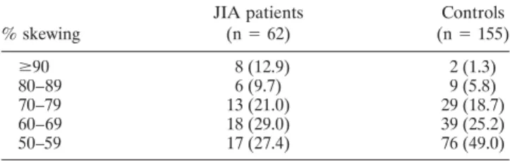
Findings on XCI status were informative in 62 of the 81 JIA patients (76.5%) and in 155 of the 211 controls (73.5%). The individuals whose alleles could not be distinguished adequately were not included in the densitometric analysis and were categorized as having noninformative results. Skewed XCI (ⱖ80% skewing) was observed in 14 of the 62 patients with informative results (22.6%), and in 11 of the 155 controls with informative results (7.1%) (P ⫽ 0.0036). Extremely skewed XCI ratios (ⱖ90% skewing) were seen in 8 of the 62 patients (12.9%), but in only 2 of the controls (1.3%) (P ⫽ 0.0008) (Table 1). Studies in large cohorts have firmly established that extremely skewed XCI is rare in the general population (10).
It has been reported that years after exposure to an immunosuppressive agent, XCI ratios in feline hema-topoietic cells may be skewed (11). We therefore inves-tigated correlations between XCI ratios and treatment
regimens in the patients. All of the patients were receiv-ing some treatment at the time of sample collection. Fourteen were receiving nonsteroidal antiinflammatory drugs (NSAIDs), 11 were receiving methotrexate (MTX), 10 were receiving MTX plus NSAIDs, and 9 were receiving intraarticular corticosteroids plus NSAIDs. The remaining 18 were receiving various com-binations of these and other drugs. Clinical details on the 62 patients are shown in Supplementary Table 1, available on the Arthritis & Rheumatism Web site at http://www3.interscience.wiley.com/journal/76509746/ home. Among the patients with skewed XCI, 7 (50%) had received treatment with immunosuppressive agents for 6–10 years, and 1 (7%) had received immunosup-pressive treatment for 2 years. The remaining 6 had received antiinflammatory treatment alone. Among the patients with random patterns of XCI, 27 (56.3%) had received immunosuppressive treatment for⬎2 years. At any stage of their treatment, suspended leukopenia or bone marrow ablation was not observed in any of the patients. These results, along with previous observations (6,8), indicate that it is unlikely that immunosuppressive therapy caused skewed XCI in the patients.
DISCUSSION
Autoimmune disorders are complex and affect ⬃5% of the world population (12). A diverse group of autoimmune diseases, including rheumatoid arthritis and JIA, affects females more frequently than males (2). In this study, we observed skewed XCI patterns in peripheral blood mononuclear cells of a significant proportion of female subjects with JIA, with 22.6% in theⱖ80% skewing range (versus 7.1% of controls) and 12.9% in the ⱖ90% skewing range (versus 1.3% of controls). These results support the hypothesis that loss of XCI mosaicism in females may constitute a risk factor for the development of JIA.
At present, the nature of the association between skewed XCI and breakdown of self tolerance, as exem-plified in this study on JIA and previous observations on scleroderma (6) and autoimmune thyroiditis (7,8,13), is not known. We propose that deleterious X-linked mu-tations could influence the survival of cells that inacti-vate a normal X chromosome and leave the mutant X transcriptionally active. Since cells that inactivate the mutant X would be immune to the deleterious effects of the putative mutations, females could tolerate them by losing their mosaic status for X-linked gene expression (14,15). An interesting aspect of extremely skewed XCI is that it does not lead to the breakdown of self tolerance Table 1. Proportion of the juvenile idiopathic arthritis (JIA) patients
and controls with skewed X chromosome inactivation*
% skewing JIA patients (n⫽ 62) Controls (n⫽ 155) ⱖ90 8 (12.9) 2 (1.3) 80–89 6 (9.7) 9 (5.8) 70–79 13 (21.0) 29 (18.7) 60–69 18 (29.0) 39 (25.2) 50–59 17 (27.4) 76 (49.0)
* Values are the number (%). Forⱖ80% skewing, P ⫽ 0.0036 (odds ratio 3.81 [95% confidence interval 1.65–8.83]); forⱖ90% skewing,
P ⫽ 0.0008 (odds ratio 11.33 [95% confidence interval 2.62–48.48]), by
Fisher’s 2-tailed exact test.
in all females. This suggests that breakdown of self tolerance may require 2 distinct events on the X chro-mosome: first, a lethal mutation leading to loss of mosaicism, and second, “heterozygosity” for the nonsyn-onymous variants of the putative critical genes. To the best of our knowledge, this is the first study that has shown an association between skewed XCI and a pedi-atric form of an autoimmune disease, i.e., JIA.
AUTHOR CONTRIBUTIONS
All authors were involved in drafting the article or revising it critically for important intellectual content, and all authors approved the final version to be published. Dr. Ozcelik had full access to all of the data in the study and takes responsibility for the integrity of the data and the accuracy of the data analysis.
Study conception and design. Ozcelik.
Acquisition of data. Uz, Mustafa, Topaloglu, Bilginer, Dursun, Kasap-copur, Ozen, Bakkaloglu.
Analysis and interpretation of data. Uz, Mustafa, Topaloglu, Bilginer, Dursun, Kasapcopur, Ozen, Bakkaloglu, Ozcelik.
REFERENCES
1. Petty RE, Southwood TR, Manners P, Baum J, Glass DN, Goldenberg J, et al. International League of Associations for Rheumatology classification of juvenile idiopathic arthritis: second revision, Edmonton, 2001. J Rheumatol 2004;31:390–2.
2. Borchers AT, Selmi C, Cheema G, Keen CL, Schoenfeld Y, Gershwin ME. Juvenile idiopathic arthritis. Autoimmun Rev 2006;5:279–98.
3. Wihlborg CE, Babyn PS, Schneider R. The association between Turner’s syndrome and juvenile rheumatoid arthritis. Pediatr Radiol 1999;29:676–81.
4. Cutolo M, Capellino S, Sulli A, Serioli B, Secchi ME, Villaggio B,
et al. Estrogens and autoimmune diseases. Ann N Y Acad Sci. 2006;1089:538–47.
5. Adams Waldorf KM, Nelson JL. Autoimmune disease during pregnancy and the microchimerism legacy of pregnancy. Immunol Invest 2008;37:631–44.
6. Ozbalkan Z, Bagislar S, Kiraz S, Akyerli CB, Ozer HT, Yavuz S, et al. Skewed X chromosome inactivation in blood cells of women with scleroderma. Arthritis Rheum 2005;52:1564–70.
7. Brix TH, Knudsen GPS, Kristiansen M, Kyvik KO, Orstavik KH, Hegedus L. High frequency of skewed X-chromosome inactivation in females with autoimmune thyroid disease: a possible explana-tion for the female predisposiexplana-tion to thyroid autoimmunity. J Clin Endocrinol Metab 2005;90:5949–53.
8. Ozcelik T, Uz E, Akyerli CB, Bagislar S, Mustafa CA, Gursoy A, et al. Evidence from autoimmune thyroiditis of skewed X-chro-mosome inactivation in female predisposition to autoimmunity. Eur J Hum Genet 2006;14:791–7.
9. Allen RC, Zoghbi HY, Moseley AB, Rosenblatt HM, Belmont JW. Methylation of HpaII and HhaI sites near the polymorphic CAG repeat in the human androgen-receptor gene correlates with X chromosome inactivation. Am J Hum Genet 1992;51:1229–39. 10. Amos-Landgraf JM, Cottle A, Plenge RM, Friez M, Schwartz CE, Longshore J, et al. X chromosome inactivation patterns of 1,005 phenotypically unaffected females. Am J Hum Genet 2006;79: 493–9.
11. Abkowitz JL, Linenberger ML, Persik M, Newton MA, Guttorp P. Behaviour of feline hematopoietic stem cells years after busulfan exposure. Blood 1993;82:2096–103.
12. World Health Organization. World Health Report 2008: Primary health care—now more than ever. Geneva: World Health Orga-nization; 2008.
13. Yin X, Latif R, Tomer Y, Davies TF. Thyroid epigenetics: X chromosome inactivation in patients with autoimmune thyroid disease. Ann N Y Acad Sci 2007;1110:193–200.
14. Ozcelik T. X-linked clonality testing and autoimmune diseases [letter]. Blood 2007;110:2769.
15. Ozcelik T. X chromosome inactivation and female predisposition to autoimmunity. Clin Rev Allergy Immunol 2008;34:348–51.