Abstract
The aim of this study was to investigate the neuroprotective effects of caffeic acid phenethyl ester (CAPE) on phosphodiesterase 4 (PDE4) mRNA isoenzymes, oxidant and antioxidant defence in ischemia/reperfusion (I/R) injured rat brains. Twenty-one rats were randomly divided into three equal groups: sham-control, ischemia/reperfusion (I/R) and I/R+CAPE. Rats in sham-control group underwent only surgical intervention without bilateral common carotid artery occlusion. Ischemia/reperfusion was induced by bilateral common carotid artery occlusion with atraumatic clips for 30 min, followed by artery reopening. The I/R+CAPE group was subjected to the same surgical procedure as I/R group, but CAPE was administered intraperitoneally at the dose of 15 µmol kg-1 twice, 1 h before occlusion and at 12th h of reperfusion.
The rats were sacrificed 24 h after I/R. The cAMP concentration was analyzed by ELISA and PDE4 isozyme mRNA transcriptions were evaluated by qRT-PCR methodology in the brain cortex. Ischemia-induced NO production was significantly attenuated by CAPE in the cerebral cortex. CAPE significantly enhanced GSH-Px activity, while SOD, CAT and XO activities non-significantly changed, as compared to the I/R group. CAPE significantly decreased PDE4A and PDE4B transcripts, without changing cAMP levels compared to I/R group. Ischemia-induced neurologic deficit scores were reduced by CAPE. These results suggest that CAPE slightly modulates the antioxidant defense system and NO release in rat brain during global cerebral ischemia/reperfusion injury. In addition, CAPE treatments produce the neuroprotective effect by reducing the levels of some PDE4 transcriptions.
Keywords: CAPE, Brain, Ischemia/reperfusion, Antioxidant activity, cAMP-phosphodiesterase 4, Neuroprotective effect, Rat
Rat Beyinlerinde Global İskemi-Reperfüzyon Hasarı Üzerine
Kafeik Asit Fenetil Esterin Nöroprotektif Etkisi
Özet
Bu çalışma iskemi-reperfüzyon (I/R) hasarlı rat beyinlerinde fosfodiesteraz 4 (PDE4) mRNA izoenzimleri, oksidant ve antioksidant savunma sistemi üzerine kafeik asit fenetil ester (KAFE)’in nöroprotektif etkilerini araştırmak amacıyla yapıldı. Yirmi bir adet rat rastgele üç eşit gruba ayrıldı. Sham-kontrol, iskemi/reperfüzyon (I/R) ve I/R+KAFE. Sham-kontrol grubundaki ratlara bilateral common carotid arter oklüzyonu yapılmaksızın sadece cerrahi müdahalede bulunuldu. İskemi/reperfüzyon (I/R) bilateral common carotid arterlerin atravmatik klempler ile 30 dakika oklüzyonu ve takiben arter klempleri açılarak reperfüzyonu ile sağlandı. I/R+KAFE grubu I/R grubu ile aynı cerrahi usüle tabi tutuldu fakat oklüzyondan 1 saat önce ve reperfüzyondan 12 saat sonra iki defa 15 µmol kg-1 dozunda intraperitoneal KAFE verildi. Ratlar iskemi/
reperfüzyondan 24 saat sonra sakrifiye edildi. Beyin korteksindeki cAMP düzeyi ELISA ile, PDE4 mRNA izoenzim transkripsiyonları ise qRT-PCR ile değerlendirildi. KAFE iskemi ile uyarılan beyin korteksindeki NO üretimini önemli oranda azalttı. I/R grubu ile karşılaştırıldığında SOD, CAT ve XO aktivitelerini KAFE anlamlı düzeyde değiştirmezken, GSH-Px aktivitesini önemli oranda arttırdı. KAFE cAMP düzeyini değiştirmeksizin PDE4A ve PDE4B düzeyini önemli oranda azalttı. İskemi ile uyarılan nörolojik hasar skorları KAFE tarafından azaltıldı. Bu sonuçlar KAFE’nin global beyin iskemi/reperfüzyon hasarı sırasında rat beyinlerinde antioksidant savunma sistemini ve NO salınımını hafifce dengelediğini önerir. Ayrıca KAFE bazı PDE4 izoenzim düzeylerini azaltarak nöroprotektif etki sağlar.
Anahtar sözcükler: KAFE, Beyin, İskemi/reperfüzyon, Antioksidant aktivite, cAMP-fosfodiesteraz 4, Nöroprotektif etki, Rat
The Neuroprotective Effect of Caffeic Acid Phenethyl Ester on
Global Ischemia-Reperfusion Injury in Rat Brains
[1]Muhammed Enes ALTUĞ
1
İsmet M. MELEK
2Suat ERDOĞAN
3Vesile DÜZGÜNER
4Atakan ÖZTÜRK
5Altuğ KÜÇÜKGÜL
6[1] 1 2 3 4 5 6
This study was presented as poster presentation at the XXVth International Symposium on Cerebral Blood Flow, Metabolism and
Function and the Xth International Conference on Quantification of Brain Function with PET. May 24-28, 2011, Barcelona, Spain
Department of Surgery, Faculty of Veterinary Medicine, Mustafa Kemal University, TR-31040 Hatay - TURKEY Department of Neurology, Tayfur Ata Sökmen Medical School, Mustafa Kemal University, TR-31034 Hatay - TURKEY Department of Medical Biochemistry, School of Medicine, Zirve University, TR-27260 Gaziantep - TURKEY
School of Health Sciences, Ardahan University, TR-75000 Ardahan - TURKEY
Department of Physiology, Tayfur Ata Sökmen Medical School, Mustafa Kemal University, TR-31034 Hatay - TURKEY Department of Biochemistry, Faculty of Veterinary Medicine, Mustafa Kemal University, TR-31040 Hatay - TURKEY
İletişim (Correspondence) +90 326 2455845/1512
[email protected]
INTRODUCTION
Reactive oxygen radicals (ROS) are likely participants in the pathogenesis of cerebral ischemia-reperfusion (I/R) injury. Transient focal and global cerebral I/R triggers a plethora of cellular and molecular events that promotes neuronal cell death in several regions of the brain due to glutamate excitotoxicity, oxidative stress, inflammation
and apoptosis [1-4]. Studies show that nitric oxide (NO)
has beneficial properties to I/R injury including increase of blood flow produced by cerebral vasodilatation and
inhibition of inflammation [1,5]. NO is also a free radical and
initiates various pathophysiological events by reaction with superoxide anion to form peroxinitrite, on the contrary of its protective effects in various models of I/R injury [1,5,6].
Mammalian cyclic nucleotide phosphodiesterases (PDEs) are composed of 21 genes and are categorized into 11 families based on sequence homology, enzymatic properties, and sensitivity to inhibitors, the enzymes that hydrolyze and inactivate cyclic adenosine monophosphate (cAMP)
and cyclic guanosine monophosphate (cGMP) [7]. As PDE4
is the major cAMP-hydrolysing family in many cells types, it represents a promising therapeutic target. The PDE4 family is composed of four subfamilies (PDE4A, PDE4B, PDE4C and PDE4D) encoded by different gene loci, and each of them has been shown to produce several mRNAs by
alternative splicing [8]. Most recently, it has demonstrated
that rolipram, selective PDE4 inhibitor, attenuates memory
deficits produced by global brain ischemia [9].
Caffeic acid phenethyl ester (CAPE), an active component of propolis, has been shown to possess anti-inflammatory, immunomodulatory, anticarcinogenic, and antioxidant
properties [1,2,10,11]. It has also been shown that CAPE
treatment significantly reduces the infarction size and neuronal damage in ischemia-induced brain injury and
suppresses cerebral lipid peroxidation [1,2,11]. To date, no
studies have been reported that how CAPE treatment affects the levels of cAMP and PDE4s after transient global brain ischemia. For this purpose, the present study was designed to evaluate the neuroprotective effects of CAPE on PDE4 mRNA isoenzymes, oxidant and antioxidant defence in transient global cerebral ischemia rat model.
MATERIAL and METHODS
Transient Global Cerebral Ischemia in Rats
Experimental procedures were approved by Mustafa Kemal University, Veterinary Faculty Ethics Committee for the use and care of laboratory animals (25.04.2007, no:09). Experiments were performed on adult male wistar rats weighing 250-275 g. They were given free access to food and tap water. All animals were maintained under a controlled temperature (21±1°C) and humidity (55-60%) throughout the experiment. Twenty-one healthy rats were
randomly divided into three equal groups: Sham-control, ischemia/reperfusion (I/R), I/R+CAPE. The experimental and surgical procedures in groups were performed as in the following: All rats were anesthetized intraperitoneally with xylazyn hydrochloride (4-5 mg/kg, Rompun, Bayer, Turkey) and ketamine HCl (40-50 mg/kg, Alfamine, Egevet, Turkey) and placed on heat blanket during surgical operation. Sham-control (sham-operated) group: Rats underwent only surgical intervention without bilateral common
carotid artery (BCCA) occlusion. Briefly, the right and left
common carotid arteries (CCAs) were isolated through a ventral midline cervical incision and separated carefully from vagosympathetic nerve. Ischemia-reperfusion (I/R) group: The common carotid arteries (CCAs) were isolated through a ventral midline cervical incision and separated carefully from vagosympathetic nerve by microsurgical procedures. Transient global cerebral ischemia was achieved by temporarily occluded (30 min) CCAs using atraumatic aneurysm clips and the opening in the skin was closed with wound clips. Reperfusion was achieved by declamping the arteries after 30 min. The surgery line was routinely closed. The rats in the sham-control and I/R groups were received intraperitoneally dimethyl sulfoxide (DMSO)
at the dose of 15 µmol kg-1 twice, 1 h before occlusion
and at 12 h of reperfusion. Ischemia-reperfusion + CAPE (I/R + CAPE) group: The rats in this group were subjected to ischemia/reperfusion with the same procedure in I/R group as mentioned above and treated with CAPE (Sigma, Germany). CAPE was dissolved in sterile DMSO (Sigma, Germany) and administered intraperitoneally at the dose
of 15 µmol kg-1 twice, 1 h before occlusion and at 12 h
of reperfusion. The CAPE doses used were chosen on the
basis of previous experiments [2,11]. Body temperature was
maintained at approximately 35.2±0.4°C with a heating pad until the animal had recovered from surgery. The rats were sacrificed 24 h after reperfusion with the anesthetic procedure reported above. Venous blood samples (5 ml) were taken by cardiac puncture into tubes with EDTA. Plasma was separated by centrifugation at 3.000 rpm for 15 min and was stored at -20°C until use. Immediately after taking blood specimen, brain was carefully removed, washed with ice-cold physiological saline and were rapidly stored at -20°C until analyses.
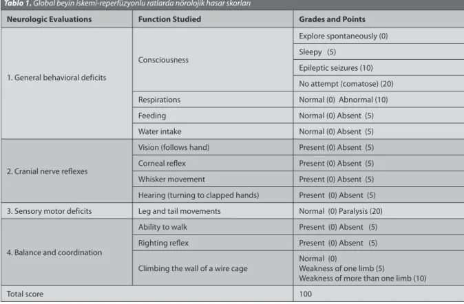
Assessments of Neurologic Deficit and Behavior
A neurologic evaluation was performed 24 h after the onset of the experiments by an investigator blinded to the study groups, using a neurologic deficit score (NDS)
as described previously, with several modifications [3,12-14].
The four categories of reactions and the functions with associated tested were: the general behavioral deficit, cranial nerve reflexes, sensory-motor deficit, balance and coordination. The presence or absence of the appropriate
reaction was scored. See Table 1 for the exact procedure
used. The NDS could range from 0 to 100, an NDS of 0 reflects normal brain function and an NDS of 100 reflects brain death.
Tissue Homogenization
Tissue samples were homogenized in a PBS buffer (pH 7.0) containing complete protease inhibitor mixture (Sigma, Germany). Homogenates were centrifuged at 4°C, 15.000 rpm for 10 min and the soluble fraction was retained. Protein concentrations of supernatants were measured
by the method of Bradford [15]using bovine serum albumin
as a standard.
The Analysis of Oxidant/Antioxidant Stress Markers
Lipid peroxidation levels were assessed by measuring malondialdehyde (MDA) concentration in tissues (μmol/
mg protein) [16]. The method was based on thiobarbituric
acid (TBA) reactivity. 2.5 mL of 20% trichloroacetic acid was added to the 0.5 mL of plasma and then 1 mL of 0.675% TBA was added. The coupling of lipid peroxide with TBA was carried out by heating at 95°C water bath for 30 min. After cooling in cold water, the resulting chromogen was extracted with 4.0 mL of n-butyl alcohol by vigorous shaking. Separating of the organic phase was facilitated by centrifugation at 3.000 rpm for 10 min and its absorbance was determined at 535 nm by spectrophotometer.
Catalase (CAT) activity was measured according to
the method of Luck [17]. One unit of CAT activity was
defined as the amount of enzyme required to decompose
1 mol of H2O2 in 1 min in a tube containing 2.95 mL of
a freshly prepared 30% H2O2 in phosphate buffer
(pH 7.0), 50 µl of tissue supernatant or plasma were added.
The rate of decomposition of H2O2 was measured
spectrophotometrically at 240 nm for 1 min. Using the reaction time (∆t) of the absorbance (A1 and A2), the following equation was generated to calculate the rate constant (k): k = (2.3/∆t)(log A1/A2). The enzyme activity was expressed as k/mg protein in tissues. Total superoxide dismutase (SOD) activity in the homogenates was determined according to the method of Sun and
colleagues [18]. The method is based on the inhibition of
nitroblue tetrazolium (NBT) reduction by the xanthine/ xanthine oxidase system as a superoxide generation. The enzyme activity was measured in the ethanol phase of the lysate after addition of 1.0 ml ethanol/ chloroform mixture (5/3, v/v) to the same amount of sample and the tubes were centrifuged. One unit of SOD was defined as the enzyme amount causing 50% inhibition in NBT reduction rate. SOD activity was expressed as U/mg protein in tissues. Nitric oxide (NO) concentration in plasma (μmol/L) and tissue (μmol/mg protein) samples were determined indirectly by measuring the nitrite
levels based on Griess reaction [19]. Samples were firstly
deproteinized with 75 mmol zinc sulphate. Total nitrite was determined by spectrophotometer at 545 nm after conversion of nitrate to nitrite by copperized cadmium granules. Xanthine oxidase (XO) activity was measured as the rate of uric acid production when xanthine was
incubated with tissue homogenates (U/g protein) [20].
Glutathione peroxidase (GSH-Px) activity was detected in the tissue homogenates by a kinetic method using a commercial kit (RANSEL by Randox Lab. UK). GSH-Px Table 1. Neurologic deficit scores (NDS) for rats with global cerebral ischemia-reperfusion
Tablo 1. Global beyin iskemi-reperfüzyonlu ratlarda nörolojik hasar skorları
Neurologic Evaluations Function Studied Grades and Points
1. General behavioral deficits
Consciousness
Explore spontaneously (0) Sleepy (5)
Epileptic seizures (10) No attempt (comatose) (20)
Respirations Normal (0) Abnormal (10)
Feeding Normal (0) Absent (5)
Water intake Normal (0) Absent (5)
2. Cranial nerve reflexes
Vision (follows hand) Present (0) Absent (5)
Corneal reflex Present (0) Absent (5)
Whisker movement Present (0) Absent (5)
Hearing (turning to clapped hands) Present (0) Absent (5) 3. Sensory motor deficits Leg and tail movements Normal (0) Paralysis (20)
4. Balance and coordination
Ability to walk Present (0) Absent (5)
Righting reflex Present (0) Absent (5)
Climbing the wall of a wire cage Normal (0)Weakness of one limb (5)
Weakness of more than one limb (10)
activity was expressed as U/mg protein. The intracellular cAMP concentration was determined in the brain homogenates (Cayman Chemical, USA) using enzyme-linked immuno-sorbent assay (ELISA).
RNA Isolation and Real-Time QRT-PCR Analyses
Transcription levels of samples were performed by a qRT-PCR system (CFX96 Touch™-USA). Total RNA from tissues was extracted using TRIZOL reagent (Sigma, USA) according to the manufacturer’s instructions. The rat primer sets (Thermo Electron Corporation, Germany) used
for PCR reactions are given in Table 2. Β-Actin was used as
endogenous control, and each sample was normalized on the basis of its β-actin content. Cycling conditions included reverse transcription at 42°C for 30 min, incubation at 94°C for 30 s and 40 cycles of 94°C for 10 s [a denature temperature of PCR profile at 95°C for 5 s, according to the manufacturer’s instructions (SYBR Green Quantitative RT-PCR Kit, Sigma)] and 60°C for 10 s for annealing and 72°C for 30 s extension step. The cycle number required to achieve a definite fluorescence signal (crossing point, CP) was calculated by the second derivative maximum method
(CFX Manager™ software, qbasePLUS).
Statistics
Statistical analyses were accomplished with the use of the SPSS computer program (version 13.0). All data were expressed as mean±S.E. The differences between groups in biochemical, molecular and neurological deficit scores were evaluated using one-way analysis of variance (ANOVA) with Tukey’s tests for post hoc comparisons. P values less than 0.05 were considered statistically significant.
RESULTS
CAPE Attenuates Ischemia-Induced Neurologic Deficit Scores
In the current study, three rats died as total, ischemia/ reperfusion (I/R) group: 2, and I/R + CAPE group: 1. No rat died in the sham-control group. Ischemia-induced neurologic deficit scores were significantly higher than the sham-operated rats (67.4±12.3, 18.5±1.3, respectively,
Fig. 1, P<0.01). CAPE administration was able to attenuate ischemia-induced neurologic deficit scores 24 h after I/R (46.4±12.1, 67.4±12.3, respectively, Fig. 1, P>0.05).
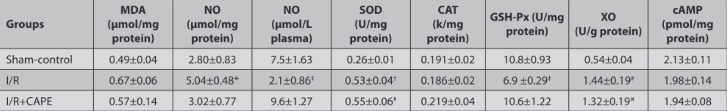
CAPE Slightly Modulates the Oxidant and Antioxidant Defense System
Transient global cerebral ischemia caused to significant increase in NO levels in brain homogenates, and this elevation was significantly inhibited by CAPE treatment
(P<0.05, Table 3). On the other hand, NO production was
decreased in plasma samples of occluded animal, but
CAPE reserved plasma NO suppression (P<0.05, Table
3). CAPE non-significantly decreased ischemia-induced
cerebral cortex MDA concentration (Table 3). The activity of
cerebral cortex XO significantly increased in the ischemic
group (P<0.05, Table 3), but CAPE non-significantly
decreased this elevation. Significant increase in cerebral
cortex SOD activity in the ischemia group (P<0.01, Table
3) was not effectively changed by CAPE. While there was
no difference in the cerebral cortex CAT activity between the groups, CAPE significantly prevented the reduction
in GSH-Px activity caused by ischemia (P<0.05, Table 3).
Table 2. List of primer sequences used for RT-PCR analyses Tablo 2. RT-PCR analizi için kullanılan primer dizileri
Gene Forward Primer (5’-3’) Reverse Primer (5’-3’)
PDE4A GCG GGA CCT AGC TGA AGA AAT TCC CAG GGT GAG TCC ACA TCG TGG
PDE4B CAG CTC ATG ACC CAG ATA AGT GG GTC TGC ACA AGT GTA CCA TGT TGC G
PDE4C ACT GAG TCT GCG CAG GAT GG CAC TCC TCT TCC TCT GCT CTC CTC
PDE4D CCC TCT TGA CTG TTA TCA TGC ACA CC GAT CCT ACA TCA TGT ATT GCA CTG GC
β-actin CAT CGT CAC CAA CTG GGA CGA C CGT GGC CAT CTC TTG CTC GAA G
Table 3. Effects of CAPE administration (15 µmol kg-1) on the antioxidant-oxidant enzyme activities and cAMP levels in cerebral cortex, and NO in plasma after 24 h ischemia-reperfusion injury
Tablo 3. İskemi-reperfüzyon hasarından 24 saat sonra plasma NO ile cerebral cortex antioksidant-oxidant enzim aktiviteleri ve cAMP düzeyleri üzerine KAFE
(15 µmol kg-1)’ in etkileri
Groups (µmol/mg MDA
protein) NO (μmol/mg protein) NO (μmol/L plasma) SOD (U/mg protein) CAT (k/mg protein) GSH-Px (U/mg
protein) (U/g protein)XO
cAMP (pmol/mg protein) Sham-control 0.49±0.04 2.80±0.83 7.5±1.63 0.26±0.01 0.191±0.02 10.8±0.93 0.54±0.04 2.13±0.11 I/R 0.67±0.06 5.04±0.48* 2.1±0.86‡ 0.53±0.04† 0.186±0.02 6.9 ±0.29‡ 1.44±0.19# 1.98±0.14 I/R+CAPE 0.57±0.14 3.02±0.77 9.6±1.27 0.55±0.06# 0.219±0.04 10.6±1.22 1.32±0.19* 1.94±0.08
Data were presented as mean ± S.E. from six rats in each group. * P<0.05, # P<0.05, † P<0.01 vs sham-control group, ‡ P<0.05 vs sham-control and I/R+CAPE group, I/R: Ischemia-reperfusion
Fig 1. Effects of CAPE on neurologic deficit scores after 24 h ischemia-reperfusion injury.
Ischemia-stimulated neurologic deficit scores were attenuated by CAPE. Data are presented as mean ± S.E. from seven rats in each group. † P<0.01 vs sham-control group. I/R: ischemia/reperfusion
Şekil 1. İskemi-reperfüzyondan 24 saat sonra nörolojik hasar skorları üzerine KAFE’in etkisi. İskemi
ile uyarılan nörolojik hasar skorları KAFE tarafından azaltıldı. Değerler herbir gruptaki 7 rat’ın ortalama ± standart hatası olarak verildi. † P<0.01, sham-kontrol grubu ile karşılaştırıldığında. I/R:
iskemi/reperfüzyon
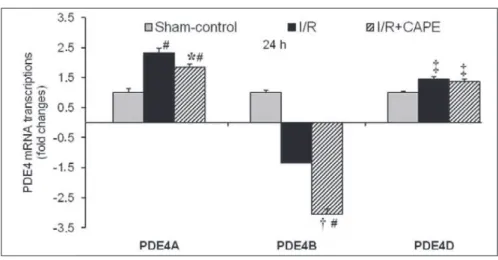
Fig 2. The fold change values of PDE4A, PDE4B and PDE4D mRNA transcriptions in qRT-PCR analyses
after 24 h ischemia-reperfusion injury.
Global cerebral I/R compared to sham-control PDE4A and PDE4D transcriptions were elevated by 2.31 and 1.44-folds after 24 h, respectively. PDE4B transcription was decreased by 2.34-fold. CAPE compared to I/R significantly decreased PDE4A and PDE4B transcriptions by 1.25 and 2.26-folds, respectively. Non-significant decrease of PDE4D transcription was detected after 24 h. Neither global cerebral I/R nor CAPE unchanged PDE4C transcription in the rat cerebral cortex (data not shown)
Data were presented as mean ± S.E. from six rats in each group. * P<0.05, † P<0.001 vs I/R group; ‡ P<0.05, # P<0.001, vs sham-control group; I/R: Ischemia/reperfusion
Şekil 2. İskemi-reperfüzyon hasarından 24 saat sonra qRT-PCR analizinde PDE4A, PDE4B ve PDE4D
mRNA kat değişim değerleri.
Sham-kontrol ile karşılaştırıldığında global beyin I/R, PDE4A ve PDE4D transkripsiyonlarını 24 saat sonra söylendiği sıra ile 2.31 ve 1.44 kat arttırdı. PDE4B transkripsiyonu 2.34 kat azaltıldı. I/R ile karşılaştırıldığında KAFE, PDE4A ve PDE4B transkripsiyonlarını söylendiği sıra ile 1.25 ve 2.26 kat önemli oranda azalttı. PDE4D transkripsiyonu 24 saat sonra önemsiz oranda azaldı. Global I/R ve KAFE rat beyin korteksinde PDE4C transkripsiyonunu değiştirmedi (veri gösterilmedi).
Değerler herbir gruptaki 6 rat’ın ortalama ± standart hatası olarak verildi. * P<0.05, † P<0.001, I/R
grubu ile karşılaştırıldığında; ‡ P<0.05, # P<0.001, sham-kontrol grubu ile karşılaştırıldığında; I/R:
CAPE Inhibits Global Cerebral
Ischemia-Induced Increases in PDE4 mRNA Expression
Ischemia-induced cerebral cortex cAMP levels were not
changed by CAPE treatment (Table 3). Global cerebral I/R
compared to sham-control raised PDE4A (P<0.001) and PDE4D (P<0.05) mRNA transcrips by 2.31 and 1.44-folds
in rat cerebral cortex 24 after I/R, respectively (Fig. 2),
and also decreased PDE4B by 2.34-fold (Fig. 2, P<0.001).
Whereas, CAPE treatment significantly decreased PDE4A (P<0.05) and PDE4B (P<0.001) expressions compared to
I/R by 1.25 and 2.26-folds 24 h after I/R, respectively (Fig.
2). CAPE has also non-significantly decreased PDE4D
transcription by 1.05-fold. Neither ischemia-reperfusion nor CAPE unchanged the PDE4C mRNA transcription in the rat cerebral cortex (data not shown).
DISCUSSION
So far, CAPE treatment has not been reported how it affects the levels of cAMP and PDE4 transcripts. We found that the global cerebral ischemia/reperfusion (I/R) and CAPE treatment non-significantly decreased in cerebral
cortex cAMP levels after 24 h reperfusion (Table 3). Our
cAMP findings are partially consistent with those of Choi
et al.[21] 24 h after I/R. As similar to that reported in an
earlier study [22], CAPE treatment may influence the cAMP
levels by increasing activity of cAMP-responsive element binding protein, and thus it may inhibit ischemia-induced
oxidative stress and inflammation. A recent study [9]has
demonstrated that cerebral ischemia led to increases in activity of PDE, primarily PDE4. However, it is still unknown whether specific PDE4 subtypes are differentially expressed after global cerebral I/R injury in rat cerebral cortex. This study firstly explains that the cerebral I/R significantly raised PDE4A and PDE4D expressions in the rat cerebral cortex at 24 h reperfusion, and also PDE4B mRNA was decreased
(Fig. 2). Accordingly, a more recent study reports increased
PDE4D expression following global cerebral ischemia [23]. In
addition, despite different experimental models, our data are in agreement with PDE4A increases newly reported by
traumatic brain injury [24]. The phosphodiesterase (PDE4) is
the predominant PDE isozyme in various leukocytes and plays an important role in the regulation of inflammatory
cell activation [7]. Consistent with our data previous studies
reported that the brain PDE4A, PDE4B and PDE4D mRNA transcripts are highly expressed, whereas PDE4C is
absent [25,26]. Furthermore, previous studies observed that
chronic antidepressant treatment increased PDE4A and PDE4B gene expression in rat cerebral cortex, but PDE4D
gene expressions were unchanged [25,27,28]. Additionally,
our results firstly shown that CAPE treatment significantly decreased PDE4A and PDE4B transcripts 24 h after I/R
compared to I/R (Fig. 2). However, PDE4D transcription
was not significantly changed (Fig. 2). The current study
clearly suggests that CAPE treatments produce the
neuro-protective effect by reducing the levels of some PDE4 isosyme transcriptions with a mechanism similar to phosphodiesterase inhibitors, and also may be useful for the treatment of cerebral ischemia.
The present study clearly showed that the cerebral cortex NO production was stimulated 24 h after global
cerebral I/R (Table 3), and this was inhibited by CAPE
treatment. CAPE, a structural derivative of flavonoids, possesses its antioxidant properties by inhibiting the gene expressions and/or catalytic activity of certain free
radical producing enzymes such as NOS [10,29]. In consistent
with the NO sera data presented here (Table 3), Tsai and
colleagues[1] noted that pretreatment with CAPE increased
NO bioavailability in plasma at 24 h of reperfusion in rats subjected to focal and global brain ischemia. CAPE is able to inhibit the reaction of NO with superoxide anion to prevent the formation of peroxinitrites which is more
toxic oxidant than either NO or superoxide anion alone [1].
It was previously reported that NO has protective effects during ischemic injury, although in a narrow concentration range, overproduction may facilitate or mediate
neuro-toxicity [5,6,30]. Indeed, NO could act with a dual action
either protective or pro-oxidant [6]. This dual effect of
NO in cerebral ischemic injury has been suggested that the effects depend on the stage of evolution of tissue damage and NOS isoforms. Immediately after induction of ischemia, NO is synthesized by endothelial NOS (eNOS) and neuronal NOS (nNOS), but later times after ischemia
NO is synthesized by iNOS [5]. As previously reported [5,6],
we suggest that an increase in NO production may be due to stimulation of all NOS subtypes expression in the brain. In addition, the increase in NO bioavailability induced by CAPE is attributed to its strong free radical scavenging ability in lipophilic environments.
Xanthine oxidase (XO) is an important oxidant enzyme
which catalyzes the reduction of O2 initiating to the
formation of superoxide anion and H2O2. In the present
study, ischemia-stimulated increases in the XO activity
could be reduced by CAPE (Table 3). A few studies have
reported similar results in the XO activity in relation to
I/R injury, which is time and dose dependent manner [1,11].
Similar to our study, it is demonstrated that ischemic circumstances lead to the accumulation of hypoxanthine
and stimulates XO activity [29]. The activity of GSH-Px, which
detoxifies H2O2 while oxidizing reduced GSH to oxidized
GSSG, was depressed during I/R [4]. In the present study,
cerebral cortex GSH-Px activity significantly decreased after 24 h ischemia. However, the decrease in the GSH-Px
activity was prevented by CAPE treatment (Table 3). The
curative effect of CAPE on GSH-Px activity could be due
to scavenging ROS produced during oxidative stress [31].
Superoxide dismutase (SOD) is an antioxidant enzyme which plays key role to convert the superoxide anion to
less toxic compound H2O2 and molecular oxygen. Toyoda
ischemic regions of brain parallel to this study (Table 3).
Horakova and colleagues [33]have reported a reduction
in GSH-Px activity, but an increase in the SOD activity in rat brain ischemia model induced by the ligation of the common carotid artery for 260 min and followed by a
reperfusion of 10 min. Another study [34] reported that
MnSOD increased in hippocampus 24, 48 and 72 h after ischemia, coincident with the marked reduction in the activity of glutathione-related enzymes. In the presented study, CAPE exhibited a slight antioxidant effect with regarding to cerebral cortex SOD and CAT enzyme activities
(Table 3). Our CAT results are in agreement with the study
of Mishra and colleagues [35]. It has also been explained
the stimulation of antioxidant enzymes in brain I/R as
the transient substrate induction [36]. Therefore, it may
be explained that free radicals produced in moderate ischemia were not intense enough to affect the catalase activity which is kept in peroxisomes.
The neurological evaluations shown that CAPE treatment could attenuate ischemia-induced cerebral neurologic
deficit scores (Fig. 1). This finding is in agreement with our
previous study showing that CAPE reduces the infarction percentage and neurological damage against focal
permanent middle cerebral artery occlusion [11]. In addition
to its antioxidant properties, this neuroprotective effect is supported further by the decrease of PDE4 isoforms in this current study. Therefore, we propose that CAPE plays a protective role for therapy against neuronal death after transient BCCA occlusion with its preconditioning and therapeutic effects.
In conclusion, these results suggest that CAPE administrations slightly modulate the antioxidant defense system and NO release in rat brain during peracute global cerebral ischemia-reperfusion injury. In addition, CAPE treatment produces the neuroprotective effect by reducing the levels of some PDE4 isosyme transcriptions.
REFERENCES
1. Tsai SK, Lin MJ, Liao PH, Yang CY, Lin SM, Liu SM, Lin RH, Chih CL, Huang SS: Caffeic acid phenethyl ester ameliorates cerebral infarction
in rats subjected to focal cerebral ischemia. Life Sci, 78, 2758-2762, 2006.
2. Irmak MK, Fadillioglu E, Sogut S, Erdogan H, Gulec M, Ozer M, Yagmurca M, Gozukara ME: Effects of caffeic acid phenethyl ester
and alpha-tocopherol on reperfusion injury in rat brain. Cell Biochem Funct, 21, 283-289, 2003.
3. Zhou Q, Cao B, Niu L, Cui X, Yu H, Liu J, Li H, Li W: Effects of permissive
hypercapnia on transient global cerebral ischemia-reperfusion injury in rats. Anesthesiology, 112, 288-297, 2010.
4. Islekel S, Islekel H, Guner G, Ozdamar N: Alterations in superoxide
dismutase, glutathione peroxidase and catalase activities in experimental cerebral ischemia-reperfusion. Res Exp Med, 199, 167-176, 1999.
5. Atochin DN, Clark J, Demchenko IT, Moskowitz MA, Huang PL:
Rapid cerebral ischemic preconditioning in mice deficient in endothelial and neuronal nitric oxide synthases. Stroke, 34, 1299-1303, 2003.
6. Gourine AV, Bulhak AA, Gonon AT, Pernow J, Sjoquist PO: Cardio
protective effect induced by brief exposure to nitric oxide before myocardial ischemia reperfusion in vivo. Nitric Oxide, 7, 210-216, 2002.
7. Omori K, Kotera J: Overview of PDEs and Their Regulation. Circ Res,
100, 309-327, 2007.
8. Houslay MD, Adams DR: PDE4 cAMP phosphodiesterases: Modular
enzymes that orchestrate signalling cross-talk, desensitization and compartmentalization. Biochem J, 370, 1-18, 2003.
9. Li LX, Cheng YF, Lin HB, Wang C, Xu JP, Zhang HT: Prevention of
cerebral ischemia-induced memory deficits by inhibition of phospho-diesterase-4 in rats. Metab Brain Dis, 26, 37-47, 2011.
10. Song YS, Park EH, Hur GM, Ryu YS, Lee YS, Lee JY, Kim YM, Jin C:
Caffeic acid phenethyl ester inhibits nitric oxide synthase gene expression and enzyme activity. Cancer Lett, 175, 53-61, 2002.
11. Altug ME, Seraslan Y, Bal R., Kontas T, Ekici F, Melek IM, Aslan M, Duman T: Caffeic acid phenethyl ester protects rabbit brains against
permanent focal ischemia by antioxidant action: A biochemical and planimetric study. Brain Res, 1201, 135-142, 2008.
12. Katz L, Ebmeyer U, Safar P, Radovsky A, Neumar R: Outcome
model of asphyxial cardiac arrest in rats. Outcome model of asphyxial cardiac arrest in rats. J Cerebr Blood F Met, 15, 1032-1039, 1995.
13. Geocadin RG, Ghodadra R, Kimura T, Lei H, Sherman DL, Hanley DF, Thakor NV: A novel quantitative EEG injury measure of global
cerebral ischemia. Clin Neurophysiol, 111, 1779-1787, 2000.
14. Brambrink AM, Koerner IP, Diehl K, Strobel G, Noppens R, Kempski O: The antibiotic erythromycin induces tolerance against transient
global cerebral ischemia in rats (pharmacologic preconditioning). Anesthesiology, 104, 1208-1215, 2006.
15. Bradford MM: A rapid and sensitive method for the quantification
of microgram quantities of protein utilizing the principle of protein-dye binding. Anal Biochem, 72, 248-254, 1976.
16. Yoshoiko T, Kawada K, Shimada T: Lipit peroxidation in maternal
and cord blood and protective mechanism aganist actived-oxygen toxicity in the blood. Am J Obstet Gynecol, 135, 372-376, 1979.
17. Luck H: Catalase. In, Bergmeyer HU (Ed): Methods in Enzyme
Analysis. Verlag-Chemic. Weinheim/Bergstrasse, Germany. 1965.
18. Sun Y, Oberley LW, Ying L: A simple method for clinical assay of
superoxide dismutase. Clin Chem, 34, 497-500, 1988.
19. Cortas NK, Wakid NW: Determination of inorganic nitrate in serum
and urine by a kinetic cadmium-reduction method. Clin Chem, 36, 440-443, 1990.
20. Prajda N, Weber G: Malignant transformation-linked imbalance:
decreased xanthine oxidase activity in hepatomas. FEBS Lett, 59, 245- 249, 1975.
21. Choi JM, Shin KS, Kim KY, Lee JH, Hong KW: Neuroprotective effect
of cilastozol against focal cerebral ischemia via antiapoptotic action in rats. J Pharm Exp Therap, 300, 787-793, 2002.
22. Montpied P, Bock F, Rondouin G, Niel G, Briant L, Courseau AS, Lerner-Natoli M, Bockaert J: Caffeic acid phenethyl ester (CAPE)
prevents inflammatory stress in organotypic hippocampal slice cultures. Brain Res Mol Brain Res, 23, 111-120, 2003.
23. He Z, He B, Behrle BL, Fejleh MPC, Cui L, Paule MG, John L: Greenfield
ischemia-induced increase in microvascular phosphodiesterase 4 d expression in rat hippocampus associated with blood brain barrier permeability: Effect of age. Neuroscience, 3, 428-432, 2012.
24. Oliva AA, Kang Y, Furones C, Alonso OF, Bruno O, Dietrich WD, Atkins CM: Phosphodiesterase isoform-specific expression induced by traumatic
brain injury. J Neurochem, 123, 1019-1029, 2012.
25. Takahashi M, Terwilliger R, Lane C, Mezes PS, Conti M, Duman RS:
Chronic antidepressant administration increases the expression of cAMP phosphodiesterase 4A and 4B isoforms. J Neurosci, 19, 610-618, 1999.
26. D’Sa C, Eisch AJ, Bolger GB, Duman RS: Differential expression
and regulation of the cAMP-selective phosphodiesterase type 4A splice vari- ants in rat brain by chronic antidepressant administration. Eur J Neurosci, 22, 1463-1475, 2005.
27. Ye Y, Conti M, Houslay MD, Faroqui SM, Chen M, O’Donnell JM:
Noradrenergic activity differentially regulates the expression of rolipram-sensitive, high-affinity cyclic AMP phosphodiesterase (PDE4) in rat
brain. J Neurochem, 69, 2397-2404, 1997.
28. Ye Y, Jackson K, O’Donnell JM: Effects of repeated antidepressant
treatment of type 4A phosphodiesterase (PDE4A) in rat brain. J Neurochem, 74, 1257-1262, 2000.
29. Hosnuter M, Gurel A, Babuccu O, Armutcu F, Kargi E, Isikdemir A:
The effect of caffeic acid phenethyl ester on lipid peroxidation and nitric oxide levels in the plasma of rats following thermal injury. Burns, 30, 121-125, 2004.
30. Dalkara T, Yoshida T, Irikura K, Moskowitz MA: Dual role of
nitric oxide in focal cerebral ischemia. Neuropharmacology, 33, 1447-1452, 1994.
31. Griffith OW: Biologic and pharmacologic regulation of mammalian
glutathione synthesis. Free Radic Biol Med, 27, 922-935, 1999.
32. Toyoda T, Lee KS: Differential induction of superoxide dismutase
in core and penumbra regions after transient focal ischemia in the rat neocortex. Neurosci Lett, 235, 29-32, 1997.
33. Horakova L, Uraz V, Ondrejickova O, Lukovic L, Juranek I: Effect of
stobadine on brain lipid peroxidation induced by incomplete ischemia and subsequent reperfusion. Biomed Biochim Acta, 50, 1019-1025, 1991.
34. Candelario-Jalil E, Mhadu NH, Al-Dalain SM, Martínez G, Leó OS:
Time course of oxidative damage in different brain regions following transient cerebral ischemia in gerbils. Neurosci Res, 41, 233-241, 2001.
35. Mishra OP, Papadopoulos MD, Wagerle LC: Anti-oxidant enzymes
in the brain of newborn piglets during ischemia followed by reperfusion. Neuroscience, 35, 211-215, 1990.
36. Stanimirovic DB, Micic DV, Markovic M, Spatz M, Mrsulja BB:
Therapeutic window for multiple drug treatment of experimental cerebral ischemia in gerbils. Neurochem Res, 19, 189-194, 1994.