Saudi J Kidney Dis Transpl 2020;31(5):1051-1056
© 2020 Saudi Center for Organ Transplantation
Original Article
The Effect of Renal Transplantation on Cardiac Functions
Kerem Can Yilmaz1, Arzu Neslihan Akgun1, Suzan Keskin1, Orcun Ciftci1, Gokhan Moray2, Haldun Muderrisoglu1, Mehmet Haberal2
1
Department of Cardiology, Başkent University Faculty of Medicine, Ankara,2Department of General Surgery, Division of Transplantation, Başkent University Faculty of Medicine,
Ankara, Turkey
ABSTRACT. Chronic renal failure is a well-known risk factor for cardiovascular poor outcome.
Despite advances in dialysis and renal transplantation, these patients still have high cardio-vascular morbidity and mortality. The aim of our study was to evaluate the changes in blood parameters and echocardiographic parameters of patients undergoing renal transplantation in our center. One hundred and eighty-three patients who underwent renal transplantation between September 2012 and January 2016 were included in the study. Pre- and postoperative hemoglobin values, lipid profiles, ejection fractions, presence of left ventricular hypertrophy, presence of diastolic dysfunction, and valve pathologies were retrospectively scanned. Data were obtained from all patients in terms of blood parameters, but we compared 92 patients’ echocardiographic data because of lack of both pre- and postoperative echocardiography records. In our study, 124 patients (67.8%) were male, and the mean age was 42.6 ± 14.4 years. Hemoglobin levels (11.2 ± 1.98, 12.7 ± 2.2 mg/dL, P <0.001) and high-density lipoprotein (HDL) values (37.6 ± 10.5, 46.6 ± 13.6 mg/dL, P <0.001) were found to be different significantly. In echocardiographic evaluation, there was no difference between pre- and postoperative ejection fractions in 92 patients. However, patients with preoperative ejection fraction <50% had a significant increase in postoperative ejection fraction (40.1 ± 6.2, 48.4% ± 9.4%, P = 0.012). Renal transplantation can improve left ventricle ejection fraction in patients with basal ejection fraction less than 50% and also provide a significant increase in hemoglobin and HDL levels in all patients. This suggests that renal transplantation may reverse the process for dilated cardiomyopathy and may improve cardiac function in patients with low ejection fraction. However, transplantation should be performed as early as possible in these patients.
Correspondence to: Dr. Kerem Can Yilmaz, Department of Cardiology,
Başkent University Faculty of Medicine, Ankara, Turkey.
E-mail: [email protected]
Introduction
Chronic renal failure is a well-known risk factor for cardiovascular poor outcome.1 Despite advances in dialysis and renal trans-plantation, these patients still have high cardio-vascular morbidity and mortality.2-4 Renal
Saudi Journal
of Kidney Diseases
and Transplantation
transplantation provides longer survival and better quality of life in long-term dialysis patients.2 Despite significant progress in the care of patients with heart failure (HF), indi-viduals with ESRD and concomitant cardiac dysfunction are generally considered less suitable candidates for kidney transplantation due to increased risk of operative morbidity and mortality. Therefore, the coexistence of HF and renal failure becomes important. In fact, some previous studies have shown that kidney transplantation improves cardiac func-tion.5-7 The aim of our study was to evaluate the changes in blood parameters and echo-cardiographic parameters of patients under-going renal transplantation in our center.
Materials and Methods
Two hundred and thirteen patients were included in the study who underwent renal transplantation between September 2012 and January 2016 in our center. Pre- and post-operative hemoglobin values, lipid profiles, ejection fractions, presence of left ventricular hypertrophy, presence of diastolic dysfunction, and mitral and tricuspid valve insufficiencies were retrospectively scanned. Thirty patients who could not reach any of the pre- or post-operative blood or echo parameters were excluded from the study. Thirty patients were excluded from the study who we could not reach one of the pre- and postoperative blood parameters; as a result, we examined 183 patients’ laboratory parameters. Likewise, since the pre- and postoperative echocardio-graphy results of 92 of these 183 patients were obtained, the data of these 92 patients were examined. The reference values for echo para-meters were defined based on the American Society of Echocardiography guidelines.8 Laboratory parameters were obtained from the most recent pretransplant and 12-month post-transplant office visits. One exception was echocardiographic data, the timing of which could not be controlled for in this retrospective study. Echocardiograms were obtained during the pretransplant evaluation, with the most recent one taken into account and the time
closest to 12–24 months posttransplantation. Continuous data were compared preope-ratively and postopepreope-ratively with K-related samples. Data are expressed as the mean ± standard deviation unless otherwise stated. Categorical data were analyzed with Fisher’s exact test and the Chi-squared test. A P <0.05 was considered statistically significant. All analyses were undertaken using the IBM SPSS Statistics version 25.0 (IBM Corp., Armonk, NY, USA).
Protocols approved by the local ethics committee were utilized throughout the study. A standardized questionnaire was used to collect clinical and demographic information, including medication history.
Results
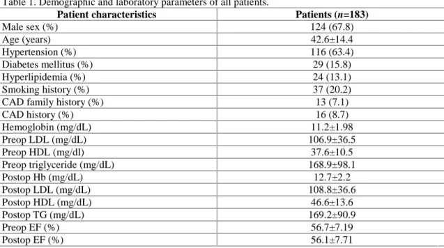
In our study, 124 patients (67.8%) were male, and the mean age was 42.6 ± 14.4 years. The demographic data of the patients in the study are given in Table 1. In terms of blood para-meters, preoperative hemoglobin levels were 11.2 ± 1.98 mg/dL, postoperative hemoglobin values were 12.7 ± 2.2 mg/dL, and this diffe-rence was statistically significant (P = 0.001). High-density lipoprotein (HDL) values, one of the lipid parameters, were significantly increased postoperatively (37.6 ± 10.5, 46.6 ± 13.6 mg/dL, respectively, P = 0.001). There was no significant change in low-density lipoprotein (LDL) and triglyceride levels.
For echo parameters, there was no significant difference between the values of 92 patients whose ejection fractions were screened pre-operatively and postpre-operatively, whereas 21 patients with a preoperative ejection fraction below 50% had a significant increase after transplantation (40.1% ± 6.2%, 48.4% ± 9.4%, P = 0.012) (Table 2).
Patients’ immunosuppressive treatment and other medical treatments are given in Table 3. All of the patients were given deltacortril. The most preferred immunosuppressive agents were mycophenolate mofetil (MMF) and tacrolimus.
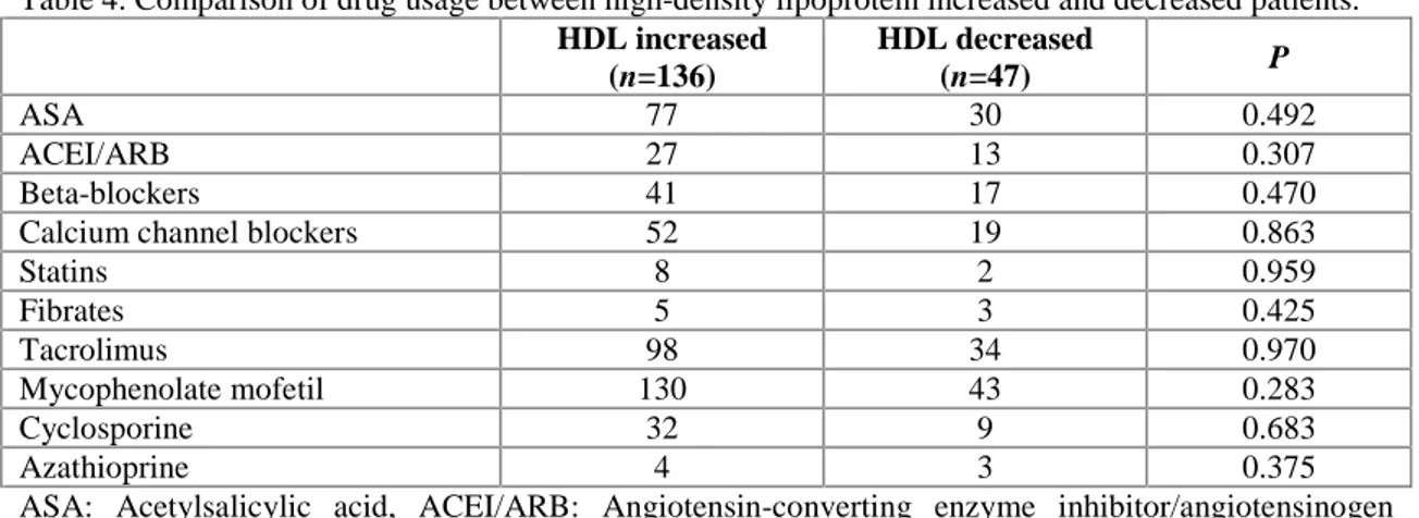
Therefore, to investigate the effect of other drugs that affect HDL increase, we compared the drugs for two groups of patients whose
Table 1. Demographic and laboratory parameters of all patients.
Patient characteristics Patients (n=183)
Male sex (%) 124 (67.8) Age (years) 42.6±14.4 Hypertension (%) 116 (63.4) Diabetes mellitus (%) 29 (15.8) Hyperlipidemia (%) 24 (13.1) Smoking history (%) 37 (20.2)
CAD family history (%) 13 (7.1)
CAD history (%) 16 (8.7) Hemoglobin (mg/dL) 11.2±1.98 Preop LDL (mg/dL) 106.9±36.5 Preop HDL (mg/dl) 37.6±10.5 Preop triglyceride (mg/dL) 168.9±98.1 Postop Hb (mg/dL) 12.7±2.2 Postop LDL (mg/dL) 108.8±36.6 Postop HDL (mg/dL) 46.6±13.6 Postop TG (mg/dL) 169.2±90.9 Preop EF (%) 56.7±7.19 Postop EF (%) 56.1±7.71
CAD: Coronary artery disease, Preop: Preoperative, Postop: Postoperative, LDL: Low-density lipoprotein, HDL: High-density lipoprotein, TG: Triglyceride, Hb: Hemoglobin, EF: Ejection fraction. Table 2. Comparison of laboratory parameters and ejection fraction in pre- and postoperative patients.
Preoperative values Postoperative values P
Hemoglobin (mg/dL) 11.2±1.98 12.7±2.2 0.001 HDL (mg/dL) 37.6±10.5 46.6±13.6 0.001 LDL (mg/dL) 106.9±36.5 108.8±36.6 0.428 Triglyceride (mg/dL) 168.8±98.1 169.1±90.6 0.919 EF (%) (n=92) 56.7±7.19 56.1±7.71 0.509 EF preoperative %50< (%) (n=21) 40.1 ± 6.2 48.4±9.4 0.012
LDL: Low-density lipoprotein, HDL: High-density lipoprotein, EF: Ejection fraction, Preop: Preoperative.
Table 3. Drug usage of all patients.
Patients (n=183)
ASA (%) 107 (57.8)
ACEI/ARB (%) 40 (21.6)
Beta-blockers (%) 58 (31.4)
Calcium channel blockers (%) 71 (38.4)
Statins (%) 10 (5.4) Fibrates (%) 8 (4.3) Tacrolimus (%) 132 (72.1) Mycophenolate mofetil (%) 173 (94.5) Cyclosporine (%) 41 (22.2) Azathioprine (%) 7 (3.8)
ASA: Acetylsalicylic acid, ACEI/ARB: Angiotensin-converting enzyme inhibitor/angiotensinogen receptor blocker.
HDL levels raised and decreased after trans-plantation, as shown in Table 4. There was no difference between the groups in terms of the drugs.
According to the other echocardiographic parameters such as left ventricular hyper-trophy, valve insufficiencies, and diastolic dysfunction, there were no significant diffe-rences between the two groups (Table 5).
Discussion
Renal transplantation decreases mortality in end-stage renal disease patients. Previously, there are several studies investigating the effects of renal transplantation on cardiac functions. In a study performed with SPECT imaging,9 renal transplant patients were com-pared with nontransplant patients, and a signi-ficant change was observed in the ejection fractions of transplanted patients (72 ± 10% vs. 67% ± 10%, P =0.001). Although the
improvement in ejection fraction was thought to be due to volume management, no signi-ficant changes were found in the left ventri-cular end-diastolic volumes between the two groups. This was thought to be due to the effective removal of toxins and the prevention of uremic cardiomyopathy rather than the effect of transplantation on volume. In the same study, electrocardiographic parameters were also screened, but no significant diffe-rence was found. In our study, there were no significant differences in ejection fractions when 92 patients were considered. However, in patients with ejection fraction of 50% and less, ejection fraction was significantly improved after transplantation (40.1 ± 6.2 vs. 48.4 ± 9.4, P = 0.012). One reason of this situation may be due to the high mean ejection fraction in pretransplant echocardiographies. In our study, the number of patients who compared their echoes was 92 and higher like the previous study. On the other hand, it can be predicted
Table 4. Comparison of drug usage between high-density lipoprotein increased and decreased patients.
HDL increased (n=136) HDL decreased (n=47) P ASA 77 30 0.492 ACEI/ARB 27 13 0.307 Beta-blockers 41 17 0.470
Calcium channel blockers 52 19 0.863
Statins 8 2 0.959 Fibrates 5 3 0.425 Tacrolimus 98 34 0.970 Mycophenolate mofetil 130 43 0.283 Cyclosporine 32 9 0.683 Azathioprine 4 3 0.375
ASA: Acetylsalicylic acid, ACEI/ARB: Angiotensin-converting enzyme inhibitor/angiotensinogen receptor blocker.
Table 5. Comparison of pre- and postoperative echocardiography parameters.
Preoperative echo parameters
Postoperative echo
parameters P
Left ventricular hypertrophy 74 65 0.05
Left ventricle diastolic dysfunction
Stage 1 46 31 0.135
Stage 2 8 7 0.272
Stage 3 1 2 0.898
Mitral regurgitation mild 43 38 0.535
Mitral regurgitation moderate 4 7 0.612
Tricuspid regurgitation mild 35 33 0.879
that patients with low ejection fraction may benefit more than patients with normal ejection fraction.
In another study,10 patients with ejection fraction below 50% before renal transplan-tation were examined, and it was found that EF was significantly improved in patients with both mild and moderate HFs after transplan-tation. In 66 patients with mild left ventricular systolic dysfunction, EF before transplantation was 41% ± 10% and 50% ± 12% after trans-plantation (P <0.0001). In patients with mode-rate dysfunction (n = 28), EF was 32% ± 7% before transplantation and 47% ± 14% after transplantation (P <0.001). This suggests that the ejection fraction is not the most important value for determining the preoperative cardiac risk. In the same study, they found that the best survival was determined in patients with normal ejection fraction. Survival of patients with more than 10% improvement in ejection fraction after transplantation was significantly better than patients with <10% improvement in ejection fraction. In our study, mortality was observed in only six patients during follow-up, so no data were compared for survival.
Omrani et al11 studied 181 patients with ejection fraction below 50% and performed echocardiography at six and 12 months. Echo-cardiography showed a significant improve-ment in ejection fractions compared to basal echoes both six and 12 months. In the same study, they found that left ventricular hyper-trophy also regressed and mitral and tricuspid valve insufficiencies decreased after transplan-tation. In another study examining left ventri-cular hypertrophy (LVH)12 post transplan-tation, 63% of renal transplant recipients showed normal echocardiographic finding of LV, while 37% of patients remained with LVH after first post-transplant year. These findings were similar to our study. In our study, left ventricular hypertrophy regressed after transplantation (P = 0.05). Unlike, there was no significant difference in valve insuffi-ciencies before and after transplantation (Table 5). This may be due to the small number of patients with severe valve insufficiency in our study cohort. There was no significant
diffe-rence between the two groups in terms of diastolic functions although they were reduced numerically.
Dyslipidemia is often seen with renal disease and is associated with increased cardiovascular mortality and morbidity.13 Immunosuppressants and steroids used after transplantation have important effects on lipid metabolism. In a comprehensive study in which these effects were investigated by drug classes,14 the highest change in total cholesterol and triglyceride was shown to be associated with cyclos-porine/sirolimus or cyclosporine/everolimus use. Moreover, it was shown that average HDL cholesterol decreased by 14% in men and 22% in women. However, in subgroup analyses, in patients who were receiving tacrolimus and mycophenolate mofetil, HDL cholesterol increased in men from 41 ± 10 mg/dL to 47 ± 12 mg/dL, in women HDL cholesterol increased from 47 ± 12 mg/dL to 56 ± 11 mg/dL. In our patient population, MMF (94.5%) and tacrolimus (72.1%) were the most commonly used drugs, and it may be the reason of increased HDL cholesterol. While LDL and triglyceride increased slightly, HDL cholesterol increased significantly after trans-plantation (37.6 ± 10.5 vs. 46.6 ± 13.6, P = 0.001). This emphasizes the importance of drug selection after transplantation. No significant difference was seen with the use of other drugs on HDL levels. In another study, serum total cholesterol and triglyceride levels tended to increase during CyA and steroid therapy among patients undergoing renal transplantation Independent of hyperlipidemia risk factors.15 Following the transplantation, the lipid profiles of the patients should be followed up during the controls and medical treatment should be kept in mind if LDL cholesterol is persistently higher which is a cardiovascular risk factor.
As a result, renal transplantation can improve left ventricle ejection fraction in patients with basal ejection fraction <50%. This suggests that the risk assessment of patients with low ejection fraction should be made carefully before transplantation because there will be improvement after transplantation in cardiac
functions. At the same time, lipid side effects of some immunosuppressive agents were observed less than others, so it could be a guide for treatment selection.
Conflict of interest: None declared.
References
1. Foley RN, Parfrey PS, Sarnak MJ. Clinical epidemiology of cardiovascular disease in chronic renal disease. Am J Kidney Dis 1998; 32:S112-9.
2. Wolfe RA, Ashby VB, Milford EL, et al. Comparison of mortality in all patients on dialysis, patients on dialysis awaiting trans-plantation, and recipients of a first cadaveric transplant. N Engl J Med 1999;341:1725-30. 3. Tonelli M, Wiebe N, Knoll G, et al. Systematic
review: Kidney transplantation compared with dialysis in clinically relevant outcomes. Am J Transplant 2011;11:2093-109.
4. Jardine AG, Gaston RS, Fellstrom BC, Holdaas H. Prevention of cardiovascular disease in adult recipients of kidney transplants. Lancet 2011;378:1419-27.
5. Wali RK, Wang GS, Gottlieb SS, et al. Effect of kidney transplantation on left ventricular systolic dysfunction and congestive heart failure in patients with end-stage renal disease. J Am Coll Cardiol 2005;45:1051-60.
6. Deng Y, Pandit A, Heilman RL, Chakkera HA, Mazur MJ, Mookadam F. Left ventricular torsion changes post kidney transplantation. J Cardiovasc Ultrasound 2013;21:171-6. 7. Namazi MH, Parsa SA, Hosseini B, et al.
Changes of left ventricular mass index among end-stage renal disease patients after renal transplantation. Urol J 2010;7:105-9.
8. Lang RM, Badano LP, Mor-Avi V, et al. Recommendations for cardiac chamber quantification by echocardiography in adults:
An update from the American Society of
Echocardiography and the European
Association of Cardiovascular Imaging. J Am Soc Echocardiogr 2015;28:1-39.e14.
9. Crosland W, Aggarwal H, Farag A, et al. The effect of renal transplantation on left ventricular function, electrocardiography, and mechanical synchrony by gated myocardial perfusion imaging. J Nucl Cardiol 2019;26: 1962-70.
10. Hawwa N, Shrestha K, Hammadah M, et al. Reverse remodelling and prognosis following kidney transplantation in contemporary patients with cardiac dysfunction. J Am Coll Cardiol 2015;66:1779-87.
11. Omrani H, Rai A, Daraei Z, Sadeghi M. Study of echocardiographic changes after kidney transplantation in end-stage renal disease patients. Med Arch 2017;71:408-11.
12. Dzemidzić J, Rasić S, Saracević A, et al. Predictors of left ventricular remodelling in kidney transplant recipents in the first posttransplant year. Bosn J Basic Med Sci 2010;10 Suppl 1:S51-5.
13. US Renal Data System: 1999 USRDS Annual Data Report. Bethesda, Md: The National Institutes of Health. National Institute of Diabetes and Digestive and Kidney Diseases; 1999.
14. Spinelli GA, Felipe CR, Park SI, Mandia-Sampaio EL, Tedesco-Silva H Jr., Medina-Pestana JO. Lipid profile changes during the first year after kidney transplantation: Risk factors and influence of the immuno-suppressive drug regimen. Transplant Proc 2011;43:3730-7.
15. Akman B, Uyar M, Afsar B, Sezer S, Ozdemir FN, Haberal M. Lipid profile during azathioprine or mycophenolate mofetil combinations with cyclosporine and steroids. Transplant Proc 2007;39:135-7