34 (2) 268-277, 1987
A SEROLOGIC SURVEY OF DOGS FOR BRUCELLA CANIS AND BRUCELLA ABORTUS AND EVALUATION OF MERCAPTOETHANOL
MICROAGGLUTINATION TEST
K. Serdar Diker1 Nejat Aydm2
Mustafa Özyurt4
Jale Erdeğer3
Köpeklerin Bruceıla canis ve Bruceıla abortus inCeksiyonlarl üzerinde serolojik bir tarama ve mercaptoethanol mikroaglütinasyon testinin değerlendirilmesi
Özet: Bu çalışmada, Brucella canis infeksi)'onlarının teşhisi için, mer-kaptoetanol mikroaglütinasyon testinin deiterlendirilmesi yapıldı. A)'7'lca, üç değişik kopek grubunda Br. canis ve Br. abortus irifeksiyonlarınıız sıklığı ince-lendi. Br. canis aglütininlerinin saptanması için Merkaptoetanol Tüp Aglü-tinasyon Testi (M E- TA T), Merkaptoetanol Mikro Aglütinasyon Testi (ME-MA T) ce Mikroaglütina.~yon Testi (AfA T) karşılaştınldı, Br. abortus irifeksi,yoııurwn teşhisi için mikroaglütitzas)'on testi kullanıldı. M E-TAT testinde 1:200 titrede pozitij reaksiyon, aktij Br. canis infeksiyonunun belirtisi olarak kabul edildi. BiL titre M E-MAT testinde 1 :40 olarak kabul edildi. M E- TA T ve M E- MAT sonuçları paralellik gösterdi. Br. canis infek-siyonlarının teşhisi bakımından AfAT güvenilir sonuçlar vermedi.
Incelenen 222 serumun 11 (% 6.3) ü 1:200 vlJ"a daha )'üksek titrede pozitif bulundu ve bunlar aktif Br. canis infeksi)'onu olarak kabul edildi. So-kak köpeklerinin % 15.6 sı, ev köpeklerinin % 4.5 inde infeksiyon saptan-masına karşın, askeri hizmet köpeklerinde irifeksiyon bulunmadı. Br. abortııs infeksiyonları )'o'nünden, birkaç serum çok düşük titrede IJozitij reaksiyon verdi ve bu durum infeksiyon belirtisi olarak kabili edilmedi.
Sunınıary: The application of micromodification of mercaptoethanol agglutination test to the serologic diagnosis qf Br. canis infection was evalZlated.
The prevalence of antibodies to Br. canis and Br. abortııs in three different
1 Dr., A.O. Veterinary Faculıy, Microbiology Department, Ankara 06110 2 Doç. Dr., 3 Araş. GÖL, A.Ü. Veterinary Faculıy, Microbiology Department .. An-kara 06110.
groups Of dogs was also inı'estigated. Sera were compared by AfA- TA T ( Mer-captoethanol Tube Agglııtination Test), ME-MAT (Mercaptoethanol Micro-Agglutination Test) MA T (Micro Agglutination Test) for agglutinins to Br. canis alZd tested h)! MA T for agglııtinins to Br. abortus. A titer of i:200 in M E- TA T was considered as indicative of active Br. canis infection. This titer corresponded to i:40 in ME-MA T. All results of ME-MAT correlated well with those of AfE- TA T. MATfor Br. canis infection did not give reliable result.
Of 222 sera examined, 14 (6.3 %) had a titer of 1:200 or more and these were considered as active Br. canis ilZfection. None of the military service dogs were positive for Br. canis whereas 15.6
%
of stray dogs and 4.5%
of pets were positiDe. In AfA T for Br. abortus, afew of sera gave positive reactionat low titer, and these were considered as negative test result.
Introduction
Brucella canis is well known as a cause of abortion and infertility in bitehes, epididymitis and testicular atrophy in male dogs (8, 16). Several studies have indicated that the disease is widcly distributed throught the world in many breeds of dogs. There is even serological evidence for infection in the wildlife population and in cat (23, 24).
i The results of scrosurveys of Br. canis antibodies in dogs indicated 30.5
%
positive reaction in Argentina (21), 2.9%
in Japan (25), 28%
in Mexico (lO), 0.3%
in Canada (5),8.2%
in Brasil (18) and i%
to 12%
in USA (4, 16, 26). In Turkey ,canine bruceliosis due to Br. calZiswas first recognized serologically in 1983 (17). Most infec-ted dogs are free of cilinical signs though many experience reproductive failure and loos vigor. Br. ahortus can also cause canine infection inf-requendy (3).The ability of Br. canis to infect human beings has also been es-tablishcd. Sporadic cases of human infection associated with labora-tory exposure to cultures and contact with infected dog have been reported (8, 20). There is serologic evidence of human infection due to Br. canis in Turkey (9). Laboratory methods are essential to iden-tify the presence of the disease as its cilinical signs may be very varied. Various serological methods have been developed for diagnosis of canine brucellosis: tube agglutination test with or without mercap-toethanol, slide agglutination test, complement fixation and agar-gel diffusion, using Br. canis or Br. ovis antigen (2, 7, LL). Since Br.
llr. canis
canis is naturally mucoid, the standart antigens and test procedures used for the diagnosis of bruceıla infeetions caused by smooth bru-cella, e.g. Br. aborlus can not be med for the diagnosis of canine disease caused by Br. canis (27).
The present report deals with tlıe application of micro-modifi-cation of ME-agglutination test to the serologic diagnosis of Br. canis infection. The prevalence of antibodies to Br. canis and Br. abarlus in three different groups of dogs has also becn investigated.
Materials and Methods
Serum samples: Blood samples were collected from 222 mature dogs, of which 64 were stray dogs, 88 were household pets and 70 were military service dogs. Sex and breed distinction was not included when a blood sample submitted. Each blood sample allowed to clot and after centrifugation, the serum was pipetted into screw-topped vials and stored at - 20 oC until med. Positive (high titer and medium titer) and negative control sera for Br. canis and Br. aborlus were inc-luded in each experiment.
Antigens: Br. canis ME- TAT antigen was kindly provided by Dr. G.M. Brown (USDA Diagnostic Reagent Section, Ames, lowa). \ Br. abarlus TATantigen was a product of Pendik Veterinary Research Institute, IstanbuL.
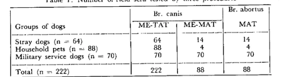
Serologic Tcsts: All control sera ,'Tre tested by Br. canis :ME--TAT, ME-MAT, MAT and Br. aborlus MAT to compare these tests and evaluate most reliable one and any cross-reaction. l'\umber of field samples investigated by Br. canis ME-TAT, :ME-MAT and Br. aborlus MAT is shown in Tablc i.
Br. canis ME-TAT: The method of "USDA diagnostic reagent ~ection" was med. Diluent for preparing the ME solution and test
Table ı. Number of field sera ıested by three procedurcs Br. abortus
Groups of dogs ME-TAT MAT
--- --- - --- ._---
---Stray dogs (n ~~ 64) Household pels (n =8B) Military service dogs (n =70)
64 88 70 14 4 70 14 4 70
---"--- ---_._-
--~--_._---Total (n =222) 222 88 88antigen was prepared by adding a ratio of 0.6 ml of formalized saline stoek solution (10
%
v/v) to 99.4 ml of 3.5%
saline solution. ME solution (O.i M) was prepared by addingaratio ofO. 715 ml 2- Mer-captoethanol (Merck) to 99.285 ml above diluent and pH was ad-justed to 8.5. Serum was diluted (two-fold beginning from i:50) in i ml of ME solution. Concentrated antigen was added (4.4 ml) to 95.6 ml of formalized 3.5%
saline, and i ml of this test antigen (final concentration = OD 0.9 at 550 nm) was added to each tube. Tubes were incubated at 37 cC for 18 hours.Br. canis ~E-MA T: All diluents and procedure were same of
ME- TAT except that sera were diluted in a total volume of O.i ml test reagents in microplate trays (two-fold beginning from 1:5) and plates were incubated at 37 oC for 24 hours and 4 cC for 3 hours. Cont-rol sera were also tested by using a more concentrated (OD 2. O at 550 nm) and compared witlı standart test antigen (OD 0.9 at 550 nm).
Br. canis MAT: All procedures wc re same of ME-MAT except
that ME was not used for preparing diluents.
Br. abortus MAT: Two-fold dilutions of sera were prepared in a volume of 0.05 ml and 0.05 ml of test antigen was added to each welL. The reaction was read after ineubation at 37 cC for 18 hours.
Positive reaction at i:200 or more di1ution in ME-TAT was considered as sero-positive. The results of other tests for Br. canis were evaluated after comparing with ME-TAT since no established criteria was available for tlıesc tests.
Results
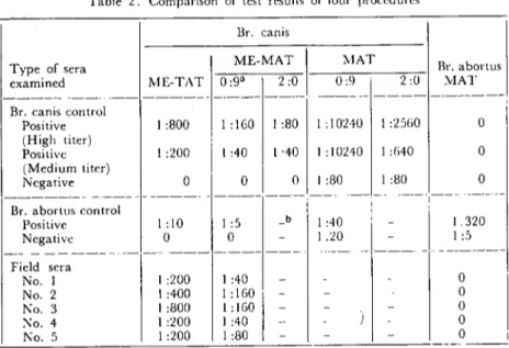
Comparison of titcrs of control sera and some of the posith"e field sera tested by three procedures for antibodies to Br. canis and one method for antibodies to Br. abortus are shown in Table 2. As compa-ring the results forBr. canis, positive rcaction at 1:200 titer in ME-TAT corresponded to 1:40 titer in ME-MAT. Positive reaction at i:40 or more in ME-MAT was considered as positive test result. All results of ME-MAT corrdated with those of ME-TAT. When comparing the antigen concentration in ME-MAT, the titer of positive result with 0.9 OD antigen was higher than wİth 2. O OD antİgen. In Br. canis MAT, posİtive control sera had very high titer (I: 10240) but negative control sera also gave high titer (I :80).
Table 2. Comparİson of test results of four procedures Br. canıs Br. a bOl'tus 2:0 YIAT Br. canis control Posiıive i :800 1 :160 i :80 i: 10210 ı:2:i(;0 (High ıiter) j Posİıive i :200 ı:40 1,40 1:10210 1:(;40 (Medium ıiıer)
i
Ncgaıive O O O 1:80 1:80 ME-MAT 1 Type of sera cxamincd l'vIE-TAT 0:9' 2:0 --- --- ..-- --- _._-- ---NIAT 0:9 , Oi
O O Br. aborltls canlrol Positive Negaıive Field sera No. ı No. 2 Ko. 3 );0. 4 Ko . .'i 1:10 O 1:200 ı:400 ı:800 1:200 1:200 ı:5 O 1:40 -i: 160, -i :160 i -1:40 , --i:80 -1:40 1.20 '.---"i--- .. _---"._-O O O O O i-_.---(a: opıieal density al 550 nm: b: Noı ıesıcd)
The incidence of sero-positive dogs of three different groups for Br. canis and Br. ahortus infection is shown in Table 3.
Table :3. Prcvalencc of sero-posiıive results İn three differenl groups of dogs for Br. cani, and Br. aborlus
Groups of dogs
i 7'io. of examined/posİtive ('Yo)
Br. canİs Br. aborlus
Stray dogs Household pet dogs Miliıary Service dogs
Toıal 64/10 (156; 88/4 ( 4.:» 70/o ( 0.0) 222[14 ( 6.3) 14/0 (0.0) 1/0 (0.0) 701o (O. O) 88/0 (0.0)
Of 222 sera examined, 14 (6.3 %) had a titer of i :i200 of more ın ME-TAT and these \Yere considered as active Br. canıs infection. None of the military service dogs were positive for agglutinins to Br. canıs whercas 15.6
%
(LO of 64) of stray dogs and 4.5%
(4 of 88) of pets were positive.Of 88 sera examined for antibodies to Br. ahortus, 4 sera (4.5 %) were positive at 1:5 titer and 4 sera (4.5 %) were positive at i: 1O titer. These reactions such a low titer not considered as positive result.
Significant cross-reaction was not obsen:ed between antigens and antisera ofBr. canis and Br. aborlus.
Discussion and Conclusion
Scrologic testing a relatively simplc method for diagnosis of Br. canis infcetian in dogs. Thcre is no complete agreement, however, on the best serologic test to use. Each author claims that his test is better than others. The ME-TAT and SAT are the most commonly v.sed procedures sİnce theyare simplc and reproducablc. The SAT is accurate when the results are negative, but lcss accurate when re-sults are positive (62.5
%
sensitive) (6). In 14.5%
of positive SAT reaction, Hubbert et al (16) failcd to confirm the results by ME-TAT. The M£- TAT enables detection of infection and eliminate most "faIse positive" results. The vcterinary use of ME-TAT for testing canin(~ serum is based on the observation that Ig?\1 antibodyin dogs is of no significance for inrcctivity (IS). Same investigators suggest an ME-TAT titer of i :100 as indicatiye ofBr. canis infection (14, 19). Others, ineluding World Health Organization Commission on bru-cellosis, reguire an M£- TA l' titer of i:200 or higher for positive re-sults (1,15). In this study it has been accepted a titer of i :200 as positive for canine bruceilosis, in accordance with the WHO commis-sion recommendation.Previous experience \,'ith microagglutination procedure led us to choose a modification of technigue previously reported (22). The result of ~1E-MAT which was modified in this study, correlated well with the results of ME-TAT. It was alsa observed that ME-MAT has some advantages. M£- TAT req uires elearing of the supematant fluid within 48 hours to be positive. In ~E-MAT this period was shortened to 24 hours. Other advantage of ME-MAT is that it needs less reagent and serum than in l\1E-TAT, to perform. The original ME-TAT requires large amount of antigen. Experiments with ME-MAT in which positive control sera for Br. aborlus were used have showed that Br. canis antigen does not cross-react with antiserum to Br. aborlus.
The reason of very high titer obtained in MAT with positive control sera for Br. canis antibodies may be non-specific agglutination. Same authors alsa pointed out that "faIse positive" reaction due to non-specific agglutination was main disadvantage of TAT (ll). Since negative control serum had also relatively high titer in this test, it is not a reliable test for diagnosis of canine brucellasis due to Br. canis.
Other purpose of the present study was to determine the pre-valenee of agglutinins to Br. (anis and to Br. aborlııs. This survey de-monstrated a prevalence of Br. (anis antibodies indicative of active infection to be approximately 6 times greater in stray dogs than in non-stray dogs. This difference is presumed to be rclated to an inc-reased oppurtinity of the stray group for exposure through multiple breedings and other contacts with infected dogs, as compared with the more restrieted mavement and decreased oppurtinity for expo-sure of the non-strays. :Most of the other workers have alsa indicatcd that the prevalence of positive serologic results in stray dogss is consi-derably higher than in non-stray dogs (12, 13).
One of the difficulties in making valid comparative evaluations of the results of Br. canis sera-survey has been the lack of standart procedures and test reagents. it is alsa difficult to compare percenta-ges reportcd, due to differences in cvaiuation of the titers obtained. Our finding of 15.6 % stray dogs with ME- TAT titer of i:200 or more is one of the largest percentage reported from all over the world. Infection rates reported for Mexico (I O) and Louisiana (16) have been greater and smaIler, respectivcly.
Although incidence of Br. caııis infection in house-hold pets is not as high as stray dogs, these animals are most likely a potential for human infection. Recent evideııce suggets that the prevalence of this disease in human, as well as its zoonotic potential, may be greater than suspected (22). The diagnosis of hu man infcetian is difficult, because routinly used Br. aborlus antigen does not cross-react with agglutinins to Br. (anis. This may Icad to misdiagnosed or un-derdiagnosed human cases. The demonstration of two cases of human bruceIlosis due to Br. (anis in Turkey shows a possible transmission from dogs to human and indicates the importance of subject (9).
Most of the sera tested wcre found negative in Br. aborlus MAT, only a few of them had law titers. These sera \vere from dogs of urban area where dogs can not feed with aborted fetuses infected with Br. aborlus. This may explain why dogs have low titers of agglutinins to Br. aborlus. On the other hand, it has alsa been reported that the lack of clinical signs produced and the variable agglutinin response after experimental infcetian indicates a marked resistance of the dogs to İnfection due to Br. aborlus (3).
Rcferences
1. Alton., G.G., Jones, L.M. and Pietz, D.E. (1975). Laboratory Techniqu£s in Brueel/osis. 2 nd Ed., p. 149-154. ,,,'olrd Health Organization, C;cneva.
2. Badakesh, F.F., Carmichael, L.L. and Douglass, J.A. (19ll2). Improred rapid slide agglutination test for presum/)lil'e diagnosis of eoni,,,, brurelfosis ..1. Clin. Microbiol., 15: 286-289.
3. Bickneıı, S.R. and Beıı, R.A. (I 979). B,ueella abortus in the biıch: subclinieal infeetion
associaled wiı/Z urhaı:)' emeıioıı ..1. Hyg., 82: 249--254.
4. Boebel, F.W., Ehrenford, F.A., Brown, G.M., Angus, R.D. and Thoen, C.O.
(1979). Aggluti'ıiııs to Brueel/a eaııis in stray dogs from eeıtoin eoımtiesilZIllilZois alZd
Wiseon-silı ..J.Am. Vet. Med. Assoc., 175: 27(;-277 .
.'i. Bosu, W.T.K. and Preseott, j.F. (1980). A serologieai suri.'ry of dogsfor Brueel/a eanis in souI!lIuslem Ontario. Can. Vet. J., 21: 198-.200.
G. Brown, Jo, Blue, j.L., Wooley, R.E., Dreesen, D.W. and Carmiehael, L.E. (1976).
A sero!ogie suTilCYof opopuloıion qf Georgia dogs for Brueel/a eanis and evaluııtionof ıhe siide aggiıılillalioıı lesi.J. Am. Vet. Med. Assoc., 169: 1214-1216.
7. Carıniehael, L.E. and Joubert, J.C. (1986). A rapid s/ide aggiutinatiolZ test for ılze sero-diognosis of Brucel/a eanis infceıion that empioyes a Vııriant (1\{-) organisli! as antigen. Cornell
Vet., 77: 3-12.
8. Currier, R.W., Raithel, W.F., Martin, R.j. and Potter, M.E. (1982). Caızine bruceliosis ..1. Am. Vet. Med. Assoc., ı80: 132 133.
9. Diker, S., İstanbulluoğlu, E., Ayhan, H. and Sosyal, G. (1984). A serosurvry qf
Bru-alla canis iıifeetioııs in man ııt Bursa distritt. :Vlikrobiyol. Bü1t., 18: 203-207 ..
10. Flores-Castro, R. and Segura, R. (1976). A serologieai a'ıd baeleriologieai survey of
caıu-ne brucel/osis in Mexieo. Corndl Vet., 60: 347-352.
ll. Flores-Castro, R. and Carmichael, L.E. (1978). CanilZe brueel/osis, curreızt status of methodsfor diagnosis. Cornell Vet., G8: Sııppl. 7: 76-88.
12. Fredriekson, L.E. and Barton, C.E. (1974). A seroiogie surveyfor mızine bmeel/osis in a metropoliton area..l.Am. Vet. :"led. Assoc., 165: 987-989.
13. Galphin, S.P. (1977). A serologic surveyfor Brueelfo eanisilZdogsaLL a miiitmy bose..J.Am.
Vet. Med. A;soc., 171: 728-729.
14. George, L.W. and Carmiehael, L.E. (1976). A piate aggiulinationlesıfor the rapid diag-'lOsis of cOlZinebmeel/osis. Am ..l.Vet. R<..'S., 35: 905-909.
15. Hoff, G.L. and Niehols, J.B. (1974). Canine brucellosis in Fiorida: seroiogie survey of /JOıl/lddogs, animill slzeiler workers (md velerinarialZs. Am.J.Epidemiol., 100: 35-39. 16. Hubbert, N.L., Beeh-Nielsen, S. and Barta, O. (ı980). Caniıze brueellosis: comparison
of clinical mıınifeslaliollS wiıh serologie lesi resulıs.J.Am. Vet. Med. Ass., 177: 168- 171. 17. İstanbulluoğlu, E. and Diker, S. (1983). Scroiogie studies on Bruceilli canis.A.D. Vet.
18. Larsson, M.H.M.A., Larsson, C.E., Mirandola, R.M.S., Yassuda, P.H. and DeGrutolla, G. (198 I). Caniııe brueellosis in Sno Paıdo: serologit sıırı-ey of keııııe! nııd slrny dogs. Int..J. Zoon., 8: 85-..90.
ı9. Lewis, G.E. (I912). A scrologicSWl'e)- of650 dogslo delecl ıilers for Ert/eella caııis..1. Am. Anim. Hosp. Assoc., 8: 102-107.
20. Monroe, P.W., Silberg, S.L. and Morgan, P.M. (1975). Seroepidemiologienl iııve.,ıi-galion of Bnl£elln canis aıııibodies in differenl humarı populaıioıı groups ..1. Clin. :Vlicrobio!., 2: 382-386.
2
ı.
Myers, D.M. and Varela-Diaz, V.M. (1980). Serological nnd bacleriological dealioıı of Brueelln caııis infecıion of slmy dogs iıı MoreılO, Argerııiııa. Corneıı Vet., 70: 258-265.22. Polt, S.S. and Schaefer, J. (19132). A microagglulinalioıı tesl for lll/maıı Brucella caııis
nıı-ıibodies. Am ..1. Clin. Paıho!., 77: 740-744.
23. Randhawa, A.S., Kelly, V.P. and Baker, E.F. (1977). Agglıııinins loCoxiella bumeıii and Brt/celln spp, u;iı!r parıieular re/ereııcelo Bruclla canis, iııwild animals 0/souıhem Te},'ns.
.1. Am. Vet. Med. Assoc., ı7 i: 939-942.
24. Randhawa, A.S., Dieterich, W.H., Hunter, C.C., Kelly, V.P., Johnson, T.C., Svoboda, B. and Wilson, D.F. (I 977). Prnnlmce qf seroposiıive rencliollSloBrııeella wl/is
iıı a limiled sıırvey0/domesıic caıs..J.Am. Vet. l'vlcd. Ass., 171: 2f:i7--2613.
25. Saegusa, J., Ueda, K., Goto, Y. and Fujiwara, K. (1978). A sıırvey of Bruce/In caııis infecıion in dogs from Tokyo area. .Jap ..1.Vet. Sci., 40: 75-80.
26. Wooley, R.E., Brown, J., Shotts, E.B., Blue, J.L, and Dreesen, D.W. (1977).
Se-rosıırvey of Brucella earıis aıııibodiesiıııırbnn aııd rural slray dogs in Ceorgia. Vet. Med. Small Anim. Clin., 72: 1581-1584.
27. Zoha, S.J. and Carrnichael, L.E. (1981). I'roperıies ,if Brueella caııis swjace nııligeııs associated wiıh coloııinl mııcoidness. Comeli Vet., 71: 428-.438.