Detection and molecular characterization of a Haemoproteus lineage
in a Tawny Owl (Strix aluco) in Turkey
Alparslan YILDIRIM1, Nuran AYSUL2, Goksel BAYRAMLI3, Abdullah INCI1, Hasan EREN2, Suleyman AYPAK2, Onder DUZLU1, Arif CILOGLU1, Zuhal ONDER1
1 Erciyes University Faculty of Veterinary Medicine, Department of Parasitology, Kayseri; 2Adnan Menderes University Faculty of Veterinary Medicine, Department of Parasitology, Aydin; 3Adnan Menderes University Faculty of Veterinary Medicine, Department
of Internal Medicine, Aydin, Turkey.
Summary: Avian blood parasites have been intensively studied using morphological methods with limited information on their host specificity and species taxonomic status. Now the analysis of gene sequences, especially the mitochondrial cytochrome b (mt-cytb) gene of the avian haemosporidian species of Plasmodium, Haemoproteus, Fallisia and Leucocytozoon, offers a new tool to review the parasite specificity and status. The material of this study was a Tawny Owl (Strix aluco) which brought to an animal hospital with broken wings in Mugla province. Meanwhile blood smears were prepared from peripheral blood sample which was taken from the wing vein. Blood sample for genetic analyses was obtained by brachial venipuncture and genomic DNA extraction was conducted. The extracted genomic DNA was analyzed by Nested Polymerase Chain Reaction (PCR) for the amplification of partial avian haemosporidian mt-cytb gene. The final PCR product was gel purified and sequenced. The obtained isolate was deposited in GenBank International Nucleotide Sequence Database with the accession number JQ768232. Intraerytrocytic stages of
Haemoproteus sp. were detected in the examination of the blood smears. The phylogenetic analyses of the amplified sequence
confirmed that the owl was infected with Haemoproteus sp. According to the phylogenetic comparisons the Haemoproteus lineage showed the highest identity (99.8%) with the “H-STAL2” lineage isolated from an owl (Strix aluco) in Germany among the
Haemoproteus lineages available in GenBank. In conclusion, this study reports the first microscopic and molecular detection of Haemoproteus infection in an owl in Turkey. The lineage characteristics and phylogenetic relationships among several Haemoproteus lineages were also evaluated in this study.
Key words: Avian, Haemoproteus, haemosporidian, owl, Turkey.
Türkiye’de bir Alaca Baykuş’ta (Strix aluco) Haemoproteus türünün saptanması ve moleküler karakterizasyonu
Özet: Kanatlı kan parazitlerinin taksonomik yeri ve konak spesifiteleri hakkında çok sınırlı bilgiler bulunmakta ve teşhis genellikle morfolojik metotlar ile yapılmaktadır. Ancak günümüzde, Plasmodium, Haemoproteus, Fallisia ve Leucocytozoon gibi kanatlı haemosporidian türlerinin özellikle mitochondrial cytochrome b (mt-cytb) gen bölgelerinin sekans analizlerinin yapılması, parazitlerin özgüllüğü ve durumları hakkında daha net bilgiler sunmaktadır. Bu çalışmanın materyalini, kanatları kırık vaziyette Muğla’daki bir hayvan hastanesine getirilen bir alaca baykuş (Strix aluco) oluşturmuştur. Baykuşun kanat altı venasından perifer kan örneği alınarak sürme preparatlar hazırlanmıştır. Genetik analizler için brachial damardan kan örneği alınmış ve genomik DNA ekstraksiyonu yapılmıştır. Elde edilen genomik DNA ekstraktı, avian haemosporidian mitokondrial (mt) DNA’sının parsiyel cyt-b gen bölgesinin amplifikasyonu için Nested PCR analizine tabii tutulmuştur. Son PCR ürünü jelden pürifiye edilerek sekanslanmıştır. Elde edilen izolat JQ768232 aksesyon numarası ile GenBank Uluslararası Nükleotid Sekans Veritabanı’na kaydedilmiştir. Kan preparatlarının mikroskobik incelemesinde Haemoproteus sp.’nin intraeritrositik formları saptanmıştır. Amplifiye edilmiş sekansın filogenetik analizi, baykuşun Haemoproteus sp. ile enfekte olduğunu doğrulamıştır. Filogenetik kıyaslamalara göre saptanan
Haemoproteus izolatı GenBank’a kayıtlı diğer Haemoproteus izolatları arasında, en yüksek identikliği (%99,8) Almanya’da bir
baykuştan (Strix aluco) izole edilen “H-STAL2” izolatı ile göstermiştir. Sonuç olarak bu çalışma, Türkiye’de bir baykuştaki
Haemoproteus enfeksiyonunun mikroskobik ve moleküler teşhisini ilk kez rapor etmektedir. Ayrıca bu çalışmada, çeşitli Haemoproteus izolatları arasındaki soy özellikleri ve filogenetik ilişkiler de değerlendirmeye alınmıştır.
Anahtar sözcükler: Kanatlı, Haemoproteus, haemosporidian, baykuş, Türkiye.
Introduction
Haemosporidian parasites are vector-born parasites in the order Haemosporida that are a peculiar and clearly phylogenetically detached group of obligate heteroxenous
protists, that inhabit the amphibians, reptiles, birds, and mammals (23). Avian haemosporidian parasites have a cosmopolitan distribution and are divided into four genera: Plasmodium, Haemoproteus, Fallisia and
Leucocytozoon (1, 2, 23). The direct extension of the information on the pathogenicity of avian haemosporidians on free-living hosts is impossible (23).
Haemoproteus species are one of the most commonly occurring avian haematozoa (4). Haemoproteus spp. develop in the representatives of the families Ceratopogonidae and Hippoboscidae. The majority of haemoproteid species were studied so far use the representatives of the genus Culicoides as vectors (23). However, in recent years, Inci et al. (12) report that two Haemoproteus lineages were sequenced from abdomen pools of two mosquito species which are Aedes vexans and Culex pipiens. Ishtiaq et al. (13) and Njabo et al. (16) report similar findings in which Haemoproteus spp. were isolated from the head and thoracic parts of culicine and anopheline mosquito species. There are a very limited number of studies on blood parasites of avian species (10, 15, 18, 19, 22) in Turkey and there is no report associated with detection and molecular characterization of Haemoproteus from avian hosts by molecular techniques. In this study it was aimed to detect the Haemoproteus infection in a Tawny Owl (Strix aluco) from Turkey by microscopic and molecular based techniques and phylogenetically characterize the obtained isolate with respect to mithocondrial cytochrome b gene region.
Materials and Methods
A Tawny Owl (Strix aluco) which was found with broken wings under a stone break machine in a stone pit was brought to the Medika-Vet Animal Hospital in Mugla province of Turkey. The owl immediately was taken into operation. Meanwhile blood samples were taken from the wing vein, blood smears were prepared and fixed in methanol for 5 min. Some blood sample was also taken by brachial venipuncture into the EDTA tubes for genetic analyses. The smears were sent to Adnan Menderes University Faculty of Veterinary Medicine, Parasitology Department. In the laboratory, the smears were stained with Giemsa solution for 45 min and examined under the immersion objective. The blood samples in EDTA tubes were sent to the Erciyes University Faculty of Veterinary Medicine, Parasitology Department in order to confirmation of the infection by molecular analyses.
Genomic DNA extraction was performed by using fully automated nucleic acid extraction system (Bioneer ExiprepTM 16, Alameda, CA, USA) and final elution was adjust to 50 µl. DNA concentration was measured by using Nano Drop Spectrophotometer (ACT Gene ASP-3700) before polymerase chain reaction (PCR) analyses in order to adjust the optimum amount of DNA used in the PCR mastermix. The extracted genomic DNA’s were stored at -20ºC until PCR analysis. Genomic DNA from blood sample was analyzed by Nested PCR for the amplification of partial mt-cytb gene of avian haemosporidians. Briefly, primers HAEMNF-HAEMNR2 were used for the first PCR and HAEMF-HAEMR2 were
used for the second PCR as described by Bensch et al. (6). The PCR products were checked on 1.5% agarose gel and visualized by ethidium bromide staining. The PCR product was gel purified by a commercial kit (High Pure PCR product purification kit, Roche) in order to obtain mt-cytb sequences. The amplified fragment was sequenced in ABI PRISM 3100 genetic analyzer (Applied Biosystems, Foster City, CA, USA) in both directions using the same primers in the second PCR. The alignment of the sequence was carried out using Clustal W method and phylogenetic analyze of isolate was performed using the neighbor-joining (NJ) method with Geneious 5.5.5 software (8). The Kimura 2 Parameter model was utilized to estimate the evolutionary distances. Bootstrap re-sampling (1,000 cycles) was performed for each method to assess tree topology.
Results
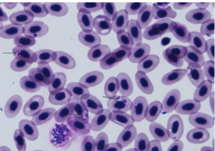
Elongated and sausage-shaped gametocytes were observed in several erythrocytes in the microscopic examination of the blood smears. The halteridial gametocytes were partially encircling the erythrocyte nucleus. The multiple, refractile, golden-brown pigment particles were observed in some of the gametocyte (Fig 1).
Figure 1. Haemoproteus sp. in the blood smear (Original, Giemsa, X1000).
Şekil 1. Sürme preparatta Haemoproteus sp (Orijinal, Giemsa, X1000).
Figure 2. Nested PCR results of the blood genomic DNA by the primers HAEMF/HAEMR2 on gel electrophoresis. M: molecular weight marker, 1, 2: The amplicons from owl blood genomic DNA (worked as duplicate), 3: Negative control.
Şekil 2. Baykuş kanından elde edilen genomik DNA örneklerinin HAEMF, HAEMR2 primerleri ile Nested PCR’ı sonucu jel elektroforezde görünümü. M: Marker, 1, 2: Baykuş kanından elde edilen amplikonlar (ikili çalışılmıştır), 3: Negatif kontrol.
Genomic DNA from the isolate yielded products of the expected size ~506 bp in the Nested PCR analysis of the mt-cytb gene (Fig 2).
Unique nucleotide sequence generated in the study was deposited in the GenBank International Nucleotide Sequence Database with the accession number: JQ768232. Several nucleotid and amino acid substations were determined among the Haemoproteus isolates based on the alignment of the partial mt-cytb gene region. The pairwise comparison and phylogenetic tree obtained from the amplified sequences confirmed that the owl was
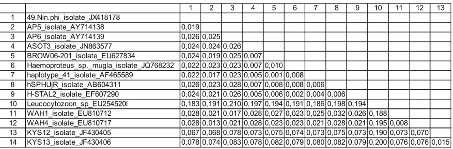
infected with Haemoproteus spp. According to the phylogenetic comparisons the Haemoproteus lineage showed the highest identity (99.8%) with the “H-STAL2” lineage isolated from an owl (Strix aluco) in Germany among the Haemoproteus lineages available in GenBank (Table 1, Fig 3).
Discussion and Conclusion
The species of Haemoproteus are some of the most common and widespread blood parasites of wild birds. Although the potential significances of the disease in
ASOT3 isolate Long-eared Owl Spain hSPHUjR isolate Humboldt Penguin Japan
Haemoproteus sp. mugla isolate Tawny Owl Turkey H-STAL2 isolate Tawny Owl Germany
BROW06-201 isolate Barred Owl USA haplotype 41 isolate Strix varia USA
AP5 isolate Yellow-billed Kingfisher Papua New Guinea AP6 isolate White-bibbed Fruit Dove Papua New Guinea WAH1 isolate White-bellied Kingfisher Gabon
WAH4 isolate Shining-blue Kingfisher Gabon 49.Nin.phi isolate Philipinne Hawk Owl Philipinnes
KYS12 isolate Cx. pipiens Turkey KYS13 isolate Ae. vexans Turkey Leucocytozoon sp EU254520 100 70 53 95 34 64 81 50 89 31 44 0.02 1 2 3 4 5 6 7 8 9 10 11 12 13 1 49.Nin.phi_isolate_JX418178 2 AP5_isolate_AY714138 0,019 3 AP6_isolate_AY714139 0,026 0,025 4 ASOT3_isolate_JN863577 0,024 0,024 0,026 5 BROW06-201_isolate_EU627834 0,024 0,019 0,025 0,007 6 Haemoproteus_sp._mugla_isolate_JQ768232 0,022 0,023 0,023 0,007 0,010 7 haplotype_41_isolate_AF465589 0,022 0,017 0,023 0,005 0,001 0,008 8 hSPHUjR_isolate_AB604311 0,026 0,023 0,028 0,007 0,008 0,008 0,006 9 H-STAL2_isolate_EF607290 0,024 0,021 0,026 0,005 0,006 0,002 0,004 0,006 10 Leucocytozoon_sp_EU254520l 0,183 0,191 0,210 0,197 0,194 0,191 0,186 0,198 0,194 11 WAH1_isolate_EU810712 0,028 0,021 0,017 0,028 0,027 0,023 0,025 0,032 0,026 0,188 12 WAH4_isolate_EU810717 0,028 0,013 0,021 0,028 0,023 0,023 0,021 0,028 0,021 0,195 0,008 13 KYS12_isolate_JF430405 0,067 0,068 0,078 0,073 0,075 0,074 0,073 0,075 0,073 0,190 0,073 0,070 14 KYS13_isolate_JF430406 0,078 0,074 0,083 0,078 0,082 0,079 0,080 0,082 0,079 0,200 0,076 0,076 0,015
Table 1. Pairwise differences of nucleotide sequence (%) in the mt-cytb gene region among Haemoproteus sp. isolates. Tablo 1. Haemoproteus sp. izolatları arasında mt-cytb gen bölgesindeki nükleotid dizilimlerinin pairwise analizleri.
Figure 3. Phylogenetic relationship among Haemoproteus sp. (JQ768232) and some other Haemoproteus sp. isolates (Neighbour Joining–Kimura 2 Parameter model). Scale bar indicates number of nucleotide substitutions per site. Leucocytozoon sp. (EU254520) was used as an outgroup.
Şekil 3. Haemoproteus sp. (JQ768232) ile diğer bazı Haemoproteus sp. izolatlarının filogenetik ilişkileri (Neighbour Joining-Kimura 2 Parameter modeli). Ölçek çizgisi yerleşim yerine göre nükleotid değişimini göstermektedir. Dış grup olarak Leucocytozoon sp. (EU254520) kullanılmıştır.
wild bird populations are largely unknown (3), the pathogenicity is generally believed to be very low and confined to domesticated birds or those kept in exotic environments (5). Haemoproteus infections have very rarely been incriminated as a cause of death, although in stressed or immunocompromised birds a severe infection can result in haemolytic anaemia, anorexia and depression (7).
The conventional diagnosis of Haemoproteus is based on the examination of the Giemsa-stained thin blood smears (3). It is possible to demonstrate the presence of erythrocytic gametocytes with prominent golden-brown or black pigment granules and absence of erythrocytic meronts that are diagnostic for Plasmodium spp. However, especially at choronic infections, species of Haemoproteus may be difficult to distinguish from avian species of Plasmodium. The number of circulating gametocytes is low and it may be difficult to determine whether the intracellular meronts characteristic of Plasmodium species is present or absent. Thus, in recent years, molecular techniques which have high sensitivity and specificity can distinguish Haemoproteus, Plasmodium and Leucocytozoon from each other (11, 12, 24). However, sequencing of PCR products and molecular characterization are necessary for identifying individual parasite lineages (3). It is also important to obtain true final sequences for molecular comparisons and carefully checking the original chromatograms is one of the crucial steps at this point. In this study, both microscopic and molecular examinations have been performed for the reliable identification and characterization of the Haemoproteus lineage.
Although Turkey has suitable geographical, climatic conditions and favorable habitats for many kind of migratory bird species, there are limited studies on blood parasites of avian species (10, 15, 18, 19, 22) based on traditional microscopic examination in which some Haemoproteus and Leucocytozooon infections were reported. There is no study regarding to molecular detection and characterization of avian haemosporidian parasites in Turkey except a molecular survey on avian malaria in vector mosquito species from Kayseri province (12). This study describes the first molecular detection along with the microscopic examination of a Haemoproteus infection in a Tawny Owl (Strix aluco) in Turkey. As the ability of microscopic examinations to detect haemosporidian infections was very low compared with molecular approaches (9, 17, 21, 24), the status of avian haemosporidiosis needs to be updated in Turkey.
Phylogenetic studies of avian haemosporidian parasites, mostly based on mitochondrial (mtDNA) cytochrome b (cyt b), have identified many parasite lineages of the genera Haemoproteus from wild birds (20). In addition this gene region has variable sections of DNA between the conserved regions which allow to
detection and identification of Haemoproteus, Plasmodium and Leucocytozoon lineages both in avian hosts and vectors (11, 24). Coding regions from the nucleus (adenylosuccinate lyase, asl), the plastid genome (caseinolytic protease, clpc) and mitochondrial cytochrome oxidase subunit I were also used for phylogenetic relationships among the haemosporidian isolates (14). In this study, the Haemoproteus sp. mugla isolate obtained from the owl was also phylogenetically analyzed with respect to mt-cytb gene region which is more common in GenBank records in order to obtain the pairwise comparison and phylogenetic tree among Haemoproteus lineages from the world. Phylogenetic analyses revealed that Haemoproteus sp. mugla isolate showed 92.1%-99.8% identity among the Haemoproteus lineages and the highest identity (99.8%) was determined with the “H-STAL2” lineage isolated from the same owl species (Strix aluco) in Germany. This finding supports the evidence (3, 5, 23) about Haemoproteus species relatively host-specific and restricted to bird species of the same family in contrast to species of avian Plasmodium which have a less host specificity and occur in several avian families by changing their character.
In conclusion, this study reports the first microscopic and molecular detection of Haemoproteus infection in a Tawny Owl (Strix aluco) owl in Turkey. The lineage characterization and phylogenetical relationships among the Haemoproteus lineages were also evaluated in this study. Further studies should be conducted in order to determine the status of avian malaria parasites and lineage diversities among both in avian hosts and vectors in Turkey.
Conflict of interest statement: None of the authors of this paper has a financial or personal relationship with other people or organizations that could inappropriately influence or bias the content of the paper.
Acknowledgement
This article was presented at 1st National Symposium
on Vectors and Vector-Borne with International Participation, 9-10 September, 2012, Avanos, Cappadocia, Nevsehir, Turkey.
References
1. Atkinson CT, van Riper III C (1991): Pathogenicity and
epizootiology of avian haematozoa: Plasmodium, Leucocytozoon, and Haemoproteus. 19-48. In: JE Loye, M
Zuk (Eds), Bird-Parasite Interactions. Ecology, Evolution, and Behavior. Oxford University Press, New York.
2. Atkinson CT (1991): Vectors, epizootiology, and
pathogenicity of avian species of Haemoproteus (Haemosporina: Haemoproteidae). Bull Soc Vector Ecol,
16, 109-126.
3. Atkinson CT (2008): Haemoproteus. 13-34. In: CT Atkinson, NJ Thomas, DB Hunter (Eds), Parasitic Diseases of Wild Birds. Wiley-Blackwell, USA.
4. Bennett GF (1987): Hematozoa. 120. EW Burr (Ed), Companion Bird Medicine. Iowa State University Press, Ames, Iowa.
5. Bennett, GF, Peirce MA, Ashford RW (1993): Avian
haematozoa: mortality and pathogenicity. J Nat Hist 27,
993-1001.
6. Bensch S, Stjernman M, Hasselquist D, Ostman O, Hansson B, Westerdahl H, Pinheiro RT (2000): Host
specificity in avian blood parasites: a study of Plasmodium and Haemoproteus mitochondrial DNA amplified from birds. Proc Biol Sci, 267, 1583-1589.
7. Campbell TW (1994): Hematology. 176-191. In: BW Ritchie, GT Harrison, LR Harrison (Eds), Avian Medicine: Principles and Application. Wingers Publishing, Lake Worth FL.
8. Drummond AJ, Ashton B, Buxton S, Cheung M, Cooper A, Duran C, Field M, Heled J, Kearse M, Markowitz S, Moir R, Stones-Havas S, Sturrock S, Thierer T, Wilson A (2013): Geneious v5.5, Available from http://www.geneious.com. Erişim Tarihi: 05.02.2013. 9. Garamszegi LZ (2010): The sensitivity of microscopy and
PCR-based detection methods affecting estimates of prevalence of blood parasites in birds. J Parasitol, 96,
1197-1203.
10. Gıcık Y, Arslan MÖ (2001): Blood parasites of wild
pigeons in Ankara district. Turk J Vet Anim Sci, 25,
169-172.
11. Hellgren O, Waldenström J, Bensch S (2004): A new
PCR assay for simultaneous studies of Leucocytozoon, Plasmodium, and Haemoproteus from avian blood. J
Parasitol, 90, 797-802.
12. Inci A, Yildirim A, Njabo KY, Duzlu O, Biskin Z, Ciloglu A (2012): Detection and molecular
characterization of avian Plasmodium from mosquitoes in central Turkey. Vet Parasitol, 188, 179-184.
13. Ishtiaq F, Guıllaumot L, Clegg SM, Phillimore AB, Black RA, Owens IP, Mundy NI, Sheldon BC (2008):
Avian haematozoan parasites and their associations with mosquitoes across Southwest Pacific Islands. Mol Ecol,
17, 4545-4555.
14. Martinsen ES, Perkins SL, Schall JJ (2008): A
three-genome phylogeny of malaria parasites (Plasmodium and closely related genera): evolution of life-history traits and host switches. Mol Phylogenet Evol, 47, 261-273.
15. Mimioğlu M, Göksu K, Sayın F (1969): Veteriner ve Tıbbi Protozooloji II. Ankara Üniv Basımevi, Ankara. 16. Njabo KY, Cornel AJ, Bonneaud C, Toffelmier E,
Sehgal RN, Valkiunas G, Russell AF, Smith TB (2011):
Nonspecific patterns of vector, host and avian malaria parasite associations in a central African rainforest. Mol
Ecol, 20, 1049-1061.
17. Ortego J, Calabuig G, Cordero PJ, Aparicio JM (2007):
Genetic characterization of avian malaria (Protozoa) in the endangered lesser kestrel, Falco naumanni. Parasitol
Res, 101, 1153-1156.
18. Özmen Ö, Halıgür M, Yukarı BA (2005): A study on the
presence of Leucocytozoonosis in wild bird of Burdur district. Turk J Vet Anim Sci, 29, 1273-1278.
19. Özmen Ö, Halıgür M, Adanır R (2009): Identification of
different protozoa species from a common buzzard (Buteo buteo). Turk J Vet Anim Sci, 33, 257-260.
20. Santiago-Alarcon D, Outlaw DC, Ricklefs RE, Parker PG (2010): Phylogenetic relationships of haemosporidian
parasites in New World Columbiformes, with emphasis on the endemic Galapagos dove. Int J Parasitol, 40, 463-470.
21. Tanigawa M, Sato Y, Ejiri H, Imura T, Chiba R, Yamamoto H, Kawaguchi M, Tsuda Y, Murata K, Yukawa M (2012): Molecular identification of avian
Haemosporidia in Wild Birds and mosquitoes on Tsushima Island, Japan. J Vet Med Sci, [Epub ahead of print].
22. Tolgay N (1972): Çeşitli kanatlıların Plasmodium,
Haemoproteus ve Leucocytozoon enfeksiyonları üzerinde araştırmalar. Ankara Univ Vet Fak, 19, 271-286.
23. Valkiunas G (2005): Avian Malaria Parasites and Other
Haemosporidia. CRC Press, Boca Raton, FL.
24. Waldenström J, Bensch S, Hasselquist D, Ostman O (2004): A new nested polymerase chain reaction method
very efficient in detecting Plasmodium and Haemoproteus infections from avian blood. J Parasitol, 90,191-194. Geliş tarihi: 28.01.2013 / Kabul tarihi: 29.03.2013
Address for correspondence:
Doç. Dr. Alparslan Yildirim
Erciyes Üniversitesi, Veteriner Fakültesi, Parazitoloji Anabilim Dalı,
38090 Melikgazi/KAYSERİ email: [email protected]