A morphological and histometric study on the distribution and
heterogeneity of mast cells found in lungs and trachea of Van Cats
Sema USLU, Mecit YÖRÜK
Yüzüncü Yıl University, Faculty of Veterinary Sciences, Departments of Histology and Embryology Van, Turkey.
Summary: This study was performed to specify the distribution and the heterogeneity of mast cells found in lungs and trachea of Van Cats. Four Van Cats were used in the study. Appropriate sized pieces were taken from the lungs and tracheas of the cats. After BLA (Basic Lead Acetate-Mota) was set, it was blocked with paraplast by doing the routine tissue follow-up. Sections taken in 6-7 μm thickness were stained with 0,5 % toluidine blue and alcian-blue safranin O combined stain. Numbers of mast cells were specified semi-quantitatively in the sections stained with toluidine blue under lens 40. Among analysed organs, the most mast cells were found in lungs. To specify the granule construction, BLA detection was determined to be suitable. In the stain of Alcian-blue safranin O, SO (+), AB (+) granulose mast cells were found; mast cells that have mixed granules were not found.
Key words: Heterogeneity, light microscope, lung, mast cell, trachea, Van Cats.
Van Kedilerinin trakeya ve akciğerlerinde bulunan mast hücrelerinin dağılımı ve heterojenitesi
üzerine morfolojik ve histometrik çalışma
Özet: Bu çalışma Van Kedilerinde trakeya ve akciğerlerde bulunan mast hücrelerinin dağılımı ve heterojenitesini belirlemek
amacıyla yapıldı. Çalışmada 4 adet Van Kedisi kullanıldı. Van Kedilerinin trakeya ve akciğerlerinden uygun büyüklükte parçalar alındı. BLA (Basic Kurşun Asetat-Mota)’da tespit edildikten sonra rutin doku takibi yapılarak paraplast ile bloklandı. 6-7 µm kalınlığında alınan kesitler % 0,5’lik toluidine blue ve alcian blue-safranin O kombine boyalarında boyandılar. Toluidine blue ile boyanan kesitlerde 40’lık objektifte mast hücre sayıları semikantitatif olarak belirlendi. İncelenen organlardan akciğerde en fazla sayıda mast hücresine rastlandı. Granül yapısını belirlemede BLA tespitinin uygun olduğu saptandı. Alcian blue- safranin O boyamasında ise trakeya ve akciğerde SO (+), AB (+) granüllü mast hücrelerine rastlandı, miks granüle sahip mast hücrelerine ise rastlanılamadı.
Anahtar sözcükler: Akciğer, heterojenite, ışık mikroskobu, mast hücresi, trakeya, Van Kedisi.
Introduction
Turkish Van Cats are endemic breed to Van province and attract public attention because of their white fur and typically colorful eyes (blue/yellow). They also behave friendly and typically like to play with water (15).
Mast cells, which are grouped mostly near blood vessels and nerves, are round or oval while in compact collagen tissues where collagen sutures are intensively found are generally in shuttle shape (10). Suitable with the shape of the cell, nucleus can be small, pale, ovoid, and be in central or excentric localization (3, 10). Mast cells are entitled as mucosal mast cell (MMC) and collagen tissue mast cell (CTMC) according to the places where they are (20). Respiratory system mast cells are particularly found in trachea and bronchus in subepithelial location, in lungs they are found in bronchial mucosa, tunica adventitia and peribronchial collagen tissues. On the other hand they were found in
the periphery of lymph follicles that belong to BALT that is found in primary and secondary bronchial lamina propria.
Mast cells granules excrete vital factors such as heparin, histamine, prostaglandin, neutral protease, B-glucuronidase, aryl-sulfatase, tryptase, eosinophil chemotactic factor of anaphylaxis (ECF-A), slow reaction substance of anaphylaxis (SRS-A) (9, 16, 19).
Because of their granule content, respiratory system mast cells participate in different illnesses and allergic reactions (34). Because most of mast cells take part actively in the response against auriferous allergens, they are found mostly in organs that contact external environment (14, 26). For most of the animals bronchial mucosa and alveoli walls are places where mast cells especially locate (1). Mast cells in the respiratory passage are important in the protection of homeostasis (29). Mast cells in bronchus and alveoli walls takes part in pathogenesis of respiratory system diseases like asthma
(34). It is also known that lung mast cells are efficient in chronic hypoxia, pulmonary hypertension (6, 7), pulmonary fibrosis (11). In the studies conducted, it was designated mast cells leading to start bronchospasm activate BALT tissue of antigen substances by excreting secretion that loosen the ties between epithelium cells (36).
This study presented aimed to specify the distribution and heterogeneity of mast cells found in lungs and trachea of Van Cats.
Material and Methods
Animal material: Four adult Van Cats weighing
2.5-3.5 kg were obtained from the Yuzuncu Yil University Veterinary Medicine Animal Clinics, Van, Turkey. Tissue samples were taken from cats that had been killed in road traffic accidents.
Taking, follow-up and examination of the tissues:
The tissue samples that were taken from proximal, medial, distal sides of the trachea and from the lungs were set in BLA (Basic Lead Acetate) (1 gr basic lead acetate, 50 ml ethanol, 50 ml distillated water, 0,5 ml glacial acetic acid) (5) for 24 hours and after the elution process and the routine tissue follow-up they were blocked with paraplast (8). 6 micron thick sections were taken from the prepared blocks. They were stained with toluidine blue (0.5% and pH=0,5) for 10 minutes (9, 17, 33) and with alcian blue 8GX-safranin O combined stains for 30 minutes (2, 32). These stained preparates were examined under examination microscope (Lecia ICC50) in terms of heterogeneity and distribution. The photos of sample sides were taken.
The number of mast cells in 1mm2 of the preparates stained with toluidine blue was evaluated semi-quantitatively as (+), (++) and (+++).
Results
The mast cells in the lower respiratory of Van Cats were distinguished in toluidine blue stain with BLA detection and in the examination under the light microscope with metachromatic coloration. In the AB/SO combined stains of the examined organs AB (+) and SO (+) stained mast cells were found. Mast cells were seen round, oval or in a shuttle shape according to their locations. In the cytoplasm of mast cells, homogeneous granulose mast cells and mast cells whose granules could be distinguished were detected. Examinations from the trachea in the lungs to the larger number of mast cells were found (Table 1).
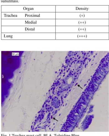
It was seen that mast cells were located in lamina propria in trachea and bronchus, around glands in the submucosa located subepithelial and near blood vessels. In the submucosa layer the mast cells were detected to have more cells than lamina propria layer (Fig 1.). It was
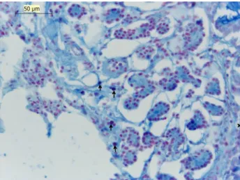
also observed that the mast cells located rarely in tunica muscularis and tunica serosa in the lungs, mast cells were found in the interalveolar septum, around blood vessels and in the subserous of the pleura (Fig 2.). In the trachea and the lung in the stain of AB/SO AB(+) and SO(+) mast cells identified (Fig 3., Fig 4., Fig 5.).
Table 1. The presentation of the density of mast cells with toluidine blue in the examined organs of Van Cats semi-quantitatively.
Tablo 1. Van Kedilerinin incelenen organlarında, toluidine blue ile belirlenen mast hücresi yoğunluğunun semi-kantitatif olarak sunulması. Organ Density Trachea Proximal (+) Medial (++) Distal (++) Lung (+++)
Fig. 1 Trachea mast cell, BLA, Toluidine Blue. Şekil 1 Trakeya mast hücreleri, BLA, Toluidin Blue.
Fig. 2 Mast cell lung intrapulmonary bronchi, BLA, Toluidine Blue.
Şekil 2 Akciğer intrapulmoner bronş mast hücreleri, BLA, Toludin Blue.
Fig. 3 Trachea AB (+) mast cell, BLA, Alcian Blue Safranin O. Şekil 3 Trakeya AB (+) mast hücreleri, BLA, Alcian Blue Safranin O.
Fig. 4 Lung AB (+) mast cell, BLA, Alcian Blue Safranin O. Şekil 4 Akciğer AB (+) mast hücreleri, BLA, Alcian Blue Safranin O.
Fig. 5 Lung intrapulmonary bronchi SO (+) mast cell, BLA, Alcian Blue Safranin O.
Şekil 5 Akciğer intrapulmoner bronş SO (+) mast hücreleri, BLA, Alcian Blue Safranin O.
Discussion and Conclusion
For the metachromatic stain and the demonstration of mast cells, basic dyes such as toluidine blue, thionin, azura A were used (8, 30). Mast cells could be distinguished easily by using the feature of metachromatic stain with toluidine blue in Van Cats.
In their study with the organs belonging to different systems of cattles, Küther et. al. (23)’s finding that mast cells especially located around blood vessels and nerve fibers is suitable with the findings of this study. In Van Cats, it was observed that mast cells located near histological construction like blood vessels and nerve fibers.
In one of their study about the respiratory passage of rats, Wilkes et. al. (35) reported that the mast cells in the trachea located subepithelial in lamina propria and near capillary vessels in tunica adventitia. Also it was observed that in the lung mast cells located near bronchial epithelium, bronchial lamina propria and bronchial muscles, in the peribronchial tissue and airways near blood capillary. Our study that found mast cells located near bronchial epithel, bronchial adventitia and the blood vessels around alveolus is in compliance with Wilkes et. al (35)’s study.
In one of their study about lower respiratory mast cells of Ankara goats, Kurtdede et. al. (22) reported that in trachea mast cells located in lamina propria and subepithelial area; and in the lung they located in inter-alveolar septum, around the bronchioles and the blood vessels. The location of mast cells in Ankara goats are similar to the location of mast cells in Van Cats.
In their study to specify the features of lower respiratory mast cells of cattles, Chen et. al. (7) indicated that the shapes of mast cells in the respiratory passage changed from round to filiform. Similarly, for Van Cats it was observed that according to their location mast cells could be round, oval or in shuttle shape and in different sizes.
Karaca (17) reported that to specify the granule construction of mast cells in chickens and quails BLA and Carnoy detection is the most suitable one. In their study about lower respiratory of geese and ducks Uslu and Yörük (33) indicated that to specify granule construction BLA detection is suitable. In this study BLA detection was also seen suitable to preserve and specify the granule construction.
In one of their research on the mast cells found in the trachea and the lung of chickens, Karaca et. al (18) concluded that there were less mast cells than the other organs examined in the other studies.
In the study on trachea, bronchus and lungs of humans, Shanahan et. al. (31) researched the effects of detections such as Carnoy, BLA and formalin on the distribution of mast cells in these tissues and they reached different conclusions. According to this study, it
is found that it changed due to the type of detection but generally there were more mast cells in bronchus. Also the same study indicated that there were more mast cells in the trachea than the lung. In this study about Van Cats identified that there were more mast cells in the lungs than the trachea.
In his study on rats Majeed (25) reported that he found rare mast cells in the lung tissue. In the study it was also observed that the number of mast cells was more in the organs which is related to external environment such as respiratory system than the other organs. In our study, contrary to this, less cells were found in the trachea although it was nearer to external environment than the lung.
In Mair et. al. (24)’s study to specify the distribution and the construction of mast cells in the respiratory passage of equine, among three parts of trachea the most mast cells were found in proximal side and the experimented parts of the lung tissue most mast cells were found near secondary bronchus and the blood vessels. The least mast cells were seen in the tissue samples taken out of the peripheral parts of the lung. In this study, different from Mair et. al. (24)’s in the proximal trachea (+), in the medial and distal trachea (++) mast cells were seen and in the lung tissue a distinguished difference was not observed that in the parts of the peripheral lungs mast cells such as parenchyma were located. In one study on the heterogeneity of mast cells in lower respiratory passage of sheep, Chen et. al. (6) used IFAA and formaline fixatively to emphasize the importance of detection to specify these cells. They concluded that IFAA was more suitable to specify the density of mast cells in subpleural parts of trachea and lungs. They also indicated that formalin sensitivity was important in mast cells heterogeneity. According to this, CTMCs are durable to formaline and MMCs are sensitive to formaline.
But CTMCs that contains formol can detect solutions. After MMC and CTMCs are detected by solutions for example carnoy that can detect both types of cells, with the staining of alcian blue/safranin O, both MMC and CTMCs are stained by alcian blue (+) while only CTMCs take safranin O stain (+) (9). Heterogeneity is not only specified by the type of stain and type of detection but also the location of cells (12). To identify the heterogeneity the presence of chymase and tryptase in mast cells of humans is effective. Mast cells found in bronchial epithelium and lung alveolar only have tryptase (30). Mast cells having tryptase in lungs are dense but two types of mast cells are found in this part (13).
Barrett and Metcalfe (4) in their study on the heterogeneity of mast cells, two types CTMC and MMCs were found in respiratory passages.
To specify the differences of the granule content of mast cells different staining can be applied. In this study
presented, to specify the heterogeneity of mast cells AB/SO stain was done. As a result of this staining AB (+), SO (+) mast cells were found in the trachea and the lung and mixed granulose mast cells could not be found.
Bachelet et. al. (1) analysed the heterogeneity of mast cells in the lower respiratory passage of rats and guinea pigs. When in the trachea, bronchus and lungs of rats AB (+), SO (+) mast cells and mixed granulose mast cells were found, in guinea pigs only AB (+) mast cells were seen. Kurtdede et. al. (22), detected only AB (+) cells after AB/SO staining in the lower respiratory passage and the lungs of Ankara goats. In their study on the heterogeneity of mast cells in lower respiratory passages of sheep and cattles Chen et. al. (6, 7) observed AB (+) cells in AB/SO staining but in the study on rats’ organs in the same system to control their previous study, they saw AB (+) and SO (+) cells.
The characteristics of mast cells of pig oviduct (27) and in the ovaries of cows (28), studies to determine the AB (+) cells were determined. These results are in parallels with our study. In these studies (27, 28) SO (+) cells were not coincident. But in our study SO (+) cells were found. These results are distinctive.
In the study on the lower respiratory passages of chickens, Koçak – Harem (21) identified AB (+), SO (+) and mixed mast cells in the lower respiratory passages of chickens in the AB/SO staining.
As a result, the lower respiratory organs of Van Cats which are local species located in Van Lake Basin were examined; the morphology, locations, distribution, heterogeneity of mast cells were tried to be specified. The results we had in this study are in conformity with the information in the literature from other studies about different animals’ respiratory systems and the organs belonging to this system. Because there were not any studies about mast cells of lower respiratory passages of Van Cats, it is thought that the findings of this study will contribute to the literature.
References
1. Bachelet CM, Bernaudin JF, Feith JF (1988): Distribution and histochemical characterization of pulmonary mast cells in the rat and guinea pig. Int Arc Allergy Appl Immunol, 87, 225-229.
2. Bancroft JD, Cook HC (1984): Manual of Histological Techniques. Churchill Livingstone, London.
3. Banks WJ (1986): Applied Veterinary Histology, pp: 86-87, Second Edition, Williams and Wilkins Co Baltimore. 4. Barett KE, Metcalfe DD (1987): Heterogeneity of mast
cells in the tissues of the respiratory tract and other organ systems. Am Rev Resp Dis, 135, 1190-1195.
5. Becker AB, Chung KF (1985): Mast cells heterogeneity in dog skin. Anat Record, 213, 477-480.
6. Chen W, Alley MR, Manktelow BW, Davey P (1990a): Mast cells in the ovine lower respiratory tract: Heterogeneity, morphology and density. Int Archs Allergy Appl Immunol, 93, 99-106.
7. Chen W, Alley MR, Manktelow BW, Slack P (1990b): Mast cells in the bovine lower respiratory tract: morphology, density and distribution. Brit Vet J, 146, 425-436.
8. Enerback L (1966b): Mast cells in the rat gastrointestinal mucosa: I. Effects of fixation. Acta Path Microb Scandinav, 66, 298-302.
9. Enerback L (1966a): Mast cells in the rat gastrointestinal mucosa II. Dye-binding and metachromatic properties. Acta Path Microb Scandinav, 66, 303-312.
10. Eren Ü, Aştı RN, Kurtdede N, Sandıkçı M, Sur E (1999): İnek uterusunda mast hücrelerinin histolojik ve histokimyasal özellikleri ve mast hücre heterojenitesi. Tr J Vet Anim Sci, 23, 193-201.
11. Fox B, Bull TB, Guz A (1981): Mast cells in the human alveolar wall: an electron microscopic study. J Clin Pathol, 34, 1333-1342.
12. Fritz FJ, Pabst R (1989): Numbers and heterogeneity of mast cells in the genital tract of the rat. Int Arch Allergy Appl Immunol, 88, 360-362.
13. Galli SJ (1990): New insights in to “The ridle of mast cells” micro environmental regulation of mast cell development and phenotypic heterogeneity. Lab Invest, 62, 5-33.
14. Guerzon GM, Pare PD, Michoud MC, Hogg JC (1979): The number and distribution of mast cells in monkey lungs. Am Rev Respir Dis, 119, 59-66.
15. Güre A (1993): Van Kedisi. In, Abdulkadiroğlu A, Yiğit M, Oğuzbaşaran B (Eds): Van Kütüğü. No: 8, Yüzüncü Yıl Üniversitesi, Van, pp. 722-726.
16. Junquiera LC, Carneiro J (2003): Temel Histoloji (Basic Histology). Çeviri Editörü: Y. Aytekin, S. Solakoğlu. pp: 101-102, Nobel Tıp Kitapevleri.
17. Karaca T (2003): Tavuk ve bıldırcınlarda sindirim sisteminde bulunan mast hücrelerinin dağılımı ve heterojenitesi üzerine morfolojik ve histometrik araştırmalar. Doktora Tezi. YYÜ Sağlık Bilimleri Enstitüsü, Van.
18. Karaca T, Yörük M, Şimşek N (2006): Age related distribution of mast cells in the trachea and lung of chicken. Indian Vet J, 83, 649-651.
19. Katz HR, Stewens RL, Austen KF (1985): Heterogeneity of mammalian mast cells differentiated in vivo and in vitro. J Allergy Clin Immunol, 76, 250-259.
20. Kitamura Y, Kanakura Y, Sonoda S, Asai H, Nakano T (1987): Mutual phenotypic changes between connective tissue type and mucosal mast cells. Int Arc Allergy Applied Immun, 82, 244-248.
21. Koçak-Harem M (2001): Tavukların alt solunum yollarındaki mast hücreleri üzerine histolojik araştırmalar. Doktora Tezi. AÜ Sağlık Bilimleri Enstitüsü, Ankara. 22. Kurtdede N, Aştı RN, Ergün L, Ergün E (2000): Ankara
keçilerinin alt solunum yolları mast hücreleri üzerine histolojik çalışmalar. AÜ Vet Fak Derg, 47, 339-349. 23. Küther K, Audige L, Kube P, Welle M (1998): Bovine
mast cells: distribution, density, heterogeneity and influence of fixation techniques. Cell Tissue Res, 293, 111-119.
24. Mair TS, Stokes CR, Bourne FJ (1988): Distribution and ultrastructure of mast cells in the equine respiratory tract. Equi Vet J, 20, 54-58.
25. Majeed SK (1994): Mast cell distribution in rats. Drug Res, 44, 3.
26. Overveld FJV, Houben LAMJ, Schmitz FEM, Bruijnzeel PLB, Raaijmakers JAM, Terpstra GK (1989): Mast cells heterogeneity in human lung tissue. Clin Sci, 77, 297-304.
27. Özen A, Bayraktaroğlu AG, Ertuğrul T, Özcan Z, Ceylan A, Özen D (2014): Domuz oviduktunda mast hücreleri üzerinde ışık ve electron mikroskobik çalışmalar. Ankara Üniv Vet Fak Derg, 61, 9-14.
28. Özen A, Ergün L, Ergün E, Şimşek N (2007): Morphological studies on ovarian mast cells in the cow. Turk J Vet Anim Sci, 31, 131-136.
29. Pesci A, Rossi GA, Bertorelli G, Aufiero A, Zanon P, Olivieri D (1994): Mast cells in the airway lumen and bronchial mucosa of patients with chronic bronchitis. Am J Resp Crit Care Med, 149, 1311-1316.
30. Schwartz LB, Austen KF (1984): Structure and function of the chemical mediators of mast cells. Prog Allergy (Karger Basel), 34, 271-321.
31. Shanahan F, Niven IM, Dyck N, Denburg JA, Binenstock J, Befus AD (1987): Human lung mast cells: distribution and abundance of histochemically distinct subpopulations. Int Archs Allergy Appl Immun, 83, 329-331.
32. Tung W (1991): Mast cells in the chick digestive tract II. fixation, distribution, histochemistry and ultrastructure. Tokai J Exp Clin Med, 16, 27-32.
33. Uslu S, Yörük M (2013): Yerli ördek (Anas Platyrhynchase) ve kaz’ın (Anser anser) alt solunum yolları ve akciğerlerinde bulunan mast hücrelerinin dağılımı ve heterojenitesi üzerine morfolojik ve histometrik araştırmalar. Kafkas Univ Vet Fak Derg, 19, 475-482.
34. Warton JM, Papadimitriou RG, Goldie RG, Paterson JW (1986): An ultrastructural study of mast cells in the alveolar wall of normal and asthmatic lung. Aust J Exp Biol Med Sci, 64, 435-44.
35. Wilkes LK, McMenamin C, Holt PG (1991): Postnatal maturation of mast cell subpopulations in the rat respiratory tract. Immunology, 75, 535-541.
36. Xaubet A, Moises JA, Agusti C, Martos JA, Picado C (1991): Identification of mast cells in bronchoalveolar lavage fluid. Allergy, 46, 222-227.
Geliş tarihi: 30.04.2014/ Kabul tarihi: 02.07.2014
Address for correspondence:
Dr. Sema Uslu
Yüzüncü Yıl University, Faculty of Veterinary Sciences,
Departments of Histology and Embryology, 65080 Kampus / VAN - TURKEY