Short Communication / Kısa Bilimsel Çalışma
Echocardiographic diagnosis and surgical correction of
aortopulmonary window in a Belgian Shepherd Dog (Malinois)
Meric KOCATURK-OCAL 1, Hakan SALCI 2, Melike CETIN2, Tolga KARAPINAR3, Zeki YILMAZ1
University of Uludağ, The Faculty of Veterinary Medicine, 1Internal Medicine Department, Animal Teaching Hospital, 2Surgery
Department, Animal Teaching Hospital, Bursa; 3Fırat University, The Faculty of Veterinary Medicine, Internal Medicine
Department, Animal Teaching Hospital, Elazığ / Turkey.
Summary: A 20-month-old, female Belgian Malinois Dog was referred for evaluation on exercise intolerance. A high-grade
machinery murmur, tachycardia, and cardiomegaly were observed. An aortopulmonary window was diagnosed by a color-Doppler echocardiography showing a turbulent blood flow shunting from the proximal part of ascendance aorta to the main pulmonary artery. This cardiac anomaly was corrected by surgery. Dog was treated by furosemide, pimobendan, and enalapril during post-operative 7 days. 1-year after the surgery, life quality of the patient was perfectly good. This is the first report of this rare congenital heart disease and its successful outcome of surgically correction in Belgian Malinois Dogs.
Key words: Aortopulmonary window, congenital heart defect, echocardiography, dog.
Bir Belçika çoban köpeğinde (Malinois) aortapulmoner pencerenin ekokardiyografik tanımlaması ve cerrahi düzeltimi
Özet: 20 aylık, dişi Belçika Malinois köpek egzersiz intoleransın değerlendirilmesi için sunulmuştur. İleri derece mekanik bir
üfürüm, taşikardi ve kardiyomegali belirlendi. Renkli Doppler ekokardiografi ile asendens aortanın proksimal kısmı ile ana pulmoner arter arasında turbulent akım belirlenmesi ile aortopulmoner window tanısı konmuştur. Kardiyak anomali cerrahi olarak düzeltildi. Post-operatif 7 gün boyunca köpek furosemid, pimobendan ve enalapril ile tedavi edildi. Operasyondan 1 yıl sonra hastanın yaşam kalitesi çok iyiydi. Bu rapor Belçika Malinois köpeklerindeki nadir görülen kongenital kalp defektinin ve cerrahi düzeltmenin başarılı sonucunun ilk sunumudur.
Anahtar sözcükler: Ekokardiografi, aortopulmoner pencere, kongenital kalp defekti, köpek.
Aortopulmonary window (APW) is a rare congenital defect between the aorta and pulmonary artery (PA) (13). There is a little information on this defect (3, 4, 7, 9, 10, 14). There are forms of APW (Type I-III) and all these types in humans are widely closed with patch techniques (5). Main clinical signs are dyspnoea and exercise intolerance. There is neither such a case reported in dogs in Turkey nor in Belgian Malinois Dogs in the literature. Thus, the aim of this case study is to present diagnostic findings and a successful surgical treatment in a dog and adds a new knowledge about this rare congenital cardiac defect.
A 20-month-old, 24 kg, female, Belgian Malinois Dog was presented to the clinic (Dept. of Internal Medicine, Faculty of Veterinary Medicine, Firat University, Elazig - Turkey) with exercise intolerance. After the diagnostic work-up, a patent ductus arteriosus (PDA) was suspected, and the dog was treated medically for left-sided heart failure by a combination of
furosemide (2 mg/kg, orally, twice daily) and enalapril (0.5 mg/kg, orally, twice daily) for 10 days. After that, to be able to confirm the diagnosis, patient was referred to the cardiology unit of small animal clinic (Dept. of Internal Medicine, The Faculty of Veterinary Medicine, University of Uludag, Bursa – Turkey) with the similar symptoms.
On physical examination, a 4/6 left basilar continuous murmur was auscultated. Capillary re-filling time (2 sec), peripheral pulse quality, respiratory rate (24
breath per min.), and temperature (38.7◦C) were within
the reference ranges. The results of complete blood count (HM5, Abaxis) and routine serum biochemistry panel (Comprehensive profile, VetScan, Abaxis) were unremarkably normal at the first day of evaluation.
On electrocardiographic (ECG) examination (bipolar standard extremity derivation; Esoate, Italy), sinus tachycardia with a heart rate of 160 bpm was detected. Left ventricular enlargement (R wave: 3.8 mV, reference:
Meric Kocaturk-Ocal - Hakan Salcı - Melike Cetin - Tolga Karapinar - Zeki Yilmaz 76
A
B
Figure 1: Electrocardiograms of pre-operative (A) and post-operative 4th day (B). Amplitude of QRS complex is 3.8 mV
(A) and 5.0 mV (B). Calibration: 10 mm/mV; 50 mm/sec. Şekil 1: Pre-operatif (A) ve post-operatif 4. gün (B) elektrokardiogramları. QRS kopleksi amplitüdü 3.8 mV(A) ve 5.0 mV (B). Kalibrasyon: 10 mm/mV; 50 mm/saniye.
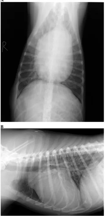
<3.5 mV/DII) was observed, as well (Figure-1). Radiological examination concluded the left atrial enlargement and appearance of the aortal arch in ventrodorsal and lateral radiograms, respectively (Figure 2A-B).
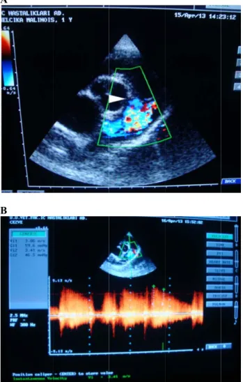
Two-dimensional (2D) echocardiography, colour flow imaging and spectral Doppler examinations were performed using a CarisPlus (Esoate, Florence, Italy) with a 7.5–10 MHz phased-array transducer. The dog was not sedated throughout the ultrasound examination and was gently restrained in the right lateral recumbent position (2). On 2D echocardiography, a defect between the ascending aorta and the main PA trunk was detected (Figure-3A). Color Doppler examination showed a turbulent blood flow shunting from the proximal part of ascending aorta to the main PA right after the pulmonary valves that was defined APW. In spite of having difficulties on measuring the connection window objectively, after measuring 5 times to have the mean measurement values, the hole was 0.65 cm. Both aortic and PA valves were intact and in their normal position. On Spectral Doppler flow evaluation presented a left to right aortapulmonary shunt. PA regurgitant jet velocity at
A
B
Figure 2. These radiographs show the left atrial enlargement (A) and appearance of the aortal arc (B).
Şekil 2: Bu röntgenlerde sol atriyal büyüme (A) ve aortik arkın şeklini göstermektedir.
early and late diastole was 3.86 m/s and 3.41 m/s and its pressure gradient was 59.6 mmHg and 46.5 mmHg, respectively, at right parasternal short axis view – pulmonary trunk level (Figure-3B). During this examination, a loud machinery continuous regurgitant flow was confirmed by phonocardiography.
The patient had a full cardiac evaluation to eliminate any other congenital defects. In M-mode echocardiographic evaluation, left atrial and left ventricular dilations, increased E point to septal separation (EPSS), and a right deviation of interventricular septum
A
B
Figure 3: Righ flow Doppler head) (A) and (3.86 m/s) betw Şekil 3: Sağ Anormal penc pulmoner arte velositesinin ( on Color flo operative fr ejection fract detected ano Doppler flow tricuspid and ventricular se General combination intratracheal 2% isofluran ventilation. P applied for thoracotomy was perform explained ao connectiona ht parasternal sh examination o high pressure g ween aorta and ğ parasternal k cerenin (beyaz er arasında yük 3.86 m/s) renkl ow Doppler ractional sho
tion (EF) was other cardiac w velocities, d aortic valve eptum. l anaesthesi of ketamin intubation, an ne and respirat Pulse oximetr monitorizat from 4th inter med respective ortal and PA about 1cmdi
hort axis view – of abnormal wi
gradient (59.6 m d main pulmona
kısa eksen gö ok ucunda) (A ksek basınç fark li akım Doppler evaluation o ortening (FS) s 63% (Table-anomaly, bas without turbu es as well as ia was ind ne HCl and naesthesia wa tion was assist ry, ECG and c ion during costal space a ely. Explorati A enlargemen iameterbetwe – aortic level. C indow (white a mmHg) and vel ary artery (B). rüntü-aortik dü A) ve aorta ile kı (59.6 mmHg r muayenesi. of the heart. ) was 35% 1). It was also ed on the no ulence, of m s intact atrial duced with diazepam. A as maintained
ted with mech capnography w
surgery. A and pericardiot
on of the ve nt as well a
eenthearterie
Color arrow locity üzey. e ana g) ve Pre-and o not ormal mitral, and the After with hanic were left tomy essels as a es. Tabl and Tabl ekok Para FS ( EF LVD LVD IVS IVS LVP LVP Ao LA( PA( FS – vent diam diast LVP LVP aorta FS-f vent inter serb duva arter Figu asce oper Nerv Şeki aras görü phre le 1: M-mode e post-operative lo 1: Pre ve p kardiografik ölç ameter Pr (%) (%) Dd (cm) Ds (cm) Sd (cm) Ss (cm) PWs (cm) PWd (cm) (cm) (cm) (cm) – fractional sho tricular diamet meter at systole tole; IVDs - i PWDd – left PWDs - left ven a; LA – left atri fraksiyonel kas trikül diyastol ç rventriküler sep est duvar diya ar sistol çapı; A r. ure 4: Surgical ending aorta (A rative image. vus phrenicus ( il 4: Assend ındaki anorma üntü. Kardiyak enicus (noktalı o echocardiograph days 1-7 post-opratif 1-7 çümler. e-operative 1 35 63 6,52 1,14 0,79 1,08 0,91 1,08 2,46 4,82 2,12 ortening; EF – e ter at diastole ; IVSd – interv nterventricular ventricular pos ntricular post-w ium; PA – pulm ılma; EF - ejek çapı; LVDs - so ptum diyastol ç astol çapı; LV Ao - aorta; LA correction of Ao) and pulmo
Ligation of ca dot arrow) dens aorta (Ao al şantın cerra k defektin lig ok) hic measureme 7. günler süre Post-operat 1th day 4th d 18 24 37 47 4,66 5,5 3,83 4,1 1,14 0,8 1,24 1,1 1,45 1,3 1,19 1,0 2,42 2,4 2,34 2,5 2,86 1,8 ejection fractio e; LVDs - l ventricular sept septum diame st-wall diamet wall diameter a monary artery. ksiyon fraksiyo ol ventrikül sist çapı; LVPWd -VPWs - sol ve - sol atriyum; f an abnormal onary artery (P ardiac defect o) ve pulmon ahi düzeltimi- gasyonu (siyah
ents during pre-esince M-mode tive days day 7th day 4 36 7 64 0 6,35 9 4,08 5 0,85 3 1,47 6 1,42 8 0,85 2 2,40 7 3,09 9 2,12 n; LVDd – left eft ventricular um diameter at eter at systole; ter at diastole; at systole; Ao – on; LVDd - sol tol çapı; IVSd -- sol ventrikül entrikül serbest PA - pulmoner shunt between PA) - an intra-(black arrow), ner arter (PA) intra operatif h ok), Nervus - e t r t ; ; – l - l t r n -, ) f s
Meric Kocaturk-Ocal - Hakan Salcı - Melike Cetin - Tolga Karapinar - Zeki Yilmaz 78
The connection between great arteries was corrected with 0 no supramid ligation (Figure-3) and thoracic incisions were closed routinely.
Cardiac examinations were repeated at 1st hour of
operation (just after the surgical correction of the cardiac
defect), and 1st, 4th and 7th days of post-operation. Right
after the surgical repairment of the abnormal shunt, shunt size was found 0.35 cm diameter. Surgical correction was confirmed by peri-operative cardiac examination; it was accepted successful based on the loss of loud machinery continuous flow (diastolic jet of PA) from PA to right ventricular out flow tract.
Day 1-4 after the operation, sinus tachycardia left itself to a normal sinus rhythm, and QRS amplitude increased to 5.0 mV/DII. In postoperative period, patient received a medical support, of pimobendan (0.5 mg/kg/
day, po; Vetmedin® 5 mg/tablet, Boehringer Ingelheim/
Istanbul), enalapril (0.5 mg/kg, twice a day, po; Enelap®
10 mg/tablet, SABA/Istanbul) and furosemide (2 mg/kg,
twice a day, po; Lasix® 40 mg/tablet, Sanofi Aventis/
Istanbul), due to left ventricular systolic dysfunction till the heart compensated and remodelled itself.
Improvements were recorded by cardio-check in 4th and
7th days (Table-1). In those days, cardiac remodelling
was observed based on the 2D, M-mode, and Doppler examinations including a decrease in LA/Ao ratio and LV diameters at systole and diastole, and an increase FS and EF, compared to their pre-operative and post-operative 1 day values (Table-1).
APW is a rare congenital anomaly, resulting from incomplete septation of the truncus arteriosus into the aorta and the PA during embryogenesis (6). According to pub-med records, there is a few report regarding with this defect in different breeds, aged from 1 month to 2 years; Labrador Retriever (6), German Shepherd (4), and mixed breed (11). This is the first case reporting diagnostic work-up, successful surgical correction and monitorization of APW in a Belgian Malinois dog, of 20-month-old.
Dogs with early stages of APW may have a few clinical signs such as exercise intolerance, dyspnoea, cyanosis, anorexia, and/or ascites (3, 4, 10). In this report, exercise situations without cyanosis and dyspnoea indicated that blood oxygen saturation might be adequate in a left-to-right shunt, as described in a previous report (11). A good auscultation is one of the most important clinical examination criteria that gives clinician the idea of cardiac abnormality (8, 12). In this case, a loud machinery murmur over the connection of aorta and PA on the left side of chest was auscultated and then based on the ECG 1) and x-ray examinations (Figure-2), PDA or APW was suspected. In parallel to our observation, APW was described in a German shepherd dog suffering from tachypnea, cardiomegaly, atrial fibrillation and tall R wave (4). In this case, APW was
confirmed by the color-flow Doppler image from the right parasternal short - axis view at PA level showed a communication between ascending aortic and pulmonary trunk by detecting a continuous flow with high pressure gradient (59.6 mmHg) just above the semilunar valves (Figure-3), in a different location in which PDA is usually seen. In this case, the pulsed-wave and the color flow Doppler tracing of mitral, tricuspid and aortic valves (data not given) showed normal flow velocities without turbulence and intact nature of inter-atrial and inter-ventricular septum, excluding other congenital heart diseases.
Despite having similarities to Truncus arterious (TA), TA has four types in a classification, APW has one widely used classification proposed three variants. According to the classification of this pathology (5), based on the echocardiographic examination, the dog presented here had type I APW which is represent the classic proximal window involving the posteromedial wall of the ascending aorta just above the left sinus of Valsalva and the adjacent wall of the main PA. Whereas the type II is located distal segment of PA and meaning to a defect between the left posterior wall of the ascending aorta and the junction of the main or right PA. The type III defect has also been called “hemitruncus”, although the use of this term has been discouraged in favor of the more descriptive “anomalous origin of the right PA from the aorta”. Anatomic localization of the abnormal shunt, reaching the operation site is difficult (5). Initial approach to the repair of APW involves, if necessity, closed techniques (suture ligation, clamping) with the successful results. Because of the development in cardiac surgery by extracorporeal system equipments, all these types in humans are widely closed with patch techniques (5).
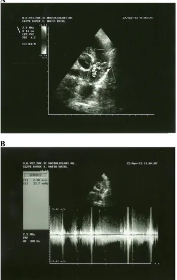
After the operation, size of the window decreases due to fibrous tissue growth and takes time to close totally. Thus full remission is not possible early days of recovery (1). In this case, a defect of 0.65 cm could be occluded to 0.35 cm. In a dog with APW, a sensible aortopulmonar communication size was reduced from 0.58 cm to 0.31 cm, which was detected by the immediate post-operative echocardiography (11), and then reduced to 0.21 cm on post-operative sixty days. Color flow Doppler examination of the abnormal window showed a pressure gradient (33.7 mmHg) and velocity (2,90 m/s) between aorta and main PA in
post-operative 4th day (Figure-5). Systolic function deficit is a
common finding in hearts after repairment of congenital defect such as PDA closure, and LV function takes from 6 months to 1 year to return to pre-closure levels (7). In this case, systolic dysfunction was detected by a decrease in FS and EF values just after the operation (Table-1), and thus pimobendan, an inodilator agent, was added to
A
B
Figure 5: Post-operative 4th day of right parasternal short axis
view – aortic level. Color flow Doppler examination of abnormal window (A) and pressure gradient (33.7 mmHg) and velocity (2,90 m/s) between aorta and main pulmonary artery (B).
Şekil 5: Post-operatif 4. gün sağ parasternal kısa eksen görüntü-aortik düzey. Anormal pencerenin (A) ve aorta ile ana pulmoner arter arasında yüksek basınç farkı (59.6 mmHg) ve velositesinin (3.86 m/s) renkli akım Doppler muayenesi.
compensate myocardial dysfunction. On cardio-check at post-operative 4 – 7 days, functional parameters of LV contractility (EF and FS) and some geometric data (IVS, LVW, Ao) returned to reference range (2), thus cardiac medical support was ceased. During postoperative 4 -7 days, left atrial diameter decreased from 4.8 cm to 2.5cm, resulting a decrease in LA/Ao ratio (from 1.95 to 0.9; reference: <1.5). Also, in the present case, LV volume overload that was characterized by an increase in LVDd, LVDs, EPSS and LA/Ao ratio as well as severe mitral regurgitation was treated by furosemide and enalapril medications. During the motorization, clinical and haematological parameters, and routine serum biochemistry profile did not changed significantly (data not shown). Post-operative x-ray (images not shown) and ECG examinations (Figure-2B) showed a normal heart size
and an adaptation process in cardiac electric activity, as well. Other researcher reported an atrial fibrillation (4), a supraventricular bigeminy, and trigeminy (10) in dogs with APW, relating with right (10) or left ventricular enlargement (4). In addition, Pascon et al. (11) reported a ST segment depression, wide QRS complexes and increment in Q wave voltage in leads I – II after the operation, suggesting biventricular enlargement.
Consequently, clinicians should be kept in mind that congenital cardiac defect, APW, might be asymptomatic for up to 20 months of age, and exercise intolerance and lethargy may be a sole complaint expressed by the owner. A Color Doppler echocardiography should be used as a gold standard to the diagnosis of APW. Surgical correction is a good choice to remove the clinical problem and enables to physiological remodelling itself in dogs with APW. The patient reported here lives healthy with no symptoms of heart failure, for 1 year after the operation.
References
1. Backer CL, Mavroudis C (2002): Surgical management
of aortopulmonary window: a 40- year experience. Euro J
Cardiothoracic Surg, 21, 773-779.
2. Boon JA (2011): Veterinary Echocardiography, Second Edition., Wiley Blackwell Publishing, USA.
3. Eyster GE, Dalley JB, Chaffee A, Beadle R, Trapp A, Cristopher WJ (1975): Aortopulmonary septal defect in a
dog. J Am Vet Med Assoc, 167, 1094-1096.
4. Guglielmini C, Pietra M, Cipone M (2001):
Aortopulmonary septal defect in a German Shepherd dog.
J Anim Hosp Assoc, 37, 433-437.
5. Sellke F, del Nido PJ, Swanson S (2010): Sabiston and
Spencer's Surgery of the Chest, 8th Edition, Saunders
Elsevier, Philadelphia, pages: 1911-1920.
6. Jung S, Orvalho J, Griffiths LG (2012): Aortopulmonary
window characterized with two- and three-dimensional echocardiogram in a dog. J Vet Cardiol, 14, 371-5.
7. Lombard CW, Knight DH, Buchanan JW, Riffle RA (1978): Clinico-pathologic conference: Aorticopulmonary
window. J Am Vet Med Assoc, 172, 75- 80.
8. Luisada AA, MacCanon DM (1965) Functional basis of
heart sounds. Am J Cardiol, 16, 631-3.
9. Mucha CJ, Belerenian G, Piella M (2002): Ventana
aortopulmonar tipo I en un canino. Rev Med Vet, 83,
90-92.
10. Nelson AW (1986): Aortopulmonary window in a dog. J Am Vet Med Assoc, 188: 1055- 1058.
11. Pascon JPE, Ondani AC, Junior DP, Andrade JNM, Camacho AA (2010): Aorticopulmonary septal defect in a
dog: case report. Arq Bras Med Vet Zootec, 62, 564-569.
12. Smetzer DL, Breznock EM (1972): Auscultatory
diagnosis of patent ductud arteriosus in the dog. J Am Vet
Med Assoc, 160, 80-4.
13. Tkebuchava T, Von Segesser LK, Vogt PR, Bauersfeld U, Jenni R, Künzli A, Lachat M, Turina M (1997):
Congenital aortopulmonary window: diagnosis, surgical technique and long- term results. Eur J Cardiothoracic
Meric Kocaturk-Ocal - Hakan Salcı - Melike Cetin - Tolga Karapinar - Zeki Yilmaz 80
14. Will JA (1969): Subvalvular pulmonary stenosis and
aortopulmonary septal defect in the cat. J Am Vet Med
Assoc, 154, 913-916.
Geliş tarihi: 10.02.2014/ Kabul tarihi: 27.06.2014
Adress for correspondance;
Meric Kocaturk-Öcal, PhD
Internal Medicine Department, Animal Teaching Hospital, The Faculty of Veterinary Medicine, University of Uludağ, Bursa, Turkey.