https://doi.org/10.1007/s11033-020-05912-7
ORIGINAL ARTICLE
Hypericum perforatum L.: a medicinal plant with potential as a curative
agent against obesity‑associated complications
Hilal Büşra Tokgöz1 · Filiz Altan1Received: 24 July 2020 / Accepted: 10 October 2020 / Published online: 22 October 2020 © Springer Nature B.V. 2020
Abstract
Obesity is a low-grade inflammatory disease that is getting increasingly common among adults and children and causes differ-ent complications. Insulin resistance, Type II diabetes, atherosclerosis, metabolic syndrome and hypertension are among the major health problems, that are associated with obesity. Some medications are used to treat obese individuals and metabolic surgery is recommended, if appropriate, for individuals with a BMI ≥ 40. Due to the fact that medications and metabolic surgery are not tolerated by all, researchers focus on alternative therapies. Medicinal plants comprise the most important group of these alternative treatments. Hypericum perforatum L. is the medicinal plant, which we focused on in this study.
Hypericum perforatum L. has been recognized as a medicinally valuable plant for over 2000 years. It has been used for
generations to treat anxiety, depression, insomnia, gastritis, hemorrhoids, wounds, and burns. Recent studies have indeed shown promising effects for the treatment of obesity. In this study, 3T3-L1 adipocytes were used to mimic the adipocyte dif-ferentiation associated with obesity in cellular terms. Lipoprotein lipase (Lpl), Diacylglycerol-O-acyltransferase 1 (Dgat1), Fatty acid synthase (Fasn) markers were used to study the lipid accumulation, and Collagen V (ColV) was used to study cell elasticity to investigate the relationship of the effects of the administration of Hypericum perforatum L. with obesity.
Keywords Hypericum perforatum L. · Insulin resistance · Inflammation · Obesity · Medicinal plants
Abbreviations
BMI Body mass index
Dgat1 Diacylglycerol-O-acyltransferase
ColV Collagen V
H. perforatum Hypericum perforatum
qPCR Quantitative PCR
HDL High-density lipoprotein
CPT 1 Carnitine palmitoyltransferase 1
IL-6 Interleukin-6
Lpl Lipoprotein lipase
Fasn Fatty acid synthase
VLDL Very low-density lipoprotein
CRP C-reactive protein
IBMX Isobuthylmethilxsantin
FATP1 Fatty acid transport protein 1
TNF-α Tumor necrosis factor-α
FABP Fatty acid binding protein
Introduction
Obesity is a well characterized mild chronic inflammatory disease, which plays an important role in the pathogene-sis of several chronic diseases, such as Type II diabetes, hypertension, atherosclerosis, fatty liver, cancer, asthma and sleep apnea [1]. Obesity is positively correlated with an increase or expansion of adipocyte cells [2, 3]. In most living organisms, triglycerides are the major molecules, where the metabolic energy and fatty acids are deposited. However, the over-accumulation of triglycerides correlates with dis-eases such as obesity and diabetes mellitus. The enzymes that are mainly involved in the catalytic reactions related to the synthesis of the triglycerides within a cell are the diacylglycerol-O-acyltransferase 1 (Dgat1) enzymes.
Harris et al. [4] showed that the presence of Dgat1 con-tributes significantly to the formation of lipid droplets and triglycerides, and that the triglyceride formation can only occur in the activity of these two Dgat enzymes (Dgat1 and Dgat2) [4]. One of the main characteristics of obesity is the deposition of over-accumulated triglycerides in white adipose tissues and it is often associated with an abnormal storage of triglycerides in tissues such as skeletal muscle and
* Filiz Altan [email protected]
1 Department of Molecular Biology and Genetics, Faculty
of Science, Muğla Sıtkı Koçman University, 48000 Kötekli, Muğla, Turkey
liver. Since the over-expression of Dgat1 will increase the accumulation of triglycerides, it is expected that the elevated levels of Dgat1 directly correlates with obesity [5]. The lipo-protein lipase (Lpl) has an important effect on adipocyte metabolism by hydrolyzing the circulating triglycerides to fatty acids. Thus, a decrease in Lpl activity contributes to the decrease in total body fat stores [6]. The Lpl levels in adipose tissue increases with the insulin release and after meals, but decreases in the fasting state. Studies have shown that the Lpl protein levels per adipocyte are elevated in obese humans and rodents. Intrestingly however, in obesity, the response of Lpl to insulin and nutrition is reduced [7–10]. The key lipogenic enzyme responsible from the biosynthesis of the long chain fatty acids from the acetyl-CoA precursors is known as the Fatty acid synthase (Fasn) [11].
It was observed that this enzyme increased in mice fed with a high fat diet. There is a positive correlation between the adipocyte differentiation and the elevated Fatty acid syn-thase expression in obesity [12]. It is known that the levels of Collagen V, an extracellular matrix component known to have an elasticity-reducing effect on adipose tissue, increase upon adipocyte differentiation [13]. The 3T3-L1 cell line is the in vitro model system of choice to understand the underlying mechanisms of diseases such as obesity, meta-bolic syndrome, and diabetes, due to its ability to differenti-ate into mature adipocytes.
Hypericum perforatum L. (H. perforatum L.), also known
as St. John’s wort, is used by the public for many purposes. The pharmacological studies on showed the anti-inflammatory, antidepressant, antimicrobial and antiviral activities of the H.
perforatum L. extracts [14–18]. We, therefore, wanted to deter-mine whether this plant can be used as a natural alternative for the treatment of obesity and insulin resistance related to obesity. H. perforatum contains a wide variety of secondary metabolites, including alkaloids, terpenes and phenolics [19]. Hernández-Saavedra et al. investigated the effects of these secondary metabolites on obesity. It was suggested that the hypolipidemic effect of this plant may be related to the pres-ence of some of the secondary metabolites [20]. Accordingly, he epigallocatechine gallate may stimulate thermogenesis [21] and reduce fat accumulation, which may be associated with its ability to inhibit pancreatic lipase [22] function. In addi-tion, the epigallocatechine gallate was shown to decrease the C-reactive protein (CRP) secretion by reducing the production of the reactive oxygen species from vascular smooth muscle cells [23]. The anti-obesity effect of rutin was associated with the ability to reduce the accumulation of triglycerides in the adipose tissue thereby improving the lipid profile [24]. In addi-tion, hypericin, hyperforin and adperforin, which are consid-ered to be the main bioactive compounds in H. perforatum, were identified [25]. Hyperforin, a major bioactive component of H. perforatum, was reported to protect against the cytokine-induced cell damage, thereby decreasing the loss of β-cell
function observed in diabetes and increasing survival, which could potentially be valuable for preventing or limiting β-cell damage [26]. Husain et al. [27] observed that H. perforatum extracts with standardized amounts of hypericin and hyper-forin showed promising healing effects with respect to obesity related complications in rats fed with a high-fat diet [27].
In our study, to investigate the putative curative role of H.
perforatum on molecular level, we focused on the changes of
some genetic markers that are regulated by obesity and other molecular events associated with it, such as alterations in the extracellular matrix composition or the lipid metabolism. Therefore, we analyzed the expression of the genes such as ColV, Fasn, Lpl and Dgat1 in 3T3-L1 cells treated without and with the plant for different time courses and doses by qPCR.
Materials and methods
Plant materialH. perforatum L. was collected in Muğla and all collected
samples were taxonomically identified by Prof. Dr. Güven Görk and Dr. Olcay Ceylan. The plants were dried for two weeks in a cool and moisture-free environment. The above-ground parts of the dried plants were above-ground to powder. The dried whole plant of H. perforatum L. was suspended in absolute ethanol. After the extract was filtered, the solvent was removed by evaporation [28]. The stock solutions were dissolved in ethanol to a final concentration of 20 mg/mL and stored at − 20 °C until use.
Cell culture and differentiation
3T3-L1 cells (ATCC) were cultured in Dulbecco’s Modi-fied Eagles Medium (DMEM) (L0102-500) containing 10% Newborn Calf Serum (Capricorn, NCS-1B) and 1% Penicillin–Streptomycin (Multicell, 450-201-EL) in 5% CO2 and 95% air at 37 °C. 10% Fetal Bovine Serum (Capri-corn, FBS-12B) and 1% Penicillin–Streptomycin containing DMEM was used to differentiate cells to adipocytes. Pre-differentiation medium containing 0.5 mM IBMX (Sigma, STBF6061V), 1 μM dexamethasone (Sigma, BCBV5460) and 1 μg/mL insulin (Sigma, SLBV1793) was applied for 48 h, followed by the application of the differentiation medium containing 1 μg/mL insulin to induce 3T3-L1 cells to adipocytes.
Evaluation of changes in morphology and viability of cells
The cells were placed onto petri dishes and incubated within a media containing 50 μg/mL, 100 μg/mL and 150 μg/mL H. perforatum extracts for 24-, 48- and 72 h
[29]. The adherence and the morphological characteristics of the cells on petri dishes were evaluated using the light microscopy.
Staining of 3T3‑L1 cells with Oil Red O Dye
The differentiation of 3T3-L1 adipocytes was performed as described previously. Oil Red O staining was done accord-ing to the manufacturer’s instructions, usaccord-ing the Biovision Oil Red O Staining kit (Biovision, K580-24).
Total RNA isolation and Real Time Reverse Transcription‑Polymerase Chain Reaction (qPCR) Total RNA from adipocyte cells was extracted using the RiboEx™ (Cat. No. 301-001) total RNA isolation solu-tion from GeneAll (Cat. No. 301-902). Ultraviolet light spectrophotometry followed by formaldehyde- agarose gel electrophoresis was used to determine the quantity and the quality of the isolated RNA, respectively. 500 nanograms of total RNA was reverse transcribed using oligo- dT primers with EasyScript Plus cDNA synthesis kit from ABM Alfagen (Cat. No. G236). The amplifica-tion of the reverse-transcribed RNA was achived using Ampliqon RealQ Plus 2 × Master Mix Green in the pres-ence of 0.3 mM gene-specific forward and reverse prim-ers by a thermocycling on a Roche Light Cycler 96 Real Time PCR machine for 45 cycles. The temperatures and durations for denaturation, annealing and extension were 95 °C for 30 s; 55 °C to 58 °C for 30 s and 72 °C for 30 s, respectively. The normalization of the differences in indi-vidual samples was done by the use of the amplified 18S expression as a standard control. The list of the mouse Dgat1, Lpl, Fasn, Col V and 18S RNA primer sequences
are given in Table 1. The information about the
prim-ers and their accession numbprim-ers were obtained from The
National Center for Biotechnology Information https ://
www.ncbi.nlm.nih.gov/genba nk/ [30]. Statistical analysis
The comparison of the groups with respect to the continious variables and the comparison of baseline and posttreatment measurements were done by using one-way ANOVA, fol-lowed by the Tukey’s multiple comparison test. The signifi-cance level was set at p < 0.05.
Results
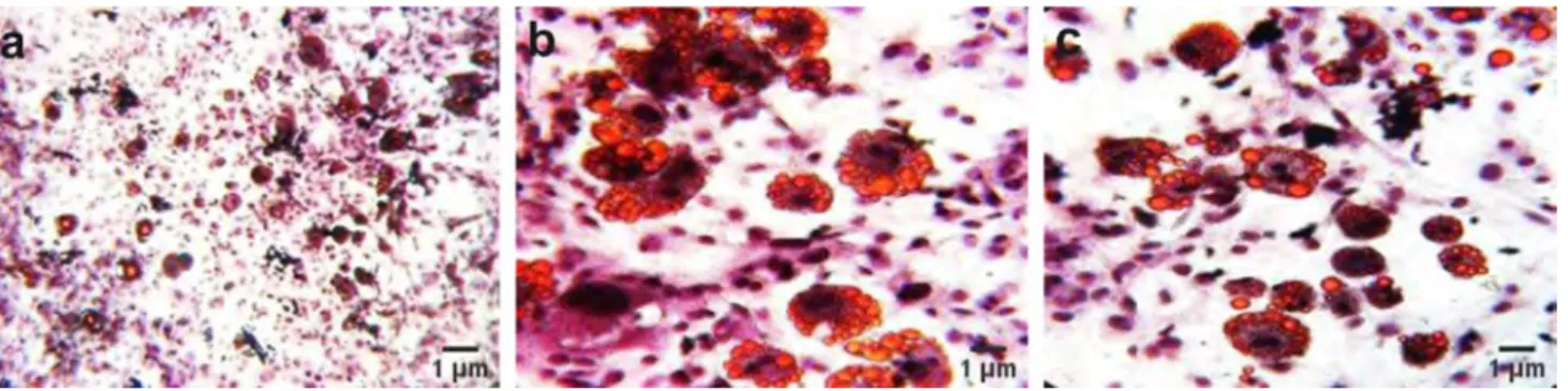
In this study, 3T3-L1 cells with fibroblastic morphology and preadipocyte nature were used. When 3T3-L1 cells are stimulated with chemicals such as insulin, dexametasone and IBMX, they become fully differentiated adipocytes. Oil Red O staining was used to evaluate lipid droplet formation at the end of a time course of two weeks Fig. 1. In our experi-ments with 3T3-L1 preadipocytes, the 24- and 48 h applica-tion of 50-, 100- and 150 μg/mL extracts of H. perforatum (HypE) dissolved in ethanol was determined to be suitable for the cell viability. Therefore, these doses were used for the experiments shown in Fig. 2.
RNA was extracted from the 3T3-L1 cells with RiboEx™, followed by cDNA synthesis. Electrophoretic and spectro-photometric analyses were performed to determine the qual-ity and the quantqual-ity of extracted RNA samples. The mRNA expression experiments of Fasn, ColV, Dgat1 and Lpl molecular markers, which were designed on the Primer3, were performed by quantitative Real Time Reverse Tran-scription-Polymerase Chain Reaction (qPCR) using samples of 3T3-L1 preadipocytes treated with H. perforatum and nontreated control cells. In addition, the expression levels of ColV, Fasn, Lpl and Dgat1 in adipocytes were analyzed by qPCR. The administration of H.perforatum extracts to adi-pocytes led to the downregulation of the ColV, Fasn, Dgat1 and Lpl genes Figs. 3, 4. The time courses and doses of
Table 1 Sequences and NCBI accession number for primers designed for Real Time PCR reactions
GeneBank accesion number Gene Primer sequence
NM_010046.3 Mus musculus Dgat1;
(Diacylglycerol-O-acyltransferase) (F): 5′-CCT CAG CCT TCT TCC ATG AG-3′(R): 5′-ACT GGG GCA TCG TAG TTG AG-3′
NM_008509.2 Mus musculus Lpl;
(Lipoprotein lipase) (F): 5′-ACT CGC TCT CAG ATG CCC TA-3′(R): 5′-TTG TGT TGC TTG CCA TTC TC-3′
NM_007988.3 Mus musculus Fasn;
(Fatty acid synthase) (F): 5′-CTG AGA TCC CAG CAC TTC TTGA-3′(R): 5′-GCC TCC GAA GCC AAA TGA G-3′
NM_015734.2 Mus musculus ColV
(Collagen V) (F): 5′-CTC AGG GGT AAC GAA AAC CA-3′(R): 5′-GGA GAA GTC CTC GGG AAA AC-3′
NR_003278.3 Mus musculus 18S ribosomal RNA (Rn18s) (F): 5′-TTC GAA CGT CTG CCC TAT CAA-3′
H. perforatum extracts used in these studies were 24 h and
48 h application of 50 μg/mL, 100 μg/mL and 150 μg/mL respectively (Table 2).
Discussion
Obesity is the most common nutritional disease and a grow-ing public health problem worldwide [31]. Obesity is a dis-ease with increasing prevalence dramatically in adult and pediatric populations and that causes different complications [2, 3].
The prevalence of diseases, such as obesity and related insulin resistance, and diabetes is increasing all over the world. Treatment options such as the use of oral hypoglyce-mic agents and insulin are currently available for diabetes. Glycemic drugs, which are used to treat obesity have seri-ous side effects [32]. The bariatric surgery can also be per-formed in obese individuals with severe comorbidity. Drugs are not tolerated well by every individual and the bariatric surgery may cause diarrhea, nausea and vomiting in patients [33]. The increasing prevalence of obesity even has adverse effects in countries, that have to deal with this health issue. Therefore, these lead researchers to seek for alternative treat-ment options. The anti-obesity and anti-diabetic effects of
H. perforatum was demonstrated in vivo and in vitro [27,
34, 35]. In in vivo studies, the parameters of sugar and fat intake were investigated in rats, fed with high fat diet to reveal the effects of the plant on lipid metabolic pathways. It was observed that the application of this plant reduced the levels of elevated cholesterol, triglyceride and blood glucose [20, 27].
Arokiyaraj et al. showed the effect of H. perforatum on blood sugar and blood lipid parameters in diabetic rats induced with streptozotocin [36]. Streptozotocin causes pan-creatic ß-cell damage, which results in insufficient insulin secretion and thus hyperglycemia. The excessive glucose production and decreased glucose utilization in tissues is known as hyperglycemia and is a hallmark of diabetes
mellitus. In their study, the researchers made use of the dia-betic and the non-diadia-betic Wistar rats. They administered diabetic rats H. perforatum in increasing doses and showed that the administration of H. perforatum extract reduced the blood glucose levels, which were elevated due to diabetes, back to an equal (or comparable) level with the healthy group. They reported that H. perforatum extract normal-ized this hyperglycemic condition. The elevated plasma insulin levels in streptozotocin-treated rats, compared to the normal rats, decreased by 2.5 fold upon the adminis-tration of H. perforatum. Moreover, the total cholesterol levels increased approximately fourfold in streptozotocin-treated diabetic rats, compared to normal control group. The HDL-cholesterol levels were threefold lower than normal in diabetic control, and the administration of 200 mg/kg H.
perforatum was reported to normalize the HDL levels. It
was shown that the triglyceride level increased fourfold in diabetic control and H. perforatum significantly decreased this level. These in vivo findings demonstrated the anti-obesity and the anti-diabetic properties of H. perforatum, and encouraged researchers to work at a molecular level to better understand the mechanism of action of this plant in treatment of obesity and obesity-related diseases. In our study, the genes that are associated with lipid accumulation and cell matrix components were used as markers in order to understand the relationship between H. perforatum and obesity. The elevated expression levels of ColV, Dgat1, Lpl and Fasn in adipocytes were downregulated upon the admin-istration of the plant extract, which is in line with the work by Arokiyaraj et al. [36].
In the study conducted by Husain et al. [27], the hypolipi-demic and the anti-obesity activity of H. perforatum was investigated. A high-fat and fructose diet was initially applied to rats followed by the administration of the increas-ing doses of H. perforatum. H. perforatum extract signifi-cantly reduced the daily food intake after 30 days. H.
per-foratum extract also inhibited the accumulation of adipose
tissue [27]. This study demonstrating the anti-obesity effect of H. perforatum is consistent with our study, in which we
Fig. 1 Oil Red O Staining to determine the lipid accumulation in fully differentiated 3T3-L1 cells. Cells were visualized before and after stain-ing (a, × 10), and only after stainstain-ing (b, c, × 40), respectively. Scale bars in each micrograph represent 1 µm
mimiced obesity in vitro and investigated the effects of H.
perforatum. We observed that the expressions of Dgat1, Lpl
and Fasn, which are the markers related to adipocyte differ-entiation, were downregulated as a result of the administra-tion of H. perforatum.
Tian et al. [35] investigated the effect of H. perforatum on lipid metabolic pathways [35]. In their study, the mice were fed on a high-fat diet for 14 days, after which high dose and low dose of H. perforatum were administered to these mice. The development of hypercholesterolaemia was observed in mice receiving the high-fat diet. When the levels Fig. 2 Microscopic visualization and the evaluation of morphological
changes and cell viability in 3T3-L1 cells, treated with plain solvent or H.perforatum extracts (HypE) with a dose of 50 μg/mL, 100 μg/
mL and 150 μg/mL for 24 h (a, d, g, j), 48 h (b, e, h, k), and 72 h (c,
f, i, l). Scale bars in each micrograph represent 1 µm
Fig. 3 Quantitative measurements of Dgat1 and Lpl mRNA in
L1 cells. qPCR analyses of Dgat1 (left) and Lpl (right) using 3T3-L1 adipocytes, differentiated with IBMX, Dexamethasone and insulin for 24 h. The results represent duplicate measurements in three
sepa-rate experiments (*p < 0.05, comparison with differentiation control; **p < 0.001, comparison with differentiation control. ┼p < 0.05,
com-parison with nondifferentiation control)
Ill ,._ :::J 0 .c -.:I" N Control Solvent control without extract
Non Diffc,enll•ted 50 µg/ml HypE differentiate~ Conlrol Control 50 µg/ml HypE 100 µg/ml HypE 150 µglml HypE
Extract dose treatments
C 0
I
3F
<t § ~~ E~ c.'.e...
..
-> ii ~ 100 µg/ml HypE 150 µg/ml HypE 120 100 80 60 40 20
•
Non Differentiated 50 µg/ml HypE 100 µg/ml Hy-pE
150 µg/ml HypE differentiated Control
of chosen metabolic parameters in mice were compared, it was revealed that in the groups, treated with low and high doses of H. perforatum, the total cholesterol levels decreased significantly by 16.2% and 22.2%, and the serum triglyceride levels decreased by 13.1% and 22.6% in a dose-dependent manner, respectively. The intramyocellular lipid accumula-tion in skeletal muscle was associated with insulin resistance and dyslipidemia. They reported that the triglyceride content in skeletal muscles increased significantly in mice fed with a high-fat diet and this increase was reversed with low and high doses of H. perforatum extracts (100 mg/kg, 200 mg/ kg). Gene expression studies on lipid metabolism were per-formed to understand the mechanisms of H. perforatum on dyslipidemia. Fatty acid transport protein 1 (FATP1), the main carrier of fatty acid, was reported to increase signifi-cantly in skeletal muscle in obese mice fed with a high-fat diet, and the administration of H. perforatum reduced the gene expression of FATP1. Carnitine palmitoyltransferase 1
(CPT1), an important regulator of fatty acid oxidation, was reported to increase slightly in the skeletal muscle of obese mice fed with a high-fat diet, while H. perforatum elevated the expression of CPT1 dramatically. Lpl is an enzyme that plays a role in the conversion of triglycerides to fatty acid and glycerol, and in our study, H. perforatum inhibited the elevation of adipocyte Lpl, associated with fully differ-entiated adipocytes, to the level of Lpl, characteristic for preadipocyte cells. Our results on adipocyte associated gene expressions are consistent with the study by Tian et al. [35]. Perez-Ramirez et al. [37] used healthy and obese mice in order to observe the hepatoprotective feature of H.
per-foratum. Mice in the obese group were given H. perforatum
extract prior to the administration of H. perforatum, in the obese group compared with the healthy group, an increase in body weight, lipid accumulation in the liver, increase in serum triglyceride levels and serum fatty acid levels were observed. H. perforatum extract was observed to cause a
Table 2 The change in the expression levels of Dgat1, Lpl, Fasn,
ColV in 3T3-L1 preadipocytes (nondifferentiated control), adipocytes (differentiated control) and Hypericum perforatum extract (HypE) treatment to 3T3-L1 adipocyte groups (50-, 100-, 150 µg/mL HypE
treatment to 3T3-L1 adipocytes; *p < 0.05, comparison with differen-tiation control; **p < 0.001, comparison with differendifferen-tiation control;
┼p < 0.05, comparison with nondifferentiation control)
Gene of interest Relative mRNA expression (arbitrary units) Samples
Non-differentiation
control Differentiation control 50 µg/mLHypE treatment to 3T3-L1 adipocytes 100 µg/mL HypE treatment to 3T3-L1 adipocytes 150 µg/mL HypE treatment to 3T3-L1 adipocytes Dgat1 10.71 ± 0.35 100┼ 6.70* ± 1.48 2.44** ± 1.35 19.24* ± 0.18 Lpl 4.73 + 0.21 100┼ 3.31* ± 1.20 1.64* ± 0.87 15.50* ± 0.31 Fasn 10.21 ± 0.12 100┼ 17.46* ± 0.20 7.43* ± 0.24 62.63* + 0.24 ColV 9.64 ± 0.40 100┼ 16.09* ± 0.14 4.73* ± 0.25 22.34* ± 0.68
Fig. 4 Quantitative measurements of Fasn and ColV mRNA in
L1 cells. qPCR analyses of Fasn (left) and ColV (right) using 3T3-L1 adipocytes, differentiated with IBMX, Dexamethasone and insulin
for 48 h.The results represent duplicate measurements in three sepa-rate experiments (*p < 0.05, comparison with differentiation control.
┼p < 0.05, comparison with nondifferentiation control)
C 0 ,ii ! Q. ><-wl!l < c :z :, "'?' Eg C • -i:,11.ll,l if!
!
.,
"'
120 100 80 60 40 20 0Non Diffonmllated 50 ~glml HypE
differentiated Con\f'OI Control 100 µglml HypE 150 µglml HypE 120 C 100 0 -a !! ~~ SQ
..,_
<" z:, "'i!' EE 60 ~~ u.e, ~ 40 .,..
;; a: 20Non Differentiated 50 µglml HypE
differentiated Control Control 100 µg/ml HypE 150 µg/ml HypE
significant decrease in the body weight of the mice in the obese group. It was reported that the application of H.
per-foratum extract decreased the serum fatty acid and serum
triglyceride levels. In order to observe the effect of obesity on inflammation, the expression of inflammatory agents, such as tumor necrosis factor-α (TNF-α) and interleukin-6 (IL-6) genes were investigated. It was observed that the elevated TNF-α and IL-6 expressions as a consequence of obesity were decreased with the administration of H.
perfo-ratum. In addition, researchers examined the level of
expres-sion of genes associated with the lipid metabolism, such as Fatty acid synthase, Fatty acid binding protein, Acetyl CoA carboxylase and Acetyl CoA dehydrogenase. The study reported that H. perforatum improved the effects in gene expression on the lipid metabolism [37]. In our study, we showed that the elevated Fatty acid synthase mRNA expres-sion in adipocytes were decreased upon H. perforatum extract treatment.
Our results showed that the Dgat1 gene, which plays a role in the biosynthesis of triglycerides, increased with adi-pocyte differentiation and that the extracts of H. perforatum at different doses significantly reduced Dgat1 expression, in a statistically significant manner. We observed that Lpl, which is known as an enzyme converting triglycerides to fatty acids and glycerol, increased with adipocyte differen-tiation and this elevation was reduced back to the preadipo-cyte control level with H. perforatum extract application. H.
perforatum extracts downregulated the expression of Fasn,
which is correlated with fully differentiated adipocytes. In addition, ColV, which exhibits size reducing effect on fat tissue, increases with adipocyte differentiation and this increase is inhibited by the administration of the plant.
The data obtained support previous studies with H.
per-foratum and provide guidance to understand the molecular
mechanisms of the effect of H. perforatum on obesity. In the present study, 3T3-L1 preadipocytes were transformed into fully differentiated adipocytes, followed by 50 μg/mL, 100 μg/mL, 150 μg/mL H. perforatum extract treatments for 24- and 48 h. We observed that the administration of H.
perforatum extract reduced the expression of Dgat1, Lpl,
Fasn and ColV on transcriptional level. Dgat1, Fasn, ColV and Lpl both were downregulated and downregulation of these markers was statistically significant. The fact that these markers show a significant decrease with the application of the plant extract is in accordance with the previous obesity related in vivo studies of the H. perforatum.
Conclusions
The application of H. perforatum L. at different doses down-regulated the expression of Dgat1, Fasn, ColV and Lpl in fully differentiated 3T3-L1 cells. Our results underline the
importance of the transcriptional regulation of H.
perfora-tum L. to understand its effects on obesity.
Acknowledgements This work was supported by the Scientific Resarch
Project (BAP) Grant (AR-GE 17/059) from the Muğla Sıtkı Koçman University granted to Dr. Filiz ALTAN. Authors would like to thank to Dr. Reşat ÜNAL for valuable scientific support and critical reading; to Dr. Zekiye Buket YILMAZ for critical reading and language editing; to Dr. Güven GÖRK and Dr. Olcay CEYLAN for providing the plant material and to Dr. Mehmet VAROL for his expertise and assistance in technical editing.
Compliance with ethical standards
Competing interests The authors declare that they have no competing
interest.
References
1. Ye J (2013) Mechanisms of insulin resistance in obesity. Front Med 7(1):14–24. https ://doi.org/10.1007/s1168 4-013-0262-6
2. Koren D, Taveras EM (2018) Association of sleep distur-bances with obesity, insulin resistance and the metabolic syn-drome. Metabolism 84:67–75. https ://doi.org/10.1016/j.metab ol.2018.04.001
3. Ng M, Fleming T, Robinson M, Thomson B, Graetz N, Mar-gono C, Mullany EC, Biryukov S, Abbafati C, Abera SF (2014) Global, regional, and national prevalence of overweight and obesity in children and adults during 1980–2013: a systematic analysis for the Global Burden of Disease Study 2013. The Lancet 384(9945):766–781. https ://doi.org/10.1016/S0140 -6736(14)60460 -8
4. Harris CA, Haas JT, Streeper RS, Stone SJ, Kumari M, Yang K, Han X, Brownell N, Gross RW, Zechner R (2011) DGAT enzymes are required for triacylglycerol synthesis and lipid droplets in adi-pocytes. J Lipid Res 52(4):657–667. https ://doi.org/10.1194/jlr. M0130 03
5. Yen C-LE, Stone SJ, Koliwad S, Harris C, Farese RV (2008) The-matic review series: glycerolipids. DGAT enzymes and triacylg-lycerol biosynthesis. J Lipid Res 49(11):2283–2301. https ://doi. org/10.1194/jlr.R8000 18-JLR20 0
6. Greenberg AS, Nordan RP, McIntosh J, Calvo JC, Scow RO, Jablons D (1992) Interleukin 6 reduces lipoprotein lipase activ-ity in adipose tissue of mice in vivo and in 3T3-L1 adipocytes: a possible role for interleukin 6 in cancer cachexia. Can Res 52(15):4113–4116
7. Hartman AD (1981) Lipoprotein lipase activities in adipose tis-sues and muscle in the obese Zucker rat. Am J Physiol Endo-crinol Metab 241(2):E108–E115. https ://doi.org/10.1152/ajpen do.1981.241.2.E108
8. Sadur UN, Yost TJ, Eckel RH (1984) Insulin responsiveness of adipose tissue lipoprotein lipase is delayed but preserved in obesity. J Clin Endocrinol Metab 59(6):1176–1182. https ://doi. org/10.1210/jcem-59-6-1176
9. Terrettaz J, Cusin I, Etienne J, Jeanrenaud B (1994) In vivo regu-lation of adipose tissue lipoprotein lipase in normal rats made hyperinsulinemic and in hyperinsulinemic genetically-obese (fa/ fa) rats. Int J Obes Relat Metab Disord 18(1):9–15
10. Wang H, Eckel RH (2009) Lipoprotein lipase: from gene to obe-sity. Am J Physiol Endocrinol Metab 297(2):E271–E288. https :// doi.org/10.1152/ajpen do.90920 .2008
11. Boizard M, Le Liepvre X, Lemarchand P, Foufelle F, Ferré P, Dugail I (1998) Obesity-related overexpression of fatty-acid syn-thase gene in adipose tissue involves sterol regulatory element-binding protein transcription factors. J Biol Chem 273(44):29164– 29171. https ://doi.org/10.1074/jbc.273.44.29164
12. Kim S-J, Bang C-Y, Guo Y-R, Choung S-Y (2016) Anti-obesity effects of aster spathulifolius extract in high-fat diet-induced obese rats. J Med Food 19(4):353–364. https ://doi.org/10.1089/ jmf.2015.3566
13. Spencer M, Yao-Borengasser A, Unal R, Rasouli N, Gurley CM, Zhu B, Peterson CA, Kern PA (2010) Adipose tissue macrophages in insulin-resistant subjects are associated with collagen VI and fibrosis and demonstrate alternative activation. Am J Physiol Endocrinol Metab. https ://doi.org/10.1152/ajpen do.00329 .2010
14. Butterweck V, Wall A, Liefländer-Wulf U, Winterhoff H, Nahrstedt A (1997) Effects of the total extract and fractions of Hypericum perforatum in animal assays for antidepres-sant activity. Pharmacopsychiatry 30(S2):117–124. https ://doi. org/10.1055/s-2007-97953 1
15. Axarlis S, Mentis A, Demetzos C, Mitaku S, Skaltsounis A, Marselos M, Malamas M (1998) Antiviral in vitro activity of
Hypericum perforatum L. extract on the human cytomegalovirus
(HCMV). Phytother Res 12(7):507–511
16. Reichling J, Weseler A, Saller R (2001) A current review of the antimicrobial activity of Hypericum perforatum L. Pharmacopsy-chiatry 34(1):116–118
17. Raso GM, Pacilio M, Di Carlo G, Esposito E, Pinto L, Meli R (2002) In-vivo and in-vitro anti-inflammatory effect of Echina-cea purpurea and Hypericum perforatum. J Pharm Pharmacol 54(10):1379–1383. https ://doi.org/10.1211/00223 57027 60345 464
18. Saddiqe Z, Naeem I, Maimoona A (2010) A review of the anti-bacterial activity of Hypericum perforatum L. J Ethnopharmacol 131(3):511–521
19. Çelen G, Ozkan S, Ayhan F (2008) The phenolic compounds from Hypericum perforatum and their antimicrobial activities. Hacettepe J Biol Chem 36(4):339–345
20. Hernández-Saavedra D, Pérez-Ramírez IF, Ramos-Gómez M, Mendoza-Díaz S, Loarca-Pina G, Reynoso-Camacho R (2016) Phytochemical characterization and effect of Calendula offici-nalis, Hypericum perforatum, and Salvia officinalis infusions on obesity-associated cardiovascular risk. Med Chem Res 25(1):163– 172. https ://doi.org/10.1007/s0004 4-015-1454-1
21. Dulloo A, Seydoux J, Girardier L, Chantre P, Vandermander J (2000) Green tea and thermogenesis: interactions between cat-echin-polyphenols, caffeine and sympathetic activity. Int J Obes 24(2):252–258. https ://doi.org/10.1038/sj.ijo.08011 01
22. Chen N, Bezzina R, Hinch E, Lewandowski PA, Cameron-Smith D, Mathai ML, Jois M, Sinclair AJ, Begg DP, Wark JD (2009) Green tea, black tea, and epigallocatechin modify body composi-tion, improve glucose tolerance, and differentially alter metabolic gene expression in rats fed a high-fat diet. Nutr Res 29(11):784– 793. https ://doi.org/10.1016/j.nutre s.2009.10.003
23. Reiter CE, Kim J-a, Quon MJ (2010) Green tea polyphenol epi-gallocatechin gallate reduces endothelin-1 expression and secre-tion in vascular endothelial cells: roles for AMP-activated protein kinase, Akt, and FOXO1. Endocrinology 151(1):103–114. https ://doi.org/10.1210/en.2009-0997
24. Hsu C-L, Wu C-H, Huang S-L, Yen G-C (2009) Phenolic com-pounds rutin and o-coumaric acid ameliorate obesity induced by high-fat diet in rats. J Agric Food Chem 57(2):425–431. https :// doi.org/10.1021/jf802 715t
25. Gray DE, Rottinghaus GE, Garrett H, Pallardy SG (2000) Simul-taneous determination of the predominant hyperforins and hypericins in St. John’s Wort (Hypericum perforatum L.) by liquid chromatography. J AOAC Int 83(4):944–949
26. Menegazzi M, Novelli M, Beffy P, D’Aleo V, Tedeschi E, Lupi R, Zoratti E, Marchetti P, Suzuki H, Masiello P (2008) Protec-tive effects of St. John’s wort extract and its component hyper-forin against cytokine-induced cytotoxicity in a pancreatic β-cell line. Int J Biochem Cell Biol 40(8):1509–1521. https ://doi. org/10.1016/j.bioce l.2007.11.019
27. Husain GM, Chatterjee SS, Singh PN, Kumar V (2011) Hypolip-idemic and antiobesity-like activity of standardised extract of
Hypericum perforatum L. in rats. ISRN Pharmacol. https ://doi. org/10.5402/2011/50524 7
28. Koç LY (2012) Bazı bitki ekstrelerinin antimikrobiyal, antiok-sidan ve sitotoksik etkileriyle, kanserli dokularda adenozin deami-naz enzimi üzerine etkisi, University of Ankara
29. Hatano T, Sameshima Y, Kawabata M, Yamada S, Shinozuka K, Nakabayashi T, Mizuno H (2014) St. John’s wort promotes adi-pocyte differentiation and modulates NF-κB activation in 3T3-L1 cells. Biol Pharma Bull 37(7):1132–1138. https ://doi.org/10.1248/ bpb.b13-00989
30. The National Center for Biotechnology Information. https ://www.
ncbi.nlm.nih.gov/genba nk/. Accessed 10 Jan 2018
31. Malik VS, Willett WC, Hu FB (2013) Global obesity: trends, risk factors and policy implications. Nat Rev Endocrinol 9(1):13–27.
https ://doi.org/10.1038/nrend o.2012.199
32. Gamboa-Gómez CI, Rocha-Guzmán NE, Gallegos-Infante JA, Moreno-Jiménez MR, Vázquez-Cabral BD, González-Laredo RF (2015) Plants with potential use on obesity and its complications. EXCLI J 14:809. https ://doi.org/10.17179 /excli 2015-186
33. Qasim A, Turcotte M, De Souza R, Samaan M, Champredon D, Dushoff J, Speakman J, Meyre D (2018) On the origin of obesity: identifying the biological, environmental and cultural drivers of genetic risk among human populations. Obes Rev 19(2):121–149.
https ://doi.org/10.1111/obr.12625
34. Amini Z, Boyd B, Doucet J, Ribnicky DM, Stephens JM (2009) St. John’s Wort inhibits adipocyte differentiation and induces insulin resistance in adipocytes. Biochem Biophys Res Commun 388(1):146–149. https ://doi.org/10.1016/j.bbrc.2009.07.137
35. Tian J, Tao R, Zhang X, Liu Q, He Y, Su Y, Ji T, Ye F (2015) Effect of Hypericum perforatum L. extract on insulin resistance and lipid metabolic disorder in high-fat-diet induced obese mice. Phytother Res 29(1):86–92. https ://doi.org/10.1002/ptr.5230
36. Arokiyaraj S, Balamurugan R, Augustian P (2011) Antihyper-glycemic effect of Hypericum perforatum ethyl acetate extract on streptozotocin–induced diabetic rats. Asian Pac J Trop Biomed 1(5):386–390. https ://doi.org/10.1016/S2221 -1691(11)60085 -3
37. Pérez-Ramírez IF, Gallegos-Corona MA, González-Dávalos ML, Mora O, Rocha-Guzmán NE, Reynoso-Camacho R (2019) Mechanisms associated with the effect of Hypericum perfora-tum and Smilax cordifolia aqueous extracts on hepatic steato-sis in obese rats: a lipidomic approach. Eur J Lipid Sci Technol 121(2):1800403. https ://doi.org/10.1002/ejlt.20180 0403
Publisher’s Note Springer Nature remains neutral with regard to