BRIEF REPORT
Axial psoriatic arthritis: the impact of underdiagnosed disease
on outcomes in real life
Sibel Zehra Aydin1 &Orhan Kucuksahin2&Levent Kilic3&Atalay Dogru4&Ozun Bayindir5&Cem Ozisler6&
Ahmet Omma7&Emine Figen Tarhan8&Abdulsamet Erden9&Gezmis Kimyon10&Meryem Can11&Ediz Dalkilic12&
Sule Yavuz11&Sibel Bakirci Ureyen13&Esen Kasapoglu Gunal14&Fatıma Arslan Alhussain15&Lutfi Akyol16&
Ayse Balkarli17&Sema Yilmaz18&Muhammet Cinar19&Muge Tufan Aydin20&Dilek Solmaz21&Ridvan Mercan22&
Sukran Erten2&Umut Kalyoncu23
Received: 3 April 2018 / Revised: 31 May 2018 / Accepted: 4 June 2018 / Published online: 13 June 2018 # International League of Associations for Rheumatology (ILAR) 2018
Abstract
Psoriatic arthritis (PsA) may affect different joints, including the spine. The prevalence of spinal involvement is variable depending on the definition and a subset of patients have been identified in cohorts that do not have clinical features of axial disease and yet have imaging findings. Still, there is not a consensus on how and when to screen axial disease. In this study, we aimed to investigate factors associated with being underdiagnosed for axial psoriatic arthritis (axPsA) and its impacts on outcomes. Disease features and outcomes of axPsA according to the physician (n = 415) were compared with patients with imaging findings only (sacroiliitis fulfilling the modified New York criteria, n = 112), using data from a real-life PsA registry. Patients with imaging findings only were more frequently women (83/220 (37.7%) vs 29/122 (23.8%); p = 0.008). This group also had higher peripheral disease activity (imaging only vs clinical AxPsA: mean (SD) tender joint count 5.3 (6.1) vs 3.3 (4.7), swollen joint count 1.9 (2.9) vs 1.2 (2.4); p < 0.001 for both comparisons) and was less often treated using TNF inhibitors (16.1 vs 38.2%; p < 0.001) than patients who were classified as axPsA. Patient-reported outcomes were similar in both groups. PsA patients, especially women with more severe peripheral disease, have a higher risk of being underdiagnosed for axPsA. The severity of peripheral symptoms may be a risk factor to mask the spinal features of PsA.
Keywords Axial disease . Psoriatic arthritis . Radiography
Introduction
Psoriatic arthritis (PsA) is a heterogenous disease with differ-ent subtypes of joint disease [1,2]. The frequency of axial involvement is 24–78% of PsA, depending on the definition of axial disease [3–5]. Subclinical axial involvement with spondylitic changes/sacroiliitis in the absence of clinical fea-tures is around 20–25% [5–8]. However, there is not a con-sensus on how to perform radiological screening in PsA. In our experience, most of the physicians do not perform routine imaging with the argument that subclinical disease has no effects on management decisions.
In this study, we aimed to clarify which group of pa-tients had underdiagnosed axial PsA (axPsA) and its clin-ical implications in real life including patient- (PROs) and physician-reported outcomes and treatment choices. If underdiagnosed axPsA has any impacts on the outcomes and therapies, this would highlight the importance of rou-tine screening by imaging methods.
Methods
Psoriatic Arthritis Registry of Turkey (PsART) was established in 2014 and includes 32 rheumatology centers. Ethics approv-al was obtained from Hacettepe University Ethics Board (GO 14/578) and all patients gave informed consent prior recruit-ment. PsART data are collected using a web-based system (www.trials-network.org) following the recommendations of a survey led by Group for Research and Assessment of
* Sibel Zehra Aydin [email protected]
Extended author information available on the last page of the article
Psoriasis and PsA (GRAPPA) [9]. The details of the registry including how missing data was handled had been previously explained in detail [10]. In brief summary demographics, dis-ease characteristics for psoriasis and PsA, PROs and physician-reported outcomes, comorbidities, and treatments have been collected. The presence of axial involvement was based on inflammatory back pain but a positive imaging find-ing suggestive of sacroiliitis and/or spondylitis was not a re-quirement. There are no specific imaging requirements for PsART beyond local practice for individual centers. Plain ra-diographs were sent for central reading, whenever available.
Images were scored by one experienced investigator (SZA), blinded to the clinical data. Radiological classification of sacroiliitis was made according to definitions used in the modified New York criteria for AS [11]. Although erosions, squaring, and sclerosis are included in composite scores, due to the low frequency and the poor reliability of detecting these lesions, spinal radiographs were only assessed for the pres-ence of syndesmophytes [12].
Statistical analysis
Descriptive statistics were given as frequencies (percentages) for categorical and mean (standard deviation (SD)) values for continuous variables. Comparisons were made using a chi-square test or Mann-Whitney U test, as appropriate. Demographics and disease features of patients with or without axPsA and the impact of axPsA by imaging on PROs and physician-reported outcomes were analyzed. Reliability for central reading for radiographs was tested in patients where both cervical and lumbar spine x-rays were available (n = 72) and on 28 patients for sacroiliac joints (SIJs) in 2 separate sessions, being 3 months apart. Kappa values and complete agreement rates were calculated for the presence of syndesmophytes and for the degree of sacroiliitis. In addition, interreader agreement with a second reader was analyzed using kappa agreement for the SIJs (n = 25), cervical spine (n = 17), and lumbar spine (n = 18).
SPSS V-24 was used for analysis (SPSS Inc., Chicago, IL, USA).
Results
Clinical features of patients with or without axial
disease
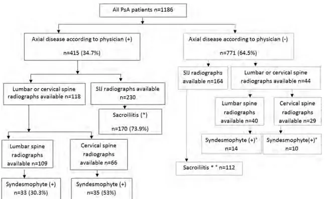
In October 2016, 1195 patients were recruited to the registry and 1186 had data on clinical subtypes for arthritis and were included in the analysis. Within these, 415 (35%) were classi-fied as having axPsA by the physician, either alone or in combination with other subtypes (Fig. 1). Subjects with
axPsA were more frequently men, were younger, were more frequently smokers, were more likely to be on TNF inhibitors (TNFis), and had more nail involvement (Table1).
Imaging findings
SIJs Of 395 SIJ radiographs, asymmetrical sacroiliitis (grade 2-3-4 on one side) was seen in 70 (17.7%) patients and 246 (62.3%) had symmetrical sacroiliitis (grade≥ 2 on both sides). Two hundred and eighty-three (71.6%) pa-tients fulfilled the sacroiliitis criteria required for classify-ing the patient as havclassify-ing AS accordclassify-ing to the modified NY criteria [11].
The spine assessment The prevalence of syndesmophytes in the cervical spine was higher than that in the lumbar spine [45/ 96 (46.9%) vs 47/150 (31.3%)]. Within 80 patients that had both cervical and lumbar spine radiographs available for cen-tral reading, 25 (31%) had syndesmophytes at either cervical or lumbar spine only. Data from 157 patients where both sa-croiliac findings and spine radiographs were available showed that patients that fulfilled the mNY criteria had more frequent syndesmophytes compared to patients that did not fulfil the criteria [58/121 (47.9%) vs 10/36 (27.8%); p = 0.04]. Overall, only 10 in 157 patients had syndesmophytes in the absence of significant sacroiliitis.
Among 71 patients that had syndesmophytes, 20 patients (28.2%) were not classified as having axPsA according to the physician. This was higher for women (15/42, 35.7%) than for men (5/29, 17.2%).
Characteristics of patients with underdiagnosed
axPsA and implications in management
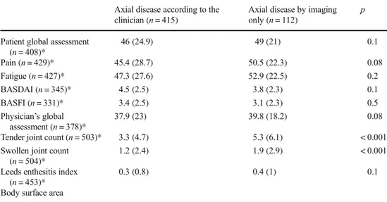
One hundred and twelve patients had sacroiliitis fulfilling the modified NY criteria, despite not being categorized as axPsA. These were mostly women [83/220 (37.7%) vs 29/122 (23.8%); p = 0.008] and were less likely to be on TNFis (16.1 vs 38.2%; p < 0.001). They also had higher tender and swollen joint counts (Table2). PROs and physician’s global
assessment for disease activity were similar in both groups, suggesting a comparable degree of disease activity according to the patients and physicians. As not all patients had routine screening by imaging, a percentage for underdiagnosed axPsA was not calculated.
Reliability
The complete agreement for detecting syndesmophytes in 2 separate readings was 70/72 (97.2%, kappa 0.971) for the cervical spine and 58/72 (80.6%, kappa 0.641) for the lumbar spine. The kappa value for the grading of sacroiliitis from 0 to 4 was 0.791 with a complete agreement in the score in 34/56
SIJs. The second reading resulted in 6/28 patients to be clas-sified differently for modified NY criteria. The interreader agreement to detect grade 2 or higher sacroiliitis showed
moderate agreement (kappa 0.451). The interreader kappa values for syndesmophytes were 0.806 for the lumbar spine and 0.870 for the cervical spine.
Fig. 1 Number of PsA patients with available data and positive for axial disease according to the clinician. The number of radiographs corresponds to the radiographs of which the quality was sufficient to be scored by the central reader, after excluding 36 sacroiliac joint (SIJ) and 5
lumbar spine radiographs. *Sacroiliitis≥ grade 3 unilateral or ≥ grade 2
bilateral. °A percentage was not calculated for these radiographs as routine imaging was not performed to all patients
Table 1 Demographics of the whole group and patients with or without axial disease according to the physician. Comparisons between patients with or without axial disease were significant at the following levels: *p < 0.001,
**p 0.04, ***p 0.01,+p 0.006,
++p 0.03,+++p 0.001. The data are
given as mean (SD) if not mentioned otherwise
Physician’s assessment for axial disease
Axial PsA (n = 415) Non-axial PsA (n = 771)
Age 44.4 (12.2)* 47.9 (13)*
Gender: women, n (%) 248 (59.8%)+ 523 (67.8%)+
Disease duration (for arthritis) (months since diagnosis) 68.7 (83.2) 66.6 (80)
Disease duration (for psoriasis) (months since the onset) 178.6 (132.6) 181.8 (139)
Nail disease, n (%) 207 (49.9)++ 333 (43.2)++
Smoking (current or past), n (%) 185 (44.6%)* 263 (34.1%)*
Body mass index 27.6 (5.1)** 28.2 (5.1)**
TNF inhibitors, n (%) 137/369 (37.1%)*** 197/670 (29.4%)***
Symmetrical polyarthritis 129 (31.1)* 343 (44.5)*
Asymmetrical oligoarthritis/monoarthritis 145 (34.9)+++ 345 (44.8)+++
Axial
Distal interphalangeal joint disease 42 (10.1) 155 (20.1)
Arthritis mutilans 3 (0.7) 6 (0.8) Plaque psoriasis 191 (69.2) 383 (74.1) Flexural 2 (0.7) 5 (1) Inverse 1 (0.4) 3 (0.6) Pustular 72 (26.1) 106 (20.5) Erytrodermic 10 (3.6) 20 (3.9)
All PsA patients n=l186
•
Axlal disease according to physician(+)n=415 (34.7%)
!
i
•
I
Lumbar or cervical .spineI
SIJ radiographs availableradiographs available n:a118 n=230
•
Sacroiliitis ( )
n=170 (73.9%)
'
Lumbar spine Cerv1ca..
l spine radlographs radfographs available n=109 available n=66i
t
Syndesmophyte (+) Syndesmophyte (+)
n=33 (30.3%) n=35 (53%)
"
Axial disease acoo rding to physician(-)
(64.5%)
n=771
•
SIJ radiographs available n=164
Lumbar or cervical spine
radiographs available n=44 T Lumb ar spine
"'
radl ographs. ble n=40 availa [ Syndes~:: hyte {+)•
' 4 Sacroiliitis • • n=ll2l I
'
"
Cervical spine radiographs available n=29'
Syndesmophyte( + )° n:10Discussion
PsA is a heterogeneous disease and the complexity of the clinical presentation makes it difficult to accurately classify patients. It is likely that there is a subset of patients with more tender and swollen joints that have imaging findings for axPsA, without clear clinical manifestations of axial disease to help the physicians for diagnosis, which is more frequent in women. In case of having other disease features, like active peripheral arthritis, both patients and physicians may be disregarding the involvement of the spine. Additionally, as recently been shown by our group, the IBP questionnaires only have moderate sensitivity in axPsA, which can be anoth-er reason for missing the axial disease [13].
In patients who had sacroiliitis on imaging despite not being classified as axPsA by the physician, the same mag-nitude of disease burden according to the patients was reflected by the PROs, which made us to describe as “underdiagnosed” instead of “subclinical” axPsA. This is an important difference as subclinical disease may not need screening unless long-term impacts on outcomes are shown. On the other hand, underdiagnosed is terminology that would suggest the need for better screening methods. In addition to the impacts on the PROs, our data show that diagnosis of axial disease not only is an academically rele-vant question but also may have an impact on the treatment. The treatment recommendations by GRAPPA and EULAR suggest that patients with axPsA and peripheral disease re-quire different treatment approaches such as peripheral dis-ease being more responsive to conventional DMARD ther-apies whereas biological therther-apies are the next medical treatment steps after the nonsteroidal anti-inflammatory drugs for axial disease [14,15]. On the other hand, patients with axial disease according to the physician have similar
disease burden with patients with axPsA by imaging only according to PROs, and yet treated less with TNFis.
Another question is which imaging modality needs to be used and which site(s) need be screened. For the SIJs, radio-graph is the initial screening method for economic concerns and for being widely available. The presence of syndesmophytes was not a part of axPsA in this study; however, 6% of patients in our data had syndesmophytes without any sacroiliitis, which was less frequent than a recent study by Jadon et al. [5]. Screening only the lumbar or cervical spine would have led to underdiagnosing 11.3% of our patients. Routine screening of SIJ and spine at least once would give a better understanding of the spectrum of the disease.
Our study has some limitations. PsA patients were consecu-tively recruited to the registry with no specific inclusion criteria; therefore, a selection bias was not applicable at the stage of recruitment. However, this is an observational study and radio-graphs were ordered in the discretion of the recruiting physician and there might have been a selection bias for the group of patients that had radiographs. There is not a widely accepted definition for axPsA for radiographs or where to draw the threshold for a positive finding. Although there are controver-sies on the effect of HLAB27 on the disease progression in axPsA [3,5], the lack of HLA-B27 limits the identification of different subsets. Despite these limitations, this registry reflects the real-life experience, and for including 32 centers across the country, the data represent the routine practice in rheumatology. In summary, PsA patients with more severe peripheral dis-ease, such as more tender/swollen joints, had higher risk of being underdiagnosed for the axPsA, which was more com-mon in women. Underdiagnosed patients were less frequently treated with biologic treatments. Axial disease needs to be ruled out using imaging modalities, even if back pain does not clinically suggest of an inflammatory pattern.
Table 2 Comparison of patient-reported outcomes and physician-based assessments in patients with axial disease according to the clinician and axial disease by imaging only. BASDAI, Bath Ankylosing Spondylitis Disease Activity Index; BASFI, Bath Ankylosing Spondylitis Function Index. Numbers are given as mean (SD)
Axial disease according to the clinician (n = 415)
Axial disease by imaging only (n = 112)
p Patient global assessment
(n = 408)* 46 (24.9) 49 (21) 0.1 Pain (n = 429)* 45.4 (28.7) 50.5 (22.3) 0.08 Fatigue (n = 427)* 47.3 (27.6) 52.9 (22.5) 0.2 BASDAI (n = 345)* 4.5 (2.5) 3.8 (2.3) 0.1 BASFI (n = 331)* 3.4 (2.5) 3.1 (2.3) 0.5 Physician’s global assessment (n = 378)* 37.9 (23) 39.8 (18.2) 0.08
Tender joint count (n = 503)* 3.3 (4.7) 5.3 (6.1) < 0.001
Swollen joint count (n = 504)*
1.2 (2.4) 1.9 (2.9) < 0.001
Leeds enthesitis index (n = 453)*
0.3 (0.8) 0.4 (1) 0.1
Body surface area
Acknowledgments We would like to thank the following collaborators for their participation: Adem Kucuk, Emel Gonullu, Emine Duygu Ersozlu Bozkir, Funda Erbasan, Gozde Yildirim Cetin, Senol Kobak, Serpil Ergullu Esmen, Servet Akar, Seval Pehlevan, Soner Senel, Tuncay Duruoz, Kenan Aksu, Yasemin Kabasakal, Timucin Kasifoglu.
We would also like to thank Drs Laure Gossec and Helena Marzo-Ortega for criticizing and helping to improve our dataset and analyzing for this manuscript.
Compliance with ethical standards
Conflict of interest Sibel Aydin has received honoraria from Abbvie,
Novartis, Pfizer, UCB, and Sanofi.
References
1. Gladman DD (2015) Clinical features and diagnostic considerations
in psoriatic arthritis. Rheum Dis Clin N Am 41:569–579.https://
doi.org/10.1016/j.rdc.2015.07.003
2. Moll JM, Wright V (1973) Psoriatic arthritis. Semin Arthritis
Rheum 3:55–78.https://doi.org/10.1016/0049-0172(73)90035-8
3. Hanly JG, Russell ML, Gladman DD (1988) Psoriatic
spondyloarthropathy: a long term prospective study. Ann Rheum
Dis 47:386–393.https://doi.org/10.1136/ard.47.5.386
4. Battistone MJ, Manaster BJ, Reda DJ, Clegg DO (1999) The
prev-alence of sacroilitis in psoriatic arthritis: new perspectives from a large, multicenter cohort. A Department of Veterans Affairs
Cooperative Study. Skeletal Radiol 28:196–201.https://doi.org/
10.1007/s002560050500
5. Jadon DR, Sengupta R, Nightingale A, Lindsay M, Korendowych
E, Robinson G, Jobling A, Shaddick G, Bi J, Winchester R, Giles JT, McHugh NJ (2017) Axial Disease in Psoriatic Arthritis study: defining the clinical and radiographic phenotype of psoriatic
spondyloarthritis. Ann Rheum Dis 76:701–707.https://doi.org/10.
1136/annrheumdis-2016-209853
6. Queiro R, Belzunegui J, Gonzalez C et al (2002) Clinically
asymp-tomatic axial disease in psoriatic spondyloarthropathy. A
retrospec-tive study. Clin Rheumatol 21:10–13.https://doi.org/10.1007/
s100670200003
7. Gladman DD (1998) Psoriatic arthritis. Rheum Dis Clin N Am 24:
829–844.https://doi.org/10.1016/S0889-857X(05)70044-2
8. Gladman DD (1994) Natural history of psoriatic arthritis.
Baillie’res Clin Rheumatol 8:379–394.https://doi.org/10.1016/
S0950-3579(94)80024-3
9. Gladman DD, Rahman P, Krueger GG, Mease PJ, Qureshi AA,
Dutz JP, Lindqvist U, Carneiro S, Helliwell PS, Ujfalussy I, Gottlieb AB, Behrens F, Ståhle M (2008) Clinical and genetic
reg-istries in psoriatic disease. J Rheumatol 35:1458–1463
10. Kalyoncu U, Bayindir O, Ferhat Oksuz M et al (2017) The Psoriatic
Arthritis Registry of Turkey: results of a multicentre registry on
1081 patients. Rheumatology (Oxford) 56:279–286.https://doi.
org/10.1093/rheumatology/kew375
11. Van Der Linden S, Valkenburg HA, Cats A (1984) Evaluation of
diagnostic criteria for ankylosing spondylitis. A proposal for mod-ification of the New York criteria. Arthritis Rheum 27:361–368.
https://doi.org/10.1002/art.1780270401
12. Aydin SZ, Kasapoglu Gunal E, Kurum E, Akar S, Mungan HE,
Alibaz-Oner F, Lambert RG, Atagunduz P, Marzo Ortega H, McGonagle D, Maksymowych WP (2017a) Limited reliability of radiographic assessment of spinal progression in ankylosing
spon-dylitis. Rheumatology (Oxford) 56:2162–2169.https://doi.org/10.
1093/rheumatology/kex321
13. Aydin SZ, Kilic L, Kucuksahin O, Ureyen SB, Kalyoncu U (2017b)
Performances of inflammatory back pain criteria in axial psoriatic
arthritis. Rheumatology (Oxford) 56:2031–2032.https://doi.org/10.
1093/rheumatology/kex307
14. Coates LC, Kavanaugh A, Mease PJ, Soriano ER, Laura
Acosta-Felquer M, Armstrong AW, Bautista-Molano W, Boehncke WH, Campbell W, Cauli A, Espinoza LR, FitzGerald O, Gladman DD, Gottlieb A, Helliwell PS, Husni ME, Love TJ, Lubrano E, McHugh
N, Nash P, Ogdie A, Orbai AM, Parkinson A, O’Sullivan D, Rosen
CF, Schwartzman S, Siegel EL, Toloza S, Tuong W, Ritchlin CT (2016) Group for Research and Assessment of Psoriasis and Psoriatic Arthritis 2015 treatment recommendations for psoriatic
arthritis. Arthritis Rheumatol 68:1060–1071.https://doi.org/10.
1002/art.39573
15. Ramiro S, Smolen JS, Landewe R et al (2016) Pharmacological
treatment of psoriatic arthritis: a systematic literature review for the 2015update of the EULAR recommendations for the
manage-ment of psoriatic arthritis. Ann Rheum Dis 75:490–498.https://doi.
org/10.1136/annrheumdis-2015-208466
Affiliations
Sibel Zehra Aydin1 &Orhan Kucuksahin2&Levent Kilic3&Atalay Dogru4&Ozun Bayindir5&Cem Ozisler6&
Ahmet Omma7&Emine Figen Tarhan8&Abdulsamet Erden9&Gezmis Kimyon10&Meryem Can11&Ediz Dalkilic12&
Sule Yavuz11&Sibel Bakirci Ureyen13&Esen Kasapoglu Gunal14&Fatıma Arslan Alhussain15&Lutfi Akyol16&
Ayse Balkarli17&Sema Yilmaz18&Muhammet Cinar19&Muge Tufan Aydin20&Dilek Solmaz21&Ridvan Mercan22&
Sukran Erten2&Umut Kalyoncu23
1 Ottawa Hospital Research Institute, University of Ottawa Faculty of
Medicine, 1967 Riverside Drive, Ottawa, ON K1H 7W9, Canada 2
Department of Internal Medicine, Division of Rheumatology, Ankara Yildirim Beyazit University, Ankara, Turkey 3
Department of Internal Medicine, Division of Rheumatology Hacettepe University Hospital, Hacettepe University, Ankara, Turkey
4 Department of Internal Medicine, Division of Rheumatology,
Suleyman Demirel University, Isparta, Turkey 5
Department of Internal Medicine, Division of Rheumatology, Ege University, Izmir, Turkey
6
Department of Internal Medicine, Division of Rheumatology, Diskapi Yildirim Beyazit Education and Research Hospital, Ankara, Turkey
7
Department of Internal Medicine, Division of Rheumatology, Ankara Numune Education and Research Hospital, Ankara, Turkey
8 Department of Internal Medicine, Division of Rheumatology, Mugla
Sitki Kocman University, Muğla, Turkey
9
Department of Internal Medicine, Division of Rheumatology, Hacettepe University, Ankara, Turkey
10
Department of Internal Medicine, Division of Rheumatology, Gaziantep University, Gaziantep, Turkey
11 Department of Internal Medicine, Division of Rheumatology,
Marmara University, Istanbul, Turkey 12
Department of Internal Medicine, Division of Rheumatology, Uludag University, Bursa, Turkey
13
Faculty of Medicine, Rheumatology, University of Ottawa, Ottawa, ON, Canada
14 Department of Internal Medicine, Division of Rheumatology,
Medeniyet University Goztepe Education and Research Hospital, Istanbul, Turkey
15
Department of Internal Medicine, Medeniyet University Goztepe Training and Research Hospital, Istanbul, Turkey
16 Department of Internal Medicine, Division of Rheumatology,
Samsun Ondokuz Mayis University, Samsun, Turkey 17
Antalya Training and Research Hospital, Rheumatology, Antalya, Turkey
18
Department of Internal Medicine, Division of Rheumatology, Selçuk Üniversity, Konya, Turkey
19 Department of Internal Medicine, Division of Rheumatology,
Gulhane Education and Research Hospital, Ankara, Turkey 20
Department of Internal Medicine, Division of Rheumatology, Baskent University, Adana, Turkey
21
Department of Internal Medicine, Division of Rheumatology, Izmir Katip Celebi University, Izmir, Turkey
22 Hatay Hospital, Rheumatology, Antakya, Turkey
23
Faculty of Medicine, Department of Internal Medicine, Division of Rheumatology, Hacettepe University, Ankara, Turkey