·
Clinical Research·
Safety of hydroimplantation in cataract surgery in
patients with pseudoexfoliation syndrome
Tevfik Oğurel, Reyhan Oğurel, Zafer Onaran, Kemal Örnek
Department of Ophthalmology, Kirikkale University School of Medicine, Kirikkale University Campus, Ankara 71100, Yahsihan, Turkey
Correspondence to: Tevfik Oğurel. Department of
Ophthal-mology, Kirikkale University School of Medicine, Kirikkale University Campus, Ankara 71100, Yahsihan, Turkey. [email protected]
Received: 2016-06-04 Accepted: 2016-12-01
Abstract
● AIM: To evaluate the safety of hydroimplantation in cataract surgery in patients with pseudoexfoliation syndrome. ● METHODS: This prospective randomized study comprised 100 eyes of 100 consecutive patients who underwent cataract surgery and implantation of foldable intraocular lens (IOL). Each eye was assigned to group 1 or group 2. Hydroimplantation without using viscoelastic agent as group 1 (n=50), and hydroxypropylmethylcellulose (Eyevisc, Biotech, India) was used in group 2 (n=50). ● RESULTS: There were no statistically significant differences in central corneal thickness (CCT) and corneal endothelial cell count (ECC) between both groups at each visit and percentage change in CCT and ECC (P>0.05). The mean intraocular pressure (IOP) at postoperative 5h increased statistically significantly in group 2 (P<0.001). There was no statistically significant difference in IOP between two groups, before and after surgery excluding the 24h postoperative IOP, but patients in group 2 had higher IOP than that in the group 1 at 24h after surgery (P=0.035). No case in either group experienced posterior capsular rupture, or zonular dialysis. Fixation of the globe during IOL implantation was better in group 1 than that in group 2.
● CONCLUSION: Hydroimplantation has advantages in terms of IOP changes and duration of the surgery and seems to be safe in patients with pseudoexfoliation syndrome. ● KEYWORDS: hydroimplantation; pseudoexfoliation; phacoemulsification
DOI:10.18240/ijo.2017.05.10
Oğurel T, Oğurel R, Onaran Z, Örnek K. Safety of hydroimplantation in cataract surgery in patients with pseudoexfoliation syndrome. Int J
Ophthalmol 2017;10(5):723-727
INTRODUCTION
O
phthalmic viscosurgical devices (OVD) are used in eye surgery commonly since first commercially viscoelastic was produced in 1980. They can be utilized to create space, prevent damage of ultrasonic radiation, maintain the ocular tonus during manuplation and other mechanical impacts on the corneal endothelium and simplify intraocular lens (IOL) implantation[1-3]. The ideal OVD should be easily removablefrom the anterior chamber at the end of the surgery because they may cause postoperative intraocular pressure (IOP) elevation and inflammation, which may result in more endothelial cell damage[4].
A new technique has been described recently. In this technique the authors used OVD only in stage of the capsulorhexis or they did not use in any stage of cataract surgery and compared advantages and disadvantages of cataract surgery with and without OVD[5-7]. In these studies, they found that contrary to
popular belief, OVD in cataract surgery was not indispensable. In our study, we evaluated the safety of IOL hydroimplantation technique in pseudoexfoliation patients who are prone to various complications in phacoemulsification surgery.
SUBJECTS AND METHODS
This prospective randomized study comprised 100 eyes of 100 consecutive patients with bilateral age related cataract scheduled for phacoemulsification cataract surgery and implantation of foldable IOL. The research was confirmed by Institutional Review Board and was conducted in accordance with the Declaration of Helsinki. All patients gave written informed consent before their participation.
The exclusion criteria consisted past ocular trauma, previous ocular surgery, subluxated cataract, coexisting corneal endothelial disease [endothelial cell count (ECC) <1500 cells/mm2], uveitis and glaucoma. The patients
randomly assigned to IOL implantation technique of either hydroimplantation or viscoimplantation. Two groups were selected. Hydroimplantation without using viscoelastic agent was group 1, consisting of 50 eyes (29 females and 21 males), and hydroxypropylmethylcellulose was used in group 2 (viscoimplantation) consisting of 50 eyes (26 females, 24 males). All patients had a similar degree of nuclear opacification (NO3, NO4 or NO5) and a similar degree of cortical opacification (C2 or C3) according to the LOCS III scale[8]. Pupil size was measured using pupil size chart
at the beginning of surgery after dilation with tropicamide 1%, cyclopentolate 1% and phenylephrine 2.5% drops. All individuals had pseudoexfoliation syndrome in both group. Pseudoexfoliation syndrome was diagnosed in the presence of evident classical scurf or flaky exfoliation materials on the pupil, lens or other ocular structures, radial pigment over the lens surface. Preoperative central corneal thickness (CCT), corneal ECC, Goldmann applanation tonometry, anterior and posterior segment examination were recorded. Postoperative IOP on the 5, 24h, 5d, 1 and 3mo after surgery were noted as were postoperative glaucoma medications. For patient safety, IOP-reducing therapies were allowed at 5h postoperative, if IOP reached 30 mm Hg. Postoperative CCT and corneal ECC were recorded on 5d, 1 and 3mo after surgery. CCT and corneal ECC were measured with a noncontact specular microscope (Cell Check SL, Konan, CA, USA). The measurements were taken in auto-aligment, auto-focus and auto-measurement mode. With Konan’s patented measurement method only one photograph is taken in each measurement. The percentage changes in ECC and CCT at each visit were calculated as follows: (preoperative value-postoperative value)/preoperative value×100.
Intraoperative parameters of ultrasound (U/S) total time and total surgery time were automatically calculated and displayed on the monitor of the PentaSys 2 phacoemulsification system. All patients were operated by the same surgeon (Oğurel T) under topical anesthesia and the quick-chop phacoemulsification technique was performed. In both groups, two side-ports and a main temporal incision were performed in all eyes. Following injection of viscoelastic 2% hydroxypropylmethylcellulose (Eyevisc, Biotech, India) in both group continuous curvilinear capsulorhexis, hydrodissection, phacoemulsification of the nuclues and cortex aspiration were performed. In group 1, no viscoelastic was injected in the eye than the irrigation cannula introduced into the anterior chamber through left side port with irrigation on. After the tip of the cartridge was put into the main port in direction of capsular bag, IOL (Eyecryl plus, Biotech,
India) was gradually injected. The aspiration cannula was inserted through the other side port and then optic and haptic were placed into the capsular bag through pressing lightly on by bimanual cannula (Figure 1). In group 2, capsular bag was expanded with assigned viscoelastic agent, and foldable acrylic IOL was implanted into the capsular bag. The viscoelastic material was aspirated from the anterior chamber, the capsule fornix, and the retrolental space using bimanual irrigation/ aspiration. Finally in both groups, all corneal incisions were hydrated. Posterior capsule rupture did not occur in any eyes. All patients were treated with prednisolone acetate for 1mo and moxifloxacin hydrochloride eye drops for 1wk five times a day postoperatively. IOP was measured at 5, 24h, 5d, 1 and 3mo after the surgery in both groups by Goldmann applanation tonometry. All patients were evaluated for facilitation of IOL implantation, posterior capsular rupture, zonular dialysis during the surgery and elevation of IOP and changes of CCT and ECC postoperatively.
Statistical Analysis Statistical analysis was done by SPSS
statistical software (SPSS for windows 16.0, Inc., Chicago, USA). Group comparisons were made with independent sample t-tests. For comparison of continuous variables in each group over time one way repeated measure analysis of variance was used followed by a Bonferroni correction. P value less than 0.05 was defined statistically significant.
RESULTS
One hundred eyes from 100 patients [45 men (45%) and 55 women (55%)] were enrolled in this study. The mean age of the patients in group 1 was 66.77±7.59y and 69.40±6.94y in group 2. Among 100 patients, 50 (29 females and 21 males) were in group 1 (hydroimplantation) and 50 (26 females and 24 males) were in group 2 (viscoimplantation). The difference between the two groups was not statistically significant in terms of age, gender, NO, pupil size, and U/S time (P>0.05) (Table 1).
The mean preoperative CCT was 530±35.2 µm in group 1 and 525±34.3 µm in group 2. The corneal thickness has mildly
Figure 1 IOL hydroimplantation technique A: IOL was introduced into capsular bag through the main corneal incision while the irrigation
decreased by 3mo, reaching 532±31.3 and 533±32.9 µm in both group, however this difference was not significant (P>0.05). There were no statistically significant differences in CCT between both groups at each visit and percentage change in CCT (P>0.05) (Table 2).
The mean IOP in group 1, preoperative and 5h postoperative, were 16±3.36 and 17±4.09 mm Hg and in group 2 they were measured as 15±2.70 and 19±6.09 mm Hg respectively. The mean IOP at 5h postoperative increased significantly in group 2 (P<0.001). There was no statistically significant difference in IOP between two groups, before and after surgery excluding the 24h postoperative IOP, but patients in group 2 had higher IOP than that in group 1 at 24h after surgery (P=0.035). Seven patients (14%) had increased IOP (higher than 30 mm Hg) at 5h postoperatively and needed brinzolamide/timolol fixed combination for IOP control. There were no patients those had IOP spikes 30 mm Hg or higher in group 1. IOP were not changed significantly from the preoperative values at each postoperative examination (P>0.05) (Table 2).
Table 2 shows the average ECC at each visit. There was no significant difference between two groups comparing the ECC and also percentage change in ECC before and after the surgery. Comparison of IOP, CCT and ECC each groups over time is showed in Table 3.
Total surgery time was shorter in group 1 when compared with group 2 where it didn’t necessitate injection and aspiration of viscoelastic material in the former (P<0.001) (Table 1). No case in either group experienced posterior capsular rupture, or zonular dialysis. Fixation of the globe during the IOL implantation was better in hydroimplantation group than viscoimplantation group.
DISCUSSION
Viscoelastics are one of the most important devices for phacoemulsification for years. They have many advantages such as protection on the corneal endothelium and facilitation of IOL implantation and capsulorhexis[2,9]. Despite their many
benefits, they can cause some side effects. One of the most side effect is elevated IOP in early postoperative process[10-11].
Pre-vious studies have shown that in the early postoperative period all ophthalmic viscosugical devices are capable of increasing IOP[12-13].The most common cause of this complication is
usually retention of viscoelastic. Although the viscoelastic is removed by irigation/aspiration at the end of the surgery, it can be retained at the capsule fornix and the retrolental space. To avoid this complication many surgeons are striving to remove viscoelastics completely from the eye[14-15]. Especially
visco-elastic behind the IOL is difficult to aspirate and it has a risk as perforating the posterior capsule. Especially this risk arise in pseudoexfoliative eyes patients because of the zonular weak-ness and non mydriatic pupilla.
Usually complete removal of the viscoelastics is not possible, it takes a time and extends the time of the surgery. Increased IOP levels in postoperative period could damage optic nerve
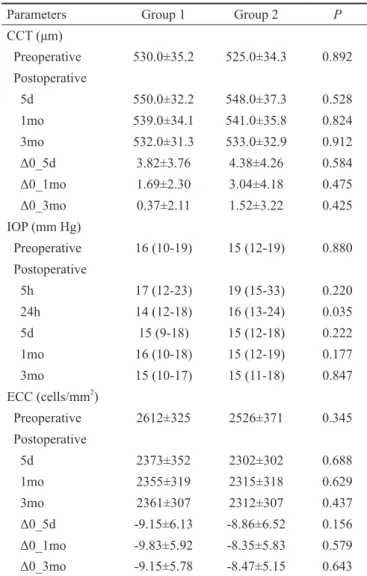
Table 2 Comparison of IOP, CCT , ECC and percentage differences in IOP, CCT and ECC between groups over time
Parameters Group 1 Group 2 P
CCT (μm) Preoperative 530.0±35.2 525.0±34.3 0.892 Postoperative 5d 550.0±32.2 548.0±37.3 0.528 1mo 539.0±34.1 541.0±35.8 0.824 3mo 532.0±31.3 533.0±32.9 0.912 Δ0_5d 3.82±3.76 4.38±4.26 0.584 Δ0_1mo 1.69±2.30 3.04±4.18 0.475 Δ0_3mo 0.37±2.11 1.52±3.22 0.425 IOP (mm Hg) Preoperative 16 (10-19) 15 (12-19) 0.880 Postoperative 5h 17 (12-23) 19 (15-33) 0.220 24h 14 (12-18) 16 (13-24) 0.035 5d 15 (9-18) 15 (12-18) 0.222 1mo 16 (10-18) 15 (12-19) 0.177 3mo 15 (10-17) 15 (11-18) 0.847 ECC (cells/mm2) Preoperative 2612±325 2526±371 0.345 Postoperative 5d 2373±352 2302±302 0.688 1mo 2355±319 2315±318 0.629 3mo 2361±307 2312±307 0.437 Δ0_5d -9.15±6.13 -8.86±6.52 0.156 Δ0_1mo -9.83±5.92 -8.35±5.83 0.579 Δ0_3mo -9.15±5.78 -8.47±5.15 0.643
Table 1 Demographic data and clinical charasteristics in each study group
Parameters Group 1 Group 2 P
Age (a) 66.77±7.59 69.40±6.94 0.135
Gender (F/M) 29 (58%)/21 (42%) 26 (52%)/24 (48%) 0.637
NO3/NO4/NO5 27/17/6 30/15/5 0.358
Pupil size 5.25±1.45 5.74±1.18 0.852
U/S time (min) 0.78±0.7 0.69±0.3 0.189
Total surgery time (min) 7.3±3.5 10.2±4.6 <0.001
and lead to ischemia especially in glaucoma patients including pseudoexfoliative eyes[16-17].
The hydroimplantation technique for inserting a foldable IOL without OVD was described by Tak[6], in his study he
compared hydroimplantation with viscoimplantation and described that depth of the anterior camera and capsular bag was similar. On the 1d postoperative there was no difference in
corneal edema. Time required for implantation of the lens was significantly less in hydroimplantation group (40 to 60s) than viscoimplantation group (2.4 to 4min).
In another study, Zia et al[18] evaluated IOP after IOL
implantation with hydroxypropylmethylcellulose 2% vs hydroimplantation and found that there was insignificant IOP elevation at postoperative 24h in hydroimplantation groups when compared with IOL implantation with hydroxypropylmethylcellulose 2%. Previous studies also stated that cleaning the interior lens capsule by irrigation with balanced salt solution could reduce the incidence of both toxic anterior segment syndrome and endophthalmitis[19-20]. Although
the viscoelastic material anterior to the IOL can easily be aspirated via irrigation and aspiration (I/A) handpiece, there could be some viscoelastic remained in the lens capsule especially behind the IOL since it could be time consuming and risky to manipulate the IOL for deeper aspirations. Here we eliminate all these mentioned factors by using hydroimplantation technique.
In our study, we compared safety of hydroimplantation and effect on postoperative IOP elevation and changes on CCT and ECC with viscoimplantation using hydroxypropy-lmethylcellulose 2%. No cases in either group experienced posterior capsular rupture, or zonular dialysis. Fixation of the globe during IOL implantation was better in hydroimplantation group than viscoimplantation group. Postoperative IOP changes were less and there was no spike of IOP elevation in hydroimplantation group. Another advantage of this technique is desired position of the IOL is very easy because of using I/A especially in toric IOLs and the surgeon will not need aspirating viscoelastic behind the IOL, and this shortens the time after nuclues and cortex aspiration.
We think that hydroimplantation is useful, safe and advantageous technique in phacoemulsification surgery. At least, it may be preferable in patients who would have the risk of viscoelastic retention in the eye.
ACKNOWLEDGEMENTS
Conflicts of Interest: Oğurel T, None; Oğurel R, None; Onaran Z, None; Örnek K, None.
REFERENCES
1 Oshika T, Bissen-Miyajima H, Fujita Y, Hayashi K, Mano T, Miyata K, Sugita T, Taira Y. Prospective randomized comparison of DisCoVisc and Healon5 in phacoemulsification and intraocular lens implantation. Eye (Lond) 2010;24(8):1376-1381.
2 Modi SS, Davison JA, Walters T. Safety, efficacy, and intraoperative characteristics of DisCoVisc and Healon ophthalmic viscosurgical devices for cataract surgery. Clin Ophthalmol 2011;5:1381-1389.
3 Hengerer FH, Dick HB, Kohnen T, Conrad-Hengerer I. Assessment of intraoperative complications in intumescent cataract surgery using 2 ophthalmic viscosurgical devices and trypan blue staining. J Cataract Refract Surg 2015;41(4):714-718.
4 Beiko GH. Endothelial cell loss after cataract phacoemulsification with Healon5 vs. I-Visc Phaco. Can J Ophthalmol 2003;38(1):52-56.
5 Studeny P, Hyndrak M, Kacerovsky M, Mojzis P, Sivekova D, Kuchynka P. Safety of hydroimplantation: a foldable intraocular lens implantation without the use of an ophthalmic viscosurgical device. Eur J Ophthalmol 2014;24(6):850-856.
6 Tak H. Hydroimplantation: foldable intraocular lens implantation without an ophthalmic viscosurgical device. J Cataract Refract Surg 2010;36(3):377-379.
7 Özyol E, Özyol P. Hydro-visco-implantation technique for wound-assisted foldable intraocular lens implantation during microinci-sion cataract surgery. J Cataract Refract Surg 2008;34(10):1748-1753. 8 Chylack LT Jr, Wolfe JK, Singer DM, Leske MC, Bullimore MA, Bailey IL, Friend J, McCarthy D, Wu SY. The lens opacities classification system III. The Longitudinal Study of Cataract Study Group. Arch Ophthalmol 1993;111(6):831-836.
9 Storr-Paulsen A, Nørregaard JC, Farik G, Tarnhøj J. The influence of viscoelastic substances on the corneal endothelial cell population during cataract surgery: a prospective study of cohesive and dispersive viscoelastics. Acta Ophthalmol Scand 2007;85(2):183-187.
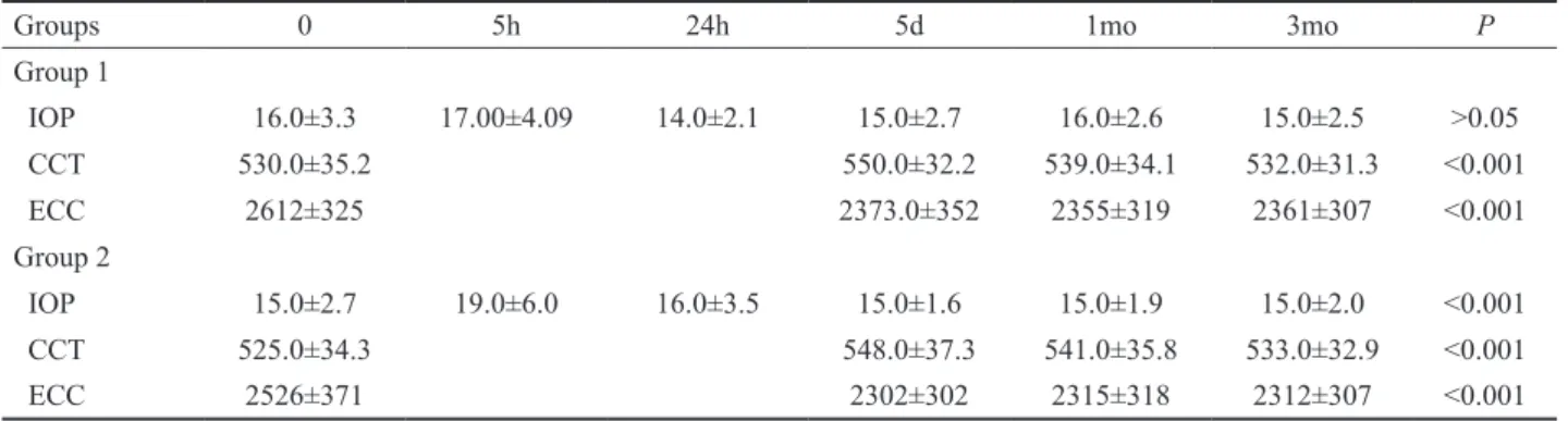
Table 3 Comparison of IOP, CCT and ECC each groups over time
Groups 0 5h 24h 5d 1mo 3mo P
Group 1 IOP 16.0±3.3 17.00±4.09 14.0±2.1 15.0±2.7 16.0±2.6 15.0±2.5 >0.05 CCT 530.0±35.2 550.0±32.2 539.0±34.1 532.0±31.3 <0.001 ECC 2612±325 2373.0±352 2355±319 2361±307 <0.001 Group 2 IOP 15.0±2.7 19.0±6.0 16.0±3.5 15.0±1.6 15.0±1.9 15.0±2.0 <0.001 CCT 525.0±34.3 548.0±37.3 541.0±35.8 533.0±32.9 <0.001 ECC 2526±371 2302±302 2315±318 2312±307 <0.001
10 Benson FG, Patterson MM, Epstein DL. Obstruction of aqueous out-flow by sodium hyaluronate in enucleated human eyes. Am J Ophthalmol 1983;95(5):668-672.
11 Morgan RK, Skuta GL. Viscoelastic-related glaucomas. Semin Ophthalmol 1994;9(4):229-234.
12 Liesegang TJ. Viscoelastic substances in ophthalmology. Surv Ophthalmol 1990;34(4):268-293.
13 Holzer MP, Tetz MR, Auffarth GU, Welt R, Völcker HE. Effect of Healon5 and 4 other viscoelastic substances on intraocular pressure and endothelium after cataract surgery. J Cataract Refract Surg 2001;27(2): 213-218. 14 Sim BW, Amjadi S, Singh R, Bhardwaj G, Dubey R, Francis IC. Assessment of adequate removal of ophthalmic viscoelastic device with irrigation/aspiration by quantifying intraocular lens 'Judders'. Clin Experiment Ophthalmol 2013;41(5):450-454.
15 Henry JC, Olander K. Comparison of the effect of four viscoelastic agents on early postoperative intraocular pressure. J Cataract Refract
Surg 1996;22(7):960-966.
16 Ornek K, Büyüktortop N, Ornek N, Oğurel R, Erbahçeci IE, Onaran Z. Effect of 1% brinzolamide and 0.5% timolol fixed combination on intraocular pressure after cataract surgery with phacoemulsification. Int J Ophthalmol 2013;18;6(6):851-854.
17 Shrivastava A, Singh K. The effect of cataract extraction on intraocular pressure. Curr Opin Ophthalmol 2010;21(2):118-122.
18 Zia S, Iqbal, Y, Ashraf KM, Mirza A. Intraocular pressure after IOL implantation with hydroxypropylmethylcellulose 2% vs hydro-implantation. Pak J Ophthalmol 2013;29(1):12-15.
19 Bodnar Z, Clouser S, Mamalis N. Toxic anterior segment syndrome: Update on the most common causes. J Cataract Refract Surg 2012; 38(11):1902-1910.
20 Roy M, Chen JC, Miller M, Boyaner D, Kasner O, Edelstein E. Epidemic Bacillus endophthalmitis after cataract surgery I: Acute presentation and outcome. Ophthalmology 1997;104(11):1768-1772.