Olgu Sunumu
©
2011
DEÜ
TIP FAKÜLTESİ DERGİSİ CİLT 25, SAYI 1, (OCAK) 2011, S: 43 – 4543
Malign Pleural Effusion In A Patient With Multiple
Myeloma
MULTİPLE MYELOMLU HASTADA GELİŞEN MALİGN PLEVRAL EFFÜZYON
Abdullah KATGI
1, Selda KAHRAMAN
1, Pınar ATACA
2, Özden PİŞKİN
1, Mehmet Ali ÖZCAN
1,
Güner Hayri ÖZSAN
1, Fatih DEMİRKAN
1, Bülent ÜNDAR
11Dokuz Eylül Üniversitesi Tıp Fakültesi, Hematoloji Anabilim Dalı 2Dokuz Eylül Üniversitesi Tıp Fakültesi, İç Hastalıkları Anabilim Dalı
Abdullah KATGI
Dokuz Eylül Üniversitesi Tıp Fakültesi
Hematoloji AD
e-posta: [email protected]
ÖZET
Malign plevral effüzyon, multiple myelomda progresyonla karekterize ve geç gelişen bir komplikasyondur. Tanıdan sonra ortalama yaşam süresi 4 aydır ve sistemik kemoterapilere oldukça dirençlidir. Biz bu makalede 49 yaşında tanıdan 4 ay sonra malign plevral effüzyon gelişen ve solunum yetmezliği nedeniyle kaybedilen bir hastayı sunuyoruz.
Anahtar sözcükler: Multiple myelom, plevral effüzyon SUMMARY
Malign pleural effusion is a late and rare complication of multiple myeloma revealing the progression. The mean survival time is four months after the diagnosis with a poor response to systemic chemotherapy.
In this paper we mentioned about a 49 year old multiple myeloma patient in her fourth month of the diagnosis with a formation of malign pleural effusion just after induction chemotherapy and died because of respiratory failure.
Key words: Multiple myeloma, Pleural effusion
Multiple Myeloma (MM) is a malignant disease defined as proliferation of neoplastic plasma cells, which replace the normal bone marrow and produce excessive amounts of immunoglobulin or light chain (1).
Pleural effusion is a rare complication develops appro‐ ximately 6% of patients with MM during the course of their disease (2).
Pleural effusions in MM are usually benign and are due to congestive heart disease, chronic renal failure,
hypoalbuminemia, cardiac amyloidosis, pulmonary in‐ farctions or infections (2). Myelomatous involment is a rare complication of a progressive disease (3).
We discussed a MM patient developed malign pleural effusion under chemotherapy in this paper.
CASE REPORT
49 year old woman approached to neurosurgery clinic four months ago with severe back pain, difficulty in walking and fatigue. An excision was made from the solid
Malign pleural effusion in a patient with multiple myeloma
44
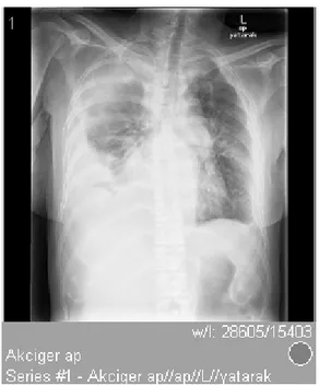
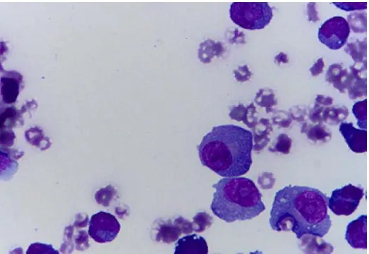
extradural mass which was detected between T7‐T10 vertebrates by thoracal MR. Pathology resulted as plasma cell neoplasia. The patient was consultated to hematology department. Monoclonal gammopaty was shown in pro‐ tein electrophoresis with serum immunoglobuline G (IgG) level as 5140 mg/dL (normal range between 552‐1631). Beta 2 macroglobuline was 8432 ng/dL with hemoglobin: 5.9 gr/dL, MCV:92 fL, leukocyte:6600, platelet 148.000, creatinin: 0.75 mg/dlt, ALT: 11 u/L AST: 16 u/L Ca: 9.2 mg/dL, Fosfor: 3.6 mg/dL, LDH: 657 U/L, CRP: 3.23 mg/dL, globulin: 6 g/dL. In bone marrow aspiration % 40 patological plasma cells was counted and the biopsy was diagnosed as Ig G Kappa multiple myeloma with stage 3A in Durie Salmon, stage 3 in ISS. No lytic lesions were detected in bone survey however radiotherapy was performed to L2‐L5, T6‐11 in case of collapsing fracture. The cyclophosphamide‐dexamethasone chemotherapy and antiresorptive zolendronic asic was prescribed. After the second cure of cyclophosphamide‐dexamethasone regimen respiratory distress was developed and massive pleural effusion detected in her right lung in the thoracal computerized tomography (Figure 1). In cytological ev‐ aluation of pleural effusion by thoracentesis pathological plasma cell infiltration was discovered (Figure 2). Under present findings the patient treated like a progressive disease with second line chemotherapy cyclophosp‐ hamide‐dexamethasone and bortezomib. The patient was dead in the fourth month of diagnosis because of the disease progression.
DISCUSSION
Involvement of the serous cavities is uncommon in MM and commonly documented sites of involvement in descending order of frequency are the pleural cavity, peritoneal cavity, cerebrospinal space and pericardium (4). Myeloma is a very rare etiology of malignant pleural effusion. The mechanisms of malign pleural effusion in myeloma have been proposed to be due to several causes: MPE results from direct pleural infiltration of myeloma cells from adjacent pulmonary or chest wall lesions, hematogenous spread and or lymphatic obstructions (5). 91 malignant myelomatous effusion cases reported until August 2005 worldwide (6). In a study analysing 57
of the cases showed that left pleural cavity was more commonly involved and IgG myeloma was the most common isotype associated with malign pleural effusion opposite to early reviews pointing out IgA predominansy (7,8). Our patient was a IgG type myelom also.
The most effective method in the diagnosis of myelo‐ matous pleural effusion is the cytological identification of malign plasma cells in pleural effusion (3). Plasma cells from the pleural fluid in our case showed typical basophilic cytoplasm with large, eccentric nuclei and prominent nucleoli.
Myelomatous pleural effusion in multiple myeloma has been thought as a late manifestation in the natural history or an expression of the aggressive behavior of the disease (9).
These patients have been found to respond poorly to available therapy and have short survivals following systemic chemotherapy. In literature, there are cases reported with transient improvement however mostly patients died in few months, the mean survival duration after a diagnosis of MPE is approximately 4 months (3,10).
Figure 1. Massive pleural effusion in right lung in the thoracal
Malign pleural effusion in a patient with multiple myeloma
45
Figure 2. In cytological evaluation of pleural effusion that plasma cell infiltration
In most of the patients MPE is detected at the time of treatment although MPE can be the initial presentation of MM. The appearance time of effusion is effecting directly on survival. In one metaanalysis showed that the mean survival in early presentation of MPE is two months on the other hand the mean survival in late presentation of MPE after MM diagnosis is 4 months (8).
In our patient myelomatous pleural effusion was
detected in the forth month of diagnosis after the induction chemotherapy and she was died after 10 days because of respiratory failure.
In conclusion, in the case of MPE, aggressive treatment including novel agents or autologous stem cell trans‐ plantation should be considered, especially if MPE is the initial presenting manifestation of MM.
REFERENCES
1. Kyle RA, Rajkumar SV. Multiple myeloma; in Goldman L, Ausiello DA (eds): Cecil Textbook of Medicine, ed 22. Philadelphia, Saunders, 2004;1184–1195.
2. John CH, May LV. Pleural effusion in multiple myeloma. Cancer 1979;44: 1150-1154.
3. Meoli A, Willsie S, Fiorella R. Myelomatous pleural effusion. South Med J 1997; 90: 65–68.
4. Palmer HE, Wilson CS, Bardales RH. Cytology and flow cytometry of malignant effusions of multiple myeloma. Diagn Cytopathol 2000; 22: 147–151.
5. Alexandrakis MG, Passam FH, Kyriakou DS and Bouros D. Pleural effusions in hematologic malignancies. Chest 2004;125:1546 – 1555.
6. VSS Attili, VP Singh, S Sundar, et al. Malignant myelomatous pleural effusion with good response to combination chemotherapy. J Assoc Physicians India. 2007;55:595-596.
7. Abbate SL, Jaff MR, Fishleder AJ, et al. Lambda light chain myeloma with pleural involvement. Cleve Clin Med 1991;58:235-239.
8. Kim YJ, Kim SJ, Min K, et al. Multiple Myeloma with Myelomatous Pleural Effusion: A Case Report and Review of the Literature. Acta Haematol 2008;120:108–111. 9. Gogia A, Agarwal PK, Jain S, Jain KP. Myelomatous
Ple-ural Effusion. J Assoc Physicians India 2005;53:734-736. 10. Kamble R, Wilson CS, Fassas A. Malignant pleural
effusion of myeloma: Prognostic factors and outcome. Leuk Lymph 2005;46:1137-1142.