Case Report
A different disease: extrasacrococcygeal
pilonidal sinuses etiopathogenesis
Fatih Çiftci1, Ibrahim Abdurrahman2
1Vocational School of Health Services, Istanbul Gelişim University, Avcılar, Istanbul, Turkey; 2Department of Inter-nal Medicine, Safa Hospital, Bagcılar, Istanbul, Turkey
Received December 28, 2014; Accepted March 26, 2015; Epub July 15, 2015; Published July 30, 2015
Abstract: Pilonidal sinuses (PS) predominantly affect young male adults, usually occurring in the sacrococcygeal region. However, PS occasionally occurs in other parts of the body, referred to as extrasacrococcygeal pilonidal sinus (ESPS). We herein evaluate ESPS ethiopathogenesis and treatment. Of a total of 949 PS cases treated between 2006 and 2011, 21 were of ESPS (2.2% of the total), which were evaluated retrospectively. The affected regions were the breast (n = 1), scalp (n = 2), sternum (n = 2), abdominal wall (n = 5), neck (n = 2), groin (n = 4), and axilla (n = 5). Lesions of the abdominal wall are rare, but less so than lesions in other regions. PS may mimic hidradenitis suppurativa histologically. To our knowledge, PS of the breast and groin have not previously been reported. Twelve of our patients reported shaving the affected region; we suggest this may have played a role in the disease patho-genesis.
Keywords: Pilonidal sinuses, extrasacrococcygeal region, etiopathogenesis Introduction
Pilonidal sinuses (PS) predominantly occur in sacrococcygeal regions in young adult males; other regions that can be affected incude the nose, umbilicus, ear, axilla, and the toe and fin-ger web spaces. PS, which may result from excessive and repetitive trauma to the affected region, may predispose the sufferer to cellulitis, abscess formation, fistula, and infrequently, squamous cell carcinoma. PS can occur in other regions of the body, where it is often linked to repetitive trauma [1]. In this retrospec-tive study we evaluated the medical records of PS patients treated between 2006 and 2011. We herein highlight cases involving atypical regions of the body and discuss the pathologi-cal findings and possible pathogenic factors. Materıals and methods
Between 2006 and 2011, we treated a total of 949 PS cases in our General Surgery Clinic. Following a retrospective review, we identified 21 ESPS cases (2.2%); medical records were evaluated by a pathology specialist, and diag-noses were confirmed histopathologically
SPSS 16.0 for Windows (SPSS Inc. Chicago, IL, USA) program was used to analyze the defini-tive statistics of the results. In the definidefini-tive sta-tistics, continuous variables were shown as mean ± standard deviations, and for the cate-gorical variables, percentages and the number of cases were used.
A PS or cyst, which involves an infected cavity under the skin, may also be characterized by a hair protruding through a small orifice; hairs and skin debris from the dermis and epidermis can provoke a foreign body reaction within the tract due to the formation of inflammed granu-lation tissue. For economic reasons, all patients were treated with surgical excision at the lesion site under local anesthesia with lidocaine (3 mg/kg) infiltration, followed by total elliptical excision and primary defect closure. All patients were discharged and returned to work on the day of the operation; oral cefuroxime axetil (500 mg) was adminstered twice.
Results
ESPS cases represented 2.2% of all of the PS cases in our series. Lesion locations and patient
Of the 21 ESPS patients, 11 were males, and 10 were females; we contacted all patients. The median follow-up period was 30 mo (range: 14-80 mo) and the patients’ median age was 25 y (range: 17-45 y). Twelve patients reported habitual shaving of the affected region; a single patient used an electric razor, another waxed, and 10 used a straight razor. All patients were treated with surgical excision (Figures 2-4). Disease recurred in two patients, variously in the sternum and inguinal region.
Dıscussion
PS is rarely reported in extrasacrococcygeal regions. However, to the best of our knowledge the incidence rate of PS has not been defined, possibly due to the diversity of locations at which it can manifest. In our series ESPS, which can be confused with cellulitis and hidradenitis suppurativa, represented 2.2% of the total PS cases. Cases involving the axilla and inguinal region were diagnosed as hidradenitis suppura-tiva; these lesions were painful but not debili-tating and did not lead to psychological discom-fort. Recurrence was reported only in lesions of the axilla [2-4]. The lesions we evaluated all included hairs, which made us confident that they were PS. Apocrinitis exhibited histopatho-logical features of folliculitis rather than hidrad-enitis suppurativa [5]. We suggest that in cer-tain cases, PS may be a component of hidradenitis suppurativa. Cellulitis was rediag-nosed in one case of scalp lesions. No alopecia scar developed, and there was no recurrence
[6, 7] indicating that cases involving the scalp also appeared to be related to PS.
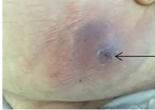
One patient who presented with an abscess-containing mass in the medial lower quadrant of the left breast was initially provided with unsuccessful medical treatment (Figure 6). Therefore, surgical excision was performed, fol-lowing which histopathological PS was diag-nosed. This patient habitually wore a tight bras-siere, which could have contributed to disease pathogenesis. PS of the periareolar region has also been characterized as an occupational hazard for hairdressers [8-11], which highlights the importance of breast surgeons’ being aware of the occupation of non-lactational breast sepsis patients, particularly when sur-gery is required. Correct identification alone is not sufficient for surgical management, in this case entailing total duct excision; the patient must also recieve advice to prevent recurrence.
PS of the scalp is rare and was observed herein in a male, Turkish patient whose sinus lesion was attributed to his habit of shaving with a straight razor. In a second patient, a cyst was detected attached to the temporal facia, in which there was a granulomatous reaction. Pseudofolliculitis is frequently observed after shaving in curly-haired individuals who may subsequently develop PS. Two of our patients presented with inguinal lesions, in one of whom a prediagnosis of sebacious cyst was rendered; hidradenitis suppurativa was diagnosed in the other case. However, histopathological diagno-sis confirmed PS. Similar histological features have been reported in cases of hidradenitis suppurativa. Wax and epilation may also be predisposing factors for pseudofolliculitis [5]. One report of ESPS involving the axilla [13] included non-hirsute healthy females between 17 and 30 years of age, in whom friction due to arm movements and persipiration was respon-sible for the etiology. Other etiological factors included shaving, maceration and minor infec-tions. Two of our patients habitually shaved the axilla (Figure 5). ESPS of the anterior abdomi-nal wall, located at umbilical and periumbilical areas, has also been reported [14, 15] in young adult male patients, most of whom exhibited poor personal hygiene, deep navels, and exces-sive hair [14, 15]. In five of our patients, anteri-or abdominal wall ESPS lesions ocurred exter-nal to the navel. Two female patients, aged 23 Figure 1. Photograph of a sinus tract with a fibrotic
dermal stoma surrounded by intense inflammation and covered by multiple layers of squamous epithe-lia. The intergrity of the epithelia is partially altered.
and 26 years, presented with abscesses in suprapubic areas. These patients also reported shaving the affected areas. Two young male adult patients presented with lesions in the hypogastric region, and a 43-year-old female patient displayed a lesion in the lower left quad-rant of the abdomen (Figure 7).
We suggest that wearing tight trousers with a belt could also have played a role in the devel-opment of inguinal lesions in our patients.
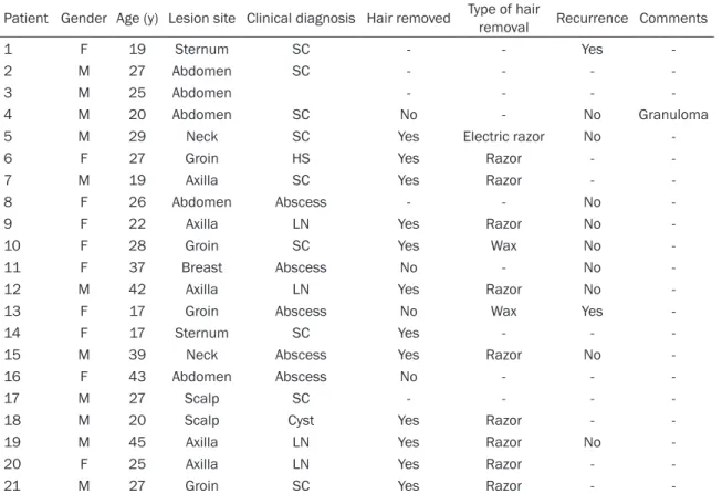
Shaving-related trauma was prevalent among our ESPS patients. During the follow-up period recurrence was observed in only two cases (one of sternal and the other of inguinal lesions) subsequent to surgical excision. We attributed sternal lesions to a combination of large breasts and tight brassieres, leading to increased friction. Considering the similar pathogenic mechanisms underying sacrococcy-geal PS and ESPS, recurrence of lesions subse-quent to surgical excision is not unexpected. Table 1. Extra-sacrococcygeal location of pilonidal sinus: clinical features
Patient Gender Age (y) Lesion site Clinical diagnosis Hair removed Type of hair removal Recurrence Comments
1 F 19 Sternum SC - - Yes
-2 M 27 Abdomen SC - - -
-3 M 25 Abdomen - - -
-4 M 20 Abdomen SC No - No Granuloma
5 M 29 Neck SC Yes Electric razor No
-6 F 27 Groin HS Yes Razor -
-7 M 19 Axilla SC Yes Razor -
-8 F 26 Abdomen Abscess - - No
-9 F 22 Axilla LN Yes Razor No
-10 F 28 Groin SC Yes Wax No
-11 F 37 Breast Abscess No - No
-12 M 42 Axilla LN Yes Razor No
-13 F 17 Groin Abscess No Wax Yes
-14 F 17 Sternum SC Yes - -
-15 M 39 Neck Abscess Yes Razor No
-16 F 43 Abdomen Abscess No - -
-17 M 27 Scalp SC - - -
-18 M 20 Scalp Cyst Yes Razor -
-19 M 45 Axilla LN Yes Razor No
-20 F 25 Axilla LN Yes Razor -
-21 M 27 Groin SC Yes Razor -
-HS, hidradenitis suppurativa; LN, lymph node; SC, sebaceous cyst.
Conclusion
Histological features of PS may resemble those of cutaneous lesions, although cutaneous lesions cannot give rise to typical
sacrococcy-geal PS. PS should be considered in the differ-ential diagnosis of extrasacrococcygeal cutane-ous lesions. The major pathogenic factor for PS is local and repetitive minor trauma in hairy areas.
Acknowledgements
The authors express their gratitutude and thanks to all participating patients and to clini-cal staff. All study participants or their legal guardians provided written informed consent prior to study enrollment.
Disclosure of conflict of Interest None.
Address correspondence to: Dr. Fatih Çiftci, Basak mah, 2.etap D-35/24 Basaksehir/Istanbul-Turkey. Tel: 90 505 616 42 48; Fax: 90 212 462 70 56; E-mail: [email protected]
References
[1] Ballas K, Psarras K, Rafailidis S, Konstantini-dis H, Sakadamis A. Interdigital piloni-dal sinus in a hairdresser. J Hand Surg Br 2006; 31: 290-1.
[2] Shah N. Hidradinitis suppurativa: a treatment challenge. Am Fam Physician 2005; 72: 1547-52.
[3] Cusack C, Buckley C. Entanercept effective in the man agement of hidradenitis suppurativa. Br J Dermatol 2006; 154: 726-9.
[4] Mendoca CO, Griffiths CE. Clindamycin and ri-fampicin combination therapy for hidradenitis suppurativa. Br J Dermatol 2006; 154: 977-9. [5] Jemec GB, Thomsen BM, Hansen U. The homo-geneity of hidradenitis suppurativa lesions. A Figure 4. Sternal region.
Figure 5. Axillary region.
Figure 6. Lower quadrant of the left breast.
histological study of intra-individual variation. APMIS 1997; 105: 378-83.
[6] Krasner BD, Hamzavi FH, Murakawa GJ, Hamzavi IH. Dissecting cellulitis treated with the long-pulse Nd: YAG laser. Dermatol Surg 2006; 32: 1039-44.
[7] Chinnaiyan P, Tena LB, Brenner MJ, Welsh JS. Modern external beam radiation therapy for refractory dissecting cellulitis of the scalp. Br J Dermatol 2006; 152: 777-9.
[8] Gannon MX, Crowson MC, Fielding JW. Peri-areolar pilonidal abscesses in a hairdresser. BMJ 1988; 297: 1641-2.
[9] Ferdinand RD, Scott DJ, Mc Lean NR. Pilonidal cyst of the breast. Br J Surgery 1997; 84: 784. [10] Kaufman PA. Pilonidal disease of the nipple.
West J Surg Obstet Gynecol 1961; 69: 9-10. [11] Rashmi L, Russell M, Mark AA, Nick CA, Anne
MP. Pilonidal abscess in the breast: a case re-port. J Surg Case Rep 2014; 6: rju061.
[12] Kelly AP. Pseudofolliculitis barbae and acne keloidalis nuchae. Dermatol Clin 2006; 21: 645-53.
[13] Ohtsuka H, Arashiro K, Watanebe T. Pilonidal sinus of the axilla: report of five patients and review of the literature. Ann Plast Surg 2006; 33: 322-5.
[14] Eryilmaz R, Sahin M, Okan I, Alimoglu O, So-may A. Umbilical pilonidal sinus disease: pre-disposing factors and treatment. World J Surg 2005; 29: 1158-60.
[15] Sion-Vardy N, Osyntsov L, Cagnano E, Osyntsov A, Vardy D, Benharroch D. Unexpected location of pilonidal sinuses. Clin Exp Dermatol 2009; 34: 599-01.