Rev Port Cardiol. 2019;38(4):269---277
ORIGINAL ARTICLE
Assessment of the relationship between preprocedural
C-reactive protein/albumin ratio and stent restenosis
in patients with ST-segment elevation myocardial
infarction
Ibrahim Rencuzogullari
a,∗, Yavuz Karaba˘g
a, Metin C
¸a˘gda¸s
a, Süleyman Karakoyun
a,
Sabri Seyis
b, Mustafa Ozan Gürsoy
c, Mahmut Yesin
d, ˙Inan¸c Arta¸c
a, Do˘gan ˙Ili¸s
a,
˙Ibrahim Halil Tanbo˘ga
ea M.D. Kafkas University Medical Faculty, Department of Cardiology, Kars, Turkey
b M.D. Istinye University Education and Research Hospital, Department of Cardiology, Istanbul, Turkey c M.D. Gaziemir State Hospital, Department of Cardiology, ˙Izmir, Turkey
d M.D. Kars Harakani State Hospital, Department of Cardiology, Kars, Turkey
e M.D. Ataturk University Medical Faculty, Department of Cardiology, Erzurum, Turkey
Received 15 December 2017; accepted 24 August 2018 Available online 16 May 2019
Abstract
Introduction: Stent restenosis remains a clinical challenge for patients with ischemic heart disease, since it is associated with repeated coronary interventions as well as higher hospital- ization rates and medical costs. Inflammation plays a significant role. Although an association between stent restenosis, increased C-reactive protein (CRP) and decreased albumin levels has been previously reported, no studies have investigated the ability of the CRP/albumin ratio to predict stent restenosis.
Methods: This retrospective study included 448 patients who had previously undergone primary percutaneous coronary intervention and who were referred for subsequent reintervention due to recurrence of anginal symptoms. The study population was divided into two groups based on whether the patient had developed stent restenosis. They were then stratified into three groups according to their CRP/albumin ratio.
Results: Out of 448 patients, stent restenosis was observed in 24.5% (n=110), as deter- mined by coronary angiography. Patients with stent restenosis had a higher CRP/albumin ratio, greater platelet distribution width (PDW), higher CRP levels, and lower levels of both high-density lipoprotein (HDL) cholesterol and serum albumin. The CRP/albumin ratio (OR: 2.289, 95% CI: 1.056-4.959; p=0.036), stent diameter, PDW and HDL cholesterol lev- els were found to be independent predictors of stent restenosis. A ROC curve comparison
∗ Corresponding author.
E-mail address: [email protected] (I. Rencuzogullari). https://doi.org/10.1016/j.repc.2018.08.008
0870-2551/© 2019 Sociedade Portuguesa de Cardiologia. Published by Elsevier Espan˜a, S.L.U.
This is an open access article under CC BY-NC-ND license.
Cardiologia
Portuguese Journal of Cardiology
www.revportcardiol.org
Revista Portuguesa de
KEYWORDS Stent restenosis; C-reactive protein/albumin ratio; ST-segment elevation myocardial infarction; Inflammationdemonstrated that the CRP/albumin ratio was a better predictor of restenosis than either albumin and CRP individually, but it was not better than PDW and HDL cholesterol.
Conclusion: As a novel inflammation-based risk score, the CRP/albumin ratio may be an easily accessible marker for assessment of stent restenosis risk.
© 2019 Sociedade Portuguesa de Cardiologia. Published by Elsevier Espan˜a, S.L.U.
This is an open access article under CC BY-NC-ND license
(http://creativecommons.org/licenses/by-nc-nd/4.0/)
Avalia¸cão pré-procedimento da rela¸cão entre a proteína C-reativa e a albumina na reestenose de stents em doentes com enfarte do miocárdio com eleva¸cão do segmento ST
Resumo
Objetivo: A reestenose de stent permanece como um desafio clínico em doentes com cardiopa- tia isquémica. A reestenose de stent está associada a interven¸cões coronárias recorrentes bem como a aumento de internamentos e de custos médicos. A inflama¸cão tem um papel signi- ficativo. Embora a associa¸cão entre reestenose de stent e o aumento dos níveis de proteína C-reativa (PCR) e a diminui¸cão dos níveis de albumina tenham sido previamente registados, nenhum estudo pesquisou a possibilidade da PCR/albumina prever um diagnóstico de reestenose de stent.
Métodos: Este estudo retrospetivo incluiu 448 doentes que tinham sido previamente submetidos a interven¸cão coronária percutânea primária e que foram referidos para reinterven¸cão posterior devido a ressurgimento dos sintomas de angina. A popula¸cão do estudo foi dividida em dois grupos com base no facto do doente ter desenvolvido reestenose de stent. Posteriormente, foram estratificados em três grupos de acordo com as rela¸cões PCR/albumina.
Resultados: De um grupo de 448 doentes, a reestenose de stent foi observada em 24,5% (n = 110), conforme determinado por avalia¸cão através de angiografia coronária. Os doentes com reestenose de stent apresentaram uma rela¸cão PCR/albumina mais elevada, varia¸cão maior do diâmetro das plaquetas (DP), níveis mais elevados de PCR e níveis mais baixos de colesterol de lipoproteínas de alta densidade (HDL) e de albumina sérica. Os níveis da rela¸cão PCR/albumina (OR: 2,289, IC 95%:1,056 --- 4,959; p = 0,036), o diâmetro do stent, a varia¸cão maior do DP e o colesterol HDL foram considerados fatores preditores independentes da restenose de stent. A compara¸cão das curvas ROC demonstrou que a rela¸cão PCR/albumina constituiu um fator preditor melhor do que a albumina e a PCR, isoladamente, não sendo, no entanto, melhor do que o DP e o colesterol HDL.
Conclusão: Como novo score de risco baseado em inflama¸cão, a rela¸cão PCR/albumina pode ser um marcador facilmente acessível para a avalia¸cão do risco de reestenose de stent. © 2019 Sociedade Portuguesa de Cardiologia. Publicado por Elsevier Espan˜a, S.L.U. Este é um
artigo Open Access sob a licença de CC BY-NC-SA (http://creativecommons.org/licenses/by-nc-
sa/4.0/).
Introduction
Deaths from ischemic heart disease have decreased in recent years due to increased use of percutaneous coronary inter- vention (PCI). Primary PCI (pPCI) is generally the first line of treatment for patients with ST-segment elevation myocar- dial infarction (STEMI), and stenting is recommended over balloon angioplasty.1 However, restenosis, a gradual re- narrowing of the stented segment, continues to be a clinical challenge. Stent restenosis decreases the patient’s quality of life and is associated with repeated coronary interven- tions as well as higher hospitalization rates and medical costs.2---4
Stent restenosis is a complex and multifactorial pro- cess that begins with stent deployment in the coronary
artery.5Inflammation plays a key role in arterial damage after stent placement, accelerating macrophage accumu- lation and neovascularization. The inflammation process also leads to neointimal tissue proliferation, thrombus formation at the stent struts and activation of neu- trophils, fibrin and platelets, all of which result in stent restenosis.6,7 Studies on stent-induced inflammation have found a significant association between C-reactive protein (CRP) and stent restenosis.5,8,9 An acute phase protein that is released from the liver, CRP is a useful marker for objectively stratifying an active inflamma- tion process. Similarly, serum albumin is a negative acute phase protein in acute inflammation and has been shown to have an inverse relationship with stent restenosis.10 PALAVRAS CHAVE Reestenose de stent; Proteína C reativa/albumina; Enfarte do miocárdio com eleva¸cão do segmento ST; Inflama¸cão
Relationship between CRP/albumin ratio and stent restenosis 271
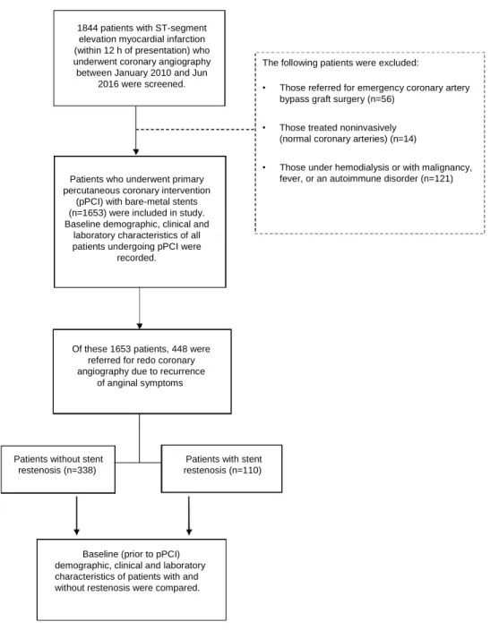
Figure 1 Flowchart of enrollment and follow-up of study patients.
The CRP/albumin ratio has been identified as an inflammation-based prognostic marker and is considered to reflect the relationship between CRP and albumin lev- els in determining the prognosis of critical diseases and malignancy.11,12 The fact that the CRP/albumin ratio is a good indicator of inflammatory status led us to investigate its relationship with stent restenosis. In this study, we aimed to assess whether the CRP/albumin ratio and levels of CRP and albumin at the time of initial pPCI are associated with subsequent stent restenosis in STEMI patients who were treated with bare-metal stents (BMS).
Methods
Study population
Between January 2010 and June 2016, 1844 STEMI patients were referred for coronary angiography and pPCI, and
these formed the basis of our study population. Of these, 191 were excluded from the study: those referred for emergency coronary artery bypass graft surgery, those treated noninvasively, those under hemodialysis, and those with malignancy, fever or an autoimmune disorder. Of the remaining 1653 patients, 448 were referred for redo coro- nary angiography due to recurrence of anginal symptoms and were therefore included in this retrospective study
(Figure 1). The first STEMI was defined based on the fol-
lowing criteria: a typical increase or decrease in cardiac biomarkers; ongoing ischemic symptoms (within 12 hours of presentation); newly developed left bundle branch block pattern or new ST elevation in two or more contiguous leads, with readings of at least 0.2 mV in leads V1, V2 and V3 or at least 0.1 mV in the other leads; or imaging evi- dence of a new loss of viable myocardium or new regional wall motion abnormality.13Stent restenosis was defined as
1844 patients with ST-segment elevation myocardial infarction (within 12 h of presentation) who underwent coronary angiography between January 2010 and Jun
2016 were screened.
The following patients were excluded:
• Those referred for emergency coronary artery bypass graft surgery (n=56)
• Those treated noninvasively (normal coronary arteries) (n=14) •
Patients who underwent primary percutaneous coronary intervention
(pPCI) with bare-metal stents (n=1653) were included in study. Baseline demographic, clinical and
laboratory characteristics of all patients undergoing pPCI were
recorded.
Those under hemodialysis or with malignancy, fever, or an autoimmune disorder (n=121)
Of these 1653 patients, 448 were referred for redo coronary angiography due to recurrence
of anginal symptoms
Patients without stent restenosis (n=338)
Patients with stent restenosis (n=110)
Baseline (prior to pPCI) demographic, clinical and laboratory characteristics of patients with and without restenosis were compared.
±
± ± the presence of clinical symptoms and/or signs of ischemia
and at least 50% narrowing in lumen diameter at follow-up angiography.14
The study protocol was reviewed and approved by the local ethics committee of Kafkas University and was con- ducted in accordance with the Declaration of Helsinki.
Data collection
All patients who were referred for coronary reintervention due to recurrence of ischemic symptoms were hospitalized. To establish the effects of the first pPCI on subsequent stent restenosis, patients’ baseline clinical, demographic, biochemical and hematological characteristics at the time of the first pPCI were recorded. Complete blood count and blood biochemical parameters were assessed in all patients on admission and prior to pPCI. CRP and serum albumin levels were determined using an automatic bio- chemical analyzer (Cobas 8000 c502, Roche Diagnostics). The CRP/albumin ratio was calculated as the ratio of CRP to albumin multiplied by 10 for ease of interpretation. The neutrophil/lymphocyte ratio was calculated from the com- plete blood count obtained on admission. Blood samples were retested for creatine kinase-myocardial band (CK-MB) every six hours until peak levels were identified. The esti- mated glomerular filtration rate (eGFR) was determined using the Cockcroft-Gault formula from the blood samples obtained on admission. Left ventricular ejection fraction (LVEF) was defined as the postprocedural ejection fraction and was assessed using a modified version of Simpson’s method.
Angiographic analysis
The standard Judkins percutaneous transfemoral technique was performed by experienced interventional cardiologists on all patients who underwent pPCI. Coronary angiograms and pPCIs were recorded using digital media (DICOM viewer, MedCom GmbH, Darmstadt, Germany). The digital angiogra- phy and PCI records of all patients who were admitted with STEMI and underwent BMS implantation (the records of the first angiography) were analyzed quantitatively in terms of lesion and intervention characteristics. Coronary blood flow patterns before and after pPCI were assessed using Throm- bolysis in Myocardial Infarction (TIMI) flow grade as defined previously, and epicardial no-reflow was defined as TIMI flow grade <3 at the target vessel lesion in the absence of spasm, thrombus, dissection and/or significant residual stenosis.15 Similarly, thrombus burden was assessed according to the TIMI thrombus grading scale as defined by Gibson et al.15The sizes of the stents used in the interventions were confirmed from hospital records. All patients had received 300 mg aspirin and a 600-mg loading dose of clopidogrel on a routine basis before the intervention and received unfractionated heparin during the intervention. The decision whether to use tirofiban was left to the operator’s discretion.
Statistical analysis
The statistical analysis was performed using IBM SPSS version 22.0 (IBM SPSS Inc., Chicago, IL). The normality
of the data was determined using the Kolmogorov-Smirnov test. With respect to data distribution and normality, contin- uous variables were expressed as mean standard deviation or median [interquartile range] (25th-75th percentiles), and the t test or Mann-Whitney U-test was conducted to compare variables between two groups. Continuous varia- bles with and without normal distribution were compared between three groups using analysis of variance and the Kruskal-Wallis H-test, respectively. Categorical variables were presented as numbers (percentages) and compared using Fisher’s exact test or the chi-square test. Multivari- ate logistic regression analysis was performed to identify independent predictors of stent restenosis using variables that showed a statistically significant association with stent restenosis in univariate analysis. Multicollinearity between the CRP/albumin ratio and CRP and albumin levels was assessed by eigenvalues and condition indices. Linearity was tested following the logarithmic transformation of each parameter. A receiver operating characteristic (ROC) curve was used to derive the best cut-off value of the CRP/albumin ratio for predicting stent restenosis using Youden’s J statistic. A p-value <0.05 was taken to indicate statistical significance.
Results
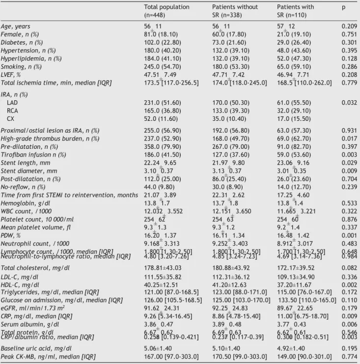
The study population consisted of 448 patients who under- went repeat coronary intervention (mean age 56 11 years; 17.8% female). The mean interval between coronary rein- tervention and the first STEMI was 21.07 3.89 months. Stent restenosis was observed in 24.5% (n=110) of the study population who underwent a second coronary angiogra- phy. The baseline clinical and angiographic characteristics of the total population and of patients with and without stent restenosis are shown in Table 1. Patients with stent restenosis had a higher CRP/albumin ratio, more frequent involvement of the left anterior descending artery (LAD), greater platelet distribution width (PDW), higher CRP lev- els, and lower levels of both high-density lipoprotein (HDL) cholesterol and serum albumin, compared to those without stent restenosis. Furthermore, patients with stent resteno- sis had higher thrombus burden, greater stent length and smaller angiographic reference lumen diameter, and had more frequently received tirofiban infusion at the time of the first pPCI. There was no difference between the two groups in terms of preprocedural TIMI flow (Table 1).
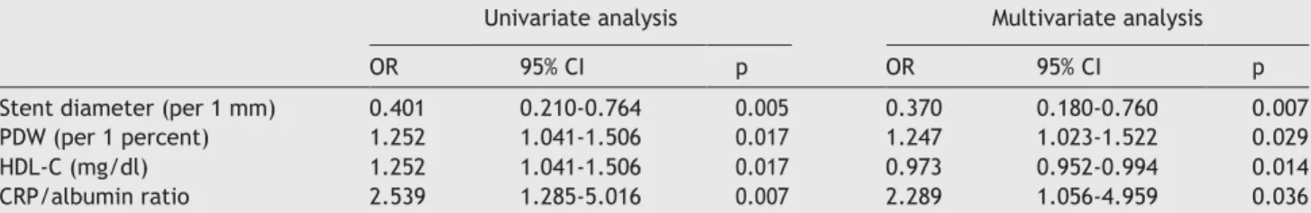
Multivariate regression analysis was used to determine the independent predictors of stent restenosis using the parameters that were associated with stent restenosis in univariate analysis. The CRP/albumin ratio (odds ratio [OR]: 2.289, 95% confidence interval [CI]: 1.056-4.959; p=0.036), stent diameter (per 1 mm, OR: 0.370, 95% CI: 0.180-0.760; p=0.007), PDW (per 1 percent, OR: 1.247, 95% CI: 1.023- 1.522; p=0.029), and HDL cholesterol (OR: 0.973, 95% CI: 0.952-0.994; p=0.014) were found to be independent pre- dictors of stent restenosis (Table 2).
The variables included in the multivariate analysis were infarct-related artery, high-grade thrombus burden, stent length, stent diameter, PDW, HDL cholesterol and CRP/albumin ratio.
Relationship between CRP/albumin ratio and stent restenosis 273 ± ± ± ± ± ± ± ± ± ± ± ± ± ± ± ± ± ± ± ± ± ± ± ± ± ± ± ± ± ± ± ± ± ± ± ± ± ± ± ± ± ±
Table 1 Demographic, clinical and laboratory characteristics of the total study population and of patients with and without
restenosis. Total population (n=448) Patients without SR (n=338) Patients with p SR (n=110) Age, years 56 11 56 11 57 12 0.209 Female, n (%) 81.0 (18.10) 60.0 (17.80) 21.0 (19.10) 0.751 Diabetes, n (%) 102.0 (22.80) 73.0 (21.60) 29.0 (26.40) 0.301 Hypertension, n (%) 180.0 (40.20) 132.0 (39.10) 48.0 (43.60) 0.395 Hyperlipidemia, n (%) 184.0 (41.10) 132.0 (39.10) 52.0 (47.30) 0.128 Smoking, n (%) 245.0 (54.70) 180.0 (53.30) 65.0 (59.10) 0.286 LVEF, % 47.51 7.49 47.71 7.42 46.94 7.71 0.208
Total ischemia time, min, median [IQR] 173.5 [117.0-256.5] 174.0 [118.0-245.0] 168.5 [110.0-262.0] 0.779
IRA, n (%)
LAD 231.0 (51.60) 170.0 (50.30) 61.0 (55.50) 0.032
RCA 165.0 (36.80) 133.0 (39.30) 32.0 (29.10)
CX 52.0 (11.60) 35.0 (10.40) 17.0 (15.50)
Proximal/ostial lesion as IRA, n (%) 255.0 (56.90) 192.0 (56.80) 63.0 (57.30) 0.931
High-grade thrombus burden, n (%) 237.0 (52.90) 168.0 (49.70) 69.0 (62.70) 0.017
Pre-dilatation, n (%) 358.0 (79.90) 267.0 (79.00) 91.0 (82.70) 0.397 Tirofiban infusion n (%) 186.0 (41.50) 127.0 (37.60) 59.0 (53.60) 0.003 Stent length, mm 22.24 9.65 21.97 9.80 23.06 9.16 0.029 Stent diameter, mm 3.10 0.37 3.13 0.37 3.01 0.35 0.009 Post-dilatation, n (%) 112.0 (25.00) 86.0 (25.40) 26.0 (23.60) 0.704 No-reflow, n (%) 44.0 (9.80) 30.0 (8.90) 14.0 (12.70) 0.239
Time from first STEMI to reintervention, months 21.07 3.89 22.31 2.62 17.25 4.60
Hemoglobin, g/dl 13.8 1.7 13.7 1.8 13.8 1.4 0.533
WBC count, /1000 12.032 3.552 12.151 3.650 11.665 3.221 0.322
Platelet count, 10 000/ml 254 62 254 63 254 60 0.876
Mean platelet volume, fl 9.3 1.3 9.3 1.2 9.2 1.4 0.337
PDW, % 16.20 1.37 16.11 1.34 16.48 1.42 0.001
Neutrophil count, /1000 9.168 3.313 9.252 3.403 8.912 3.017 0.483
Lymphocyte count, /1000, median [IQR] 1.800 [1.30-2.50] 1.800 [1.30-2.50] 1.700 [1.30-2.50] 0.648
Neutrophil-to-lymphocyte ratio, median [IQR] 4.80 [3.20-7.26] 4.85 [3.24-7.23] 4.69 [3.14-7.36] 0.984
Total cholesterol, mg/dl 178.81±43.03 180.88±43.92 172.17±39.52 0.082
LDL-C, mg/dl 111.55±35.82 112.31±36.12 109.13±34.90 0.336
HDL-C, mg/dl 40.25±12.51 41.20±12.63 37.20±11.67 0.002
Triglycerides, mg/dl, median [IQR] 121.00 [87.0-168.5] 123.00 [88.0-171.0] 115.00 [76.0-167.0] 0.172
Glucose on admission, mg/dl, median [IQR] 126.00 [105.5-168.5] 125.00 [103.0-170.0] 133.50 [110.0-165.0] 0.110
eGFR, ml/min/1.73 m2 91.62 24.31 92.25 24.83 89.67 22.65 0.179
CRP, mg/dl, median [IQR] 9.26 [5.34-16.45] 8.86 [4.78-15.40] 11.00 [6.75-18.70] 0.009
Serum albumin, g/dl 3.86 0.47 3.89 0.48 3.77 0.43 0.006
Total protein, g/dl 6.67 0.62 6.69 0.63 6.62 0.61 0.546
CRP/albumin ratio, median [IQR] 0.258 [0.139-0.421] 0.237 [0.117-0.39] 0.300 [0.182-0.51] 0.003
Baseline uric acid, mg/dl 5.06±1.40 5.10±1.40 4.92±1.40 0.195
Peak CK-MB, ng/ml, median [IQR] 167.00 [97.0-303.0] 170.50 [99.0-303.0] 149.00 [90.0-301.0] 0.774
CK-MB: creatine kinase-myocardial band; CRP: C-reactive protein; CX: circumflex artery; eGFR: estimated glomerular filtration rate; HDL- C: high-density lipoprotein cholesterol; IQR: interquartile range (25th-75th percentiles); IRA: infarct-related artery; LAD: left anterior descending artery; LDL-C: low-density lipoprotein cholesterol; LVEF: left ventricular ejection fraction; PDW: platelet distribution width; RCA: right coronary artery; SR: stent restenosis; STEMI: ST-segment elevation myocardial infarction; WBC: white blood cell.
Because no specific cut-off was identified for the CRP/albumin ratio, subjects who underwent repeat coro- nary angiography were stratified into three groups according to CRP/albumin ratio by equalizing the number of patients: tertile 1 (<0.170, n=149), tertile 2 (0.170-0.363, n=150) and tertile 3 (>0.363, n=149). There were significant differences between these tertiles. Mean CRP level, age, frequency of hypertension, total ischemia time, peak CK-MB, incidence
of proximal coronary lesion involvement, LAD involvement as the infarct-related artery, and presence of no-reflow increased progressively from tertile 1 to tertile 3, while LVEF
and albumin levels decreased progressively (Table 3). It was
also observed that stent restenosis increased progressively with the tertiles’ increasing CRP/albumin ratio (Table 3).
In ROC curve analysis, the area under the curve (AUC) of the CRP/albumin ratio was 0.601 (95% CI: 0.540-0.661;
Table 2 Univariate and multivariate logistic regression analysis of demographic, clinical, laboratory and coronary angiographic
characteristics for prediction of restenosis.
CI: confidence interval; CRP: C-reactive protein; HDL-C: high-density lipoprotein cholesterol; OR: odds ratio; PDW: platelet distribution width.
Univariate analysis Multivariate analysis
OR 95% CI p OR 95% CI p
Stent diameter (per 1 mm) 0.401 0.210-0.764 0.005 0.370 0.180-0.760 0.007
PDW (per 1 percent) 1.252 1.041-1.506 0.017 1.247 1.023-1.522 0.029
HDL-C (mg/dl) 1.252 1.041-1.506 0.017 0.973 0.952-0.994 0.014
CRP/albumin ratio 2.539 1.285-5.016 0.007 2.289 1.056-4.959 0.036
p<0.001). The cut-off value for the CRP/albumin ratio that indicated stent restenosis was 0.167 (with 80.9% sensitivity and 36.1% specificity). ROC curve comparisons were used to determine which variables best predicted stent resteno- sis. The CRP/albumin ratio was a better predictor than both serum albumin (AUC: 0.577, 95% CI: 0.516-0.638) and CRP (AUC: 0.582, 95% CI: 0.524-0.641) (p=0.050 and p=0.008,
respectively) (Figure 2), but there was no significant dif-
ference between CRP/albumin ratio and PDW (AUC: 0.604, 95% CI 0.541-0.667) and HDL cholesterol (AUC: 0.602, 95% CI: 0.538-0.665) (p=0.942 and p=0.961, respectively) (Figure 3).
Discussion
Our study demonstrated that the CRP/albumin ratio was not only associated with, but was also an independent predic- tor of stent restenosis in STEMI patients undergoing pPCI. The CRP/albumin ratio was better than both CRP and serum albumin levels, individually, in predicting stent restenosis.
pPCI is the standard treatment for patients with STEMI. Compared with balloon angioplasty alone, stenting with a BMS is associated with a lower risk of reinfarction and target vessel revascularization.1However, with a restenosis rate of 12.7% with drug-eluting stents (DES) and 20.1% with BMS, stent restenosis remains a major concern for the manage- ment of STEMI in patients treated with pPCI.16In our study, stent restenosis was observed in 24.5% of patients who were referred for repeat coronary angiography.
The etiology of stent restenosis is complex and multifac- torial. Several risk factors have been identified, including angiographic, clinical and technical factors. Consistent with the results of previous studies, patients with stent restenosis in our study had longer stents and smaller stent lumen diameter,17 higher CRP levels,5,8,9 and lower lev- els of HDL cholesterol18and serum albumin.10Additionally, the CRP/albumin ratio was significantly higher and PDW was greater in patients with restenosis than in those with- out.
Inflammation plays a central role in stent restenosis. It has been previously established in animal models that there is a significant correlation between the severity of arterial inflammation and neointimal hyperplasia following balloon angioplasty and stenting.19,20The local inflammatory reac- tion induced by the trauma of stenting leads to the release of cytokines and growth factors from macrophages and smooth muscle cells, resulting in a proliferative response.21 More importantly, higher baseline levels of acute-phase proteins
indicate the hyper-responsiveness of inflammatory cells to any inflammatory stimuli.22Hence, higher systemic CRP lev- els or lower serum albumin levels identify patients with an enhanced inflammatory response in the vessel wall to the inflammatory stimulus of stenting. In our study, base- line CRP levels were higher and serum albumin levels were significantly lower in patients with stent restenosis. These findings strongly suggest that baseline inflammatory status is a determining factor in the development of stent restenosis, in addition to the classical etiological factors.
We found that the baseline inflammatory response was exacerbated in patients with restenosis. We also observed that patients’ baseline inflammatory status was related to factors associated with a larger infarct area, including LAD involvement as the culprit lesion, no-reflow, prolonged total ischemia time and elevated peak CK-MB levels. Acute myocardial infarction triggers an inflammatory response that lasts for weeks and months, and this mid-term inflam- matory response seems to be more severe in patients with large infarcts and high baseline inflammation.23However, an increased baseline inflammatory response in patients with restenosis cannot be explained by infarct size alone. Our patients with increased CRP/albumin ratios were older and more frequently had a history of hypertension in addition to reduced eGFR and LVEF. Chronic renal insufficiency has been associated with background inflammation,24and there is a consistent correlation between heart failure (lower LVEF) and inflammation.25In addition, aging is accompanied by two- to four-fold increases in the plasma/serum lev- els of inflammatory mediators such as cytokines and acute phase proteins.26 Hypertension is also clearly associated with chronic mild inflammation.27 Although not all these factors were found to be individually associated with stent restenosis in our study, their cumulative effect might have contributed to an association between the CRP/albumin ratio and restenosis.
The various acute phase reactants may not all respond to inflammatory events to the same degree. Merging albu- min and CRP into a single index as an inflammation-based prognostic score provides stability between fluctuating CRP and albumin levels in diseases where inflammation plays an important role. The CRP/albumin ratio has been used as a parameter of inflammatory status in many studies.11,12,28,29 Consistent with previous findings, the present study showed that the CRP/albumin ratio was a better predictor of stent restenosis than either CRP or albumin, individually, in ROC curve analysis.
Relationship between CRP/albumin ratio and stent restenosis 275 ± ± ± ± ± ± ± ± ± ± ± ± ± ± ± ± ± ± ± ± ± ± ± ± ± ± ± ± ± ± ± ± ± ± ± ± ± ± ± ± ± ±
Table 3 Demographic, clinical, laboratory and coronary angiographic characteristics of the study population stratified according
to C-reactive protein/albumin ratio.
CRP/albumin ratio <0.170 (n=149) 0.170-0.363 (n=150) >0.363 p (n=149) Age 54 11 56 11 59 12 0.002 Female, n (%) 24.0 (16.10) 23.0 (15.30) 34.0 (22.80) 0.182 Diabetes, n (%) 34.0 (22.80) 33.0 (22.00) 35.0 (23.50) 0.954 Hypertension, n (%) 48.0 (32.20) 58.0 (38.70) 74.0 (49.70) 0.008 Hyperlipidemia, n (%) 59.0 (39.60) 68.0 (45.30) 57.0 (38.30) 0.418 Smoking, n (%) 77.0 (51.70) 88.0 (58.70) 80.0 (53.70) 0.459 LVEF, % 49.85 6.45 48.21 6.65 44.43 8.25 <0.001
Total ischemia time, min, median [IQR] 145.0 [112.0-223.0] 168.5 [105.0-235.0] 190.0 [137.0-308.0] <0.001 IRA, n (%)
LAD 65.0 (43.60) 78.0 (52.00) 88.0 (59.10) 0.044
RCA 66.0 (44.30) 57.0 (38.00) 42.0 (28.20)
CX 18.0 (12.10) 15.0 (10.00) 19.0 (12.80)
Proximal/ostial lesion for IRA, n (%) 73.0 (49.00) 85.0 (56.70) 97.0 (65.10) 0.020
High-grade thrombus burden, n (%) 90.0 (60.40) 97.0 (64.70) 100.0 (67.10) 0.475
Pre-dilatation, n (%) 116.0 (77.90) 117.0 (78.00) 125.0 (83.90) 0.333 Tirofiban infusion, n (%) 61.0 (40.90) 64.0 (42.70) 61.0 (40.90) 0.941 Stent length, mm 22.50 11.22 22.16 8.76 22.06 8.83 0.999 Stent diameter, mm 3.13 0.35 3.09 0.40 3.08 0.36 0.216 Post-dilatation, n (%) 31.0 (20.80) 44.0 (29.30) 37.0 (24.80) 0.235 No-reflow, n (%) 6.0 (4.00) 14.0 (9.30) 24.0 (16.10) 0.002
Time from first STEMI to reintervention (months) 21.38 2.98 21.00 3.97 20.82 4.55
Hemoglobin, g/dl 13.6 1.8 13.8 1.6 13.8 1.7 0.867
WBC count, /1000 11.737 3.373 12.046 3.339 12.312 3.917 0.428
Platelet count, 10 000/ml 255 61 250 61 257 65 0.599
Mean platelet volume, fl 9.1 1.2 9.3 1.3 9.3 1.4 0.458
PDW, % 16.22 1.22 16.34 1.39 16.05 1.47 0.747
Neutrophil count, /1000 8.914 3.041 9.223 3.078 9.368 3.774 0.579
Lymphocyte count, /1000, median [IQR] 1.700 [1.20-2.50] 1.800 [1.30-2.40] 1.800 [1.40-2.60] 0.358
Neutrophil-to-lymphocyte ratio, median [IQR] 5.00 [3.16-7.82] 4.76 [3.59-7.21] 4.65 [2.99-7.06] 0.730
Total cholesterol, mg/dl 177.04±41.82 182.73±43.21 176.61±44.08 0.313
LDL-C, mg/dl 109.01±34.43 117.01±35.81 108.56±36.81 0.107
HDL-C, mg/dl 41.24±13.46 39.42±11.74 40.08±12.30 0.756
Triglycerides, mg/dl, median [IQR] 120.00 [85.0-183.5] 124.00 [86.0-159.0] 119.0 [88.0-158.0] 0.939
Glucose on admission, mg/dl, median [IQR] 126.00 [103.0-167.0] 124.00 [103.0-168.0] 133.00 [109.0-169.0] 0.323
eGFR, ml/min/1.73 m2 95.96±26.34 90.93±23.18 87.97±22.74 0.029
CRP, mg/dl, median [IQR] 4.070 [2.88-5.33] 9.265 [7.70-12.10] 20.50 [16.40-24.50] <0.001
Serum albumin, g/dl 3.95 0.52 3.83 0.42 3.80 0.47 0.013
Total protein, g/dl 6.70 0.61 6.66 0.61 6.65 0.66 0.799
CRP/albumin ratio, median [IQR] 0.106 [0.073-0.14] 0.258 [0.204-0.31] 0.518 [0.422-0.69] <0.001
Baseline uric acid, mg/dl 4.87 1.32 5.12 1.36 5.17 1.50 0.202
Peak CK-MB, ng/ml, median [IQR] 122.00 [82.0-218.0] 160.5 [102.0-289.0] 234.0 [120.0-373.0] <0.001
Stent restenosis, n (%) 24.0 (16.10) 41.0 (27.3) 45.0 (30.2) 0.010
CK-MB: creatine kinase-myocardial band; CRP: C-reactive protein; CX: circumflex artery; eGFR: estimated glomerular filtration rate; HDL- C: high-density lipoprotein cholesterol; IQR: interquartile range (25th-75th percentiles); IRA: infarct-related artery; LAD: left anterior descending artery; LDL-C: low-density lipoprotein cholesterol; LVEF: left ventricular ejection fraction; PDW: platelet distribution width; RCA: right coronary artery; STEMI: ST-segment elevation myocardial infarction; WBC: white blood cell.
Interestingly, the present study found PDW to be an independent predictor of stent restenosis. PDW indicates variability in platelet size, which is directly related to platelet density and reactivity in circulating platelets. Changes in these variables play an important role in the
development of acute coronary syndrome.30Recently, it
has been shown that PDW is associated with acute stent thrombosis31and is an independent predictor of no-reflow and in-hospital major cardiac adverse events32 in STEMI patients. In this study, for the first time, we demon- strated that PDW is an independent predictor of stent restenosis.
Serum albumin (g/dl) AUC: 0.577 (0.516-0.638) CRP/albumin ratio AUC: 0.601 (0.540-0.661) CRP (mg/dl) AUC: 0.582 (0.524-0.641) 100
Limitations
80 60 40 20 0 0 20 40 60 80 100 100-SpecificityThe present study has several limitations. First, although the data were acquired prospectively, the study had a retrospec- tive design and was based on a registry analysis. Second, this study provided information about stent restenosis in STEMI patients treated with BMS; it did not include information on patients treated with DES, which currently are widely used. Third, high-sensitivity CRP, which has proven efficacy in demonstrating endovascular inflammation, could not be used in our study, due to the lack of this information in our subjects’ patient records. Finally, the prognostic value of the CRP/albumin ratio could not be assessed in the present study. Stent restenosis and patient prognosis may not nec- essarily correlate; therefore, the prognostic value of the CRP/albumin ratio in the development of stent restenosis should be verified by further research.
Figure 2 Receiver operating characteristic curves to detect the best variable for predicting stent restenosis. AUC: area under the curve; CRP: C-reactive protein.
100 80 60 40 20 0
Conclusion
This study suggests that baseline inflammatory status appears to be a significant factor in subsequent stent restenosis in STEMI patients. Based on the evidence that the CRP/albumin ratio is a better predictor than CRP or albu- min, individually, assessment of a patient’s preprocedural CRP/albumin ratio could be beneficial. In STEMI patients, a high CRP/albumin ratio on admission may be an early, mid- and long-term marker of inflammation that could lead to stent restenosis. Although this study only analyzed resteno- sis with BMS, which does not correspond to the majority of stents currently used in developed countries, the mecha- nisms underlying stent restenosis may be common to other types of stents, and this research may be a bridge to DES studies. Additionally, for the first time, PDW has been found to be an independent predictor of stent restenosis. There- fore, PDW may be another easily accessible marker for the
0 20 40 60 80 100
100-Specificity
risk assessment of stent restenosis.
Figure 3 Receiver operating characteristic curves comparing independent predictors of laboratory parameters to detect the best predictor of stent restenosis. AUC: area under the curve; CRP: C-reactive protein; HDL-C: high-density lipoprotein choles- terol; PDW: platelet distribution width.
In addition to inflammation-induced neointimal hyperpla- sia and vascular remodeling, oxidative stress and increased vascular levels of reactive oxygen species are also thought to be involved in the pathophysiology of stent restenosis.33,34 HDL cholesterol molecules reduce macrophage accumula- tion, promote removal of oxidized cholesterol from the arterial wall, and can inhibit monocyte activation, adhe- siveness and inflammation.35---37Previous studies have shown that lower HDL levels are an independent risk factor for stent restenosis in different patient groups.38---40Our study also showed that HDL cholesterol is an independent pre- dictor of stent restenosis. A comparison of the ROC curves showed that CRP/albumin ratio, PDW and HDL cholesterol levels were not inferior to each other in predicting stent restenosis.
Funding
The authors received no financial support for the research, authorship, and/or publication of this article.
Conflicts of interest
The authors have no conflicts of interest to declare
Acknowledgments
The authors thank www.metastata.com for their contribu- tions to the statistical analysis and trial design.
References
1. Ibanez B, James S, Agewall S, et al. 2017 ESC Guide- lines for the management of acute myocardial infarction in patients presenting with ST-segment elevation: The Task Force for the management of acute myocardial infarction in patients presenting with ST-segment elevation of the
CRA/albumin ratio AUC: 0.601 (0.540-0.661) HDL-C (mg/dl) AUC 0.602 (0.516-0.638) PDW (%) AUC: 0.604 (0.541-0.667) Se n si ti vi ty Se n si ti vi ty
Relationship between CRP/albumin ratio and stent restenosis 277
European Society of Cardiology (ESC). Eur Heart J. 2017, http://dx.doi.org/10.1093/eurheartj/ehx393.
2. Chen MS, John JM, Chew DP, et al. Bare metal stent restenosis is not a benign clinical entity. Am Heart J. 2006;151:1260---4. 3. Bainey KR, Norris CM, Graham MM, et al. APPROACH investiga-
tors. Clinical in-stent restenosis with bare metal stents: is it truly a benign phenomenon? Int J Cardiol. 2008;128:378---82. 4. Remak E, Manson S, Hutton J, et al. Cost-effectiveness
of the Endeavor stent in de novo native coronary artery lesions updated with contemporary data. EuroIntervention. 2010;5:826---32.
5. Ferrante G, Niccoli G, Biasucci LM, et al. Association between C- reactive protein and angiographic restenosis after bare metal stents: an updated and comprehensive meta-analysis of 2747 patients. Cardiovasc Revasc Med. 2008;9:156---65.
6. Komatsu R, Ueda M, Naruko T, et al. Neointimal tissue response at sites of coronary stenting in humans: macroscopic, histological, and immunohistochemical analyses. Circulation. 1998;98:224.
7. Farb A, Sangiorgi G, Carter AJ, et al. Pathology of acute and
22. Liuzzo G, Buffon A, Biasucci LM, et al. Enhanced inflamma- tory response to coronary angioplasty in patients with severe unstable angina. Circulation. 1998;98:2370---6.
23. Prabhu SD, Frangogiannis NG. The biological basis for cardiac repair after myocardial infarction: from inflammation to fibro- sis. Circ Res. 2016;119:91---112.
24. Cachofeiro V, Goicochea M, de Vinuesa SG, et al. Oxidative stress and inflammation, a link between chronic kidney disease and cardiovascular disease. Kidney Int Suppl. 2008:S4---9. 25. Shirazi LF, Bissett J, Romeo F, et al. Role of inflammation in
heart failure. Curr Atheroscler Rep. 2017;19:27.
26. Krabbe KS, Pedersen M, Bruunsgaard H. Inflammatory mediators in the elderly. Exp Gerontol. 2004;39:687---99.
27. Bautista LE, Vera LM, Arenas IA, et al. Independent asso- ciation between inflammatory markers (C-reactive protein, interleukin-6, and TNF-alpha) and essential hypertension. J Hum Hypertens. 2005;19:149---54.
28. Ranzani OT, Zampieri FG, Forte DN, et al. C-reactive pro- tein/albumin ratio predicts 90-day mortality of septic patients. PLOS ONE. 2013;8:e59321.
chronic coronary stenting in humans. Circulation. 1999;99:44. 29. C¸a˘gda¸s M, Rencüzo˘gullari I, Karakoyun S, et al. Assess- 8. Silva D, Pais de Lacerda A. High-sensitivity C-reactive protein as
a biomarker of risk in coronary artery disease. Rev Port Cardiol. 2012;31:733---45.
9. Li JJ, Ren Y, Chen KJ, et al. Impact of C-reactive protein on in-stent restenosis: a meta-analysis. Tex Heart Inst J. 2010;37:49---57.
10. Celik IE, Yarlioglues M, Kurtul A, et al. Preprocedural albumin levels and risk of in-stent restenosis after coronary stenting with bare-metal stent. Angiology. 2016;67:478---83.
11. Kinoshita A, Onoda H, Imai N, et al. The C-reactive pro- tein/albumin ratio, a novel inflammation-based prognostic score, predicts outcomes in patients with hepatocellular car- cinoma. Ann Surg Oncol. 2015;22:803---10.
12. Fairclough E, Cairns E, Hamilton J, et al. Evaluation of a mod- ified early warning system for acute medical admissions and comparison with C-reactive protein/albumin ratio as a predictor of patient outcome. Clin Med. 2009;9:30---3.
13. Steg PG, James SK, Atar D, et al. ESC guidelines for the manage- ment of acute myocardial infarction in patients presenting with ST-segment elevation. Eur Heart J. 2012;33:2569---619.
14. Dangas GD, Claessen BE, Caixeta A, et al. In-stent resteno- sis in the drug-eluting stent era. J Am Coll Cardiol. 2010;56:1897---907.
15. Gibson CM, Cannon CP, Daley WL, et al. TIMI frame count: a quantitative method of assessing coronary artery flow. Circula- tion. 1996;93:879---88.
16. Dirksen MT. Drug-eluting vs bare-metal stents in primary angio- plasty. Arch Intern Med. 2012;172:611.
17. de Feyter PJ, Kay P, Disco C, et al. Reference chart derived from post-stent-implantation intravascular ultrasound predic- tors of 6-month expected restenosis on quantitative coronary angiography. Circulation. 1999;100:1777---83.
18. Jing XD, Wei XM, Deng SB, et al. The relationship between the high-density lipoprotein (HDL)-associated sphingosine-1- phosphate (S1P) and coronary in-stent restenosis. Clin Chim Acta. 2015;446:248---52.
19. Schwartz RS, Huber KC, Murphy JG, et al. Restenosis and the proportional neointimal response to coronary artery injury: results in a porcine model. J Am Coll Cardiol. 1992;19:267---74. 20. Karas SP, Gravanis MB, Santoian EC, et al. Coronary intimal pro-
liferation after balloon injury and stenting in swine: an animal model of restenosis. J Am Coll Cardiol. 1992;20:467---74. 21. Libby P, Schwartz D, Brogi E, et al. A cascade model for resteno-
sis. A special case of atherosclerosis progression. Circulation. 1992;86:III47---52.
ment of relationship between C-reactive protein to albumin ratio and coronary artery disease severity in patients with acute coronary syndrome. Angiology. 2017, http://dx.doi.org/10.1177/0003319717743325, 3319717743325.
30. Martin JF, Plumb J, Kilbey RS, et al. Changes in volume and density of platelets in myocardial infarction. Br Med J (Clin Res Ed). 1983;287:456---9.
31. Balli M, Ta¸solar H, C¸etin M, et al. Relationship of platelet indices with acute stent thrombosis in patients with acute coronary syndrome. Postepy Kardiol Interwencyjnej. 2015;11: 224---9.
32. Celik T, Kaya MG, Akpek M, et al. Predictive value of admission platelet volume indices for in-hospital major adverse car- diovascular events in acute ST-segment elevation myocardial infarction. Angiology. 2015;66:155---62.
33. Sundaresan M, Yu ZX, Ferrans VJ, et al. Requirement for generation of H2O2 for platelet-derived growth factor signal transduction. Science. 1995;270:296---9.
34. Griendling KK, FitzGerald GA. Oxidative stress and cardiovas- cular injury: Part II: animal and human studies. Circulation. 2003;108:2034---40.
35. Murphy AJ, Chin-Dusting JP, Sviridov D, et al. The anti- inflammatory effects of high density lipoproteins. Curr Med Chem. 2009;16:667---75.
36. Hessler JR, Robertson AL Jr, Chisolm GM III. LDL-induced cytotoxicity and its inhibition by HDL in human vascular smooth muscle and endothelial cells in culture. Atherosclerosis. 1979;32:213---29.
37. Li XP, Zhao SP, Zhang XY, et al. Protective effect of high den- sity lipoprotein on endothelium-dependent vasodilatation. Int J Cardiol. 2000;73:231---6.
38. Matsumoto I, Miyake Y, Mizukawa M, et al. Impact of low-density lipoprotein cholesterol/high-density lipoprotein cholesterol ratio on long-term outcome in patients undergoing percuta- neous coronary intervention. Circ J. 2011;75:905---10.
39. Sukhija R, Aronow WS, Sureddi R, et al. Predictors of in-stent restenosis and patient outcome after percutaneous coronary intervention in patients with diabetes mellitus. Am J Cardiol. 2007;100:777---80.
40. Turak O, Canpolat U, Özcan F, et al. Usefulness of preprocedural serum uric acid level to predict restenosis of bare metal stents. Am J Cardiol. 2014;113:197---202.


![Başlık: YERLİ KÖPEK, YERLİ KEDİ VE BEYAZ YE;'\i ZEL\i\OA TAVŞA:\];,IN (ORYCTOLAGUS CC!\ICL'LUS) CI.AVICCLA'SI CZERi'\DE l\I:\KRO-A:\'ATOMİK BİR ARAŞTIRMAYazar(lar):UÇAR, Yaşar;ÖCAL, M. Kamil ;HAZIROĞLU, MerihCilt: 32 Sayı: 1 DOI: 10.1501/Vetfak_0000000346](data:image/gif;base64,R0lGODlhAQABAIAAAP///wAAACH5BAEAAAAALAAAAAABAAEAAAICRAEAOw==)