Relationship between histopathological features of aspirated
thrombi and long-term left ventricular function in patients
with ST-segment elevation myocardial infarction
ST yükselmeli miyokart enfarktüsü hastalarında çekilen pıhtının histopatolojik
özelliklerinin uzun dönem sol ventrikül fonksiyonuyla ilişkisi
1Department of Cardiology, İstanbul Medipol University, International Medical Faculty, İstanbul, Turkey 2Department of Cardiology, Kartal Koşuyolu Yüksek İhtisas Training and Research Hospital, İstanbul, Turkey
3Department of Pathology, Fatih Sultan Mehmet Training and Research Hospital, İstanbul, Turkey 4Department of Cardiology, Kayseri City Hospital, Kayseri, Turkey
5Department of Radiology (Division of CMR), Royal Brompton and Harefield NHS Foundation Trust, London, United Kingdom
Mehmet Onur Omaygenç, M.D.,1 Cem Doğan, M.D.,2 Adnan Somay, M.D.,3 Oğuz Karaca, M.D.,1
Ruken Bengi Bakal, M.D.,2 Mehmet Kamil Teber, M.D.,4 Suzan Hatipoğlu, M.D.,5
Zübeyde Bayram, M.D.,2 Cihangir Kaymaz, M.D.,2 Nihal Özdemir, M.D.2
Objective: This study was an investigation of the severity of inflammation (SOI) in aspirated material and thrombus age to examine any association with pre-discharge and long-term left ventricular (LV) function after ST-elevation myocardial infarc-tion (STEMI).
Methods: The study group comprised 25 patients with STEMI from whom an occlusive thrombus was aspirated from the in-farct-related artery with a 7-F catheter. The SOI in the aspi-rate was determined according to the mean leukocyte count in 5 high-power magnification fields and graded as mild in the presence of ≤100 leukocytes per field or significant if there were >100 leukocytes per field. The thrombi were categorized as fresh or lytic/organized (L/O) using predefined criteria. Echocardiographic assessment was performed prior to dis-charge and at 1 year. Adverse left ventricular remodeling (LVR) was defined as a 20% increase in LV end-diastolic volume in comparison with baseline values.
Results: LVR was observed in 8 patients. The mean leukocyte count of the aspirate (127.5±86.0 vs 227.2±120.7; p=0.026) and frequency of significant inflammation (35% vs 75%; p=0.046) were significantly higher in the group with LVR. The serum high-sensitivity C-reactive protein (hsCRP) level was significantly correlated with the leukocyte count of the aspi-rate (r=0.532; p=0.006). An L/O thrombus was related to better pre-discharge and long-term LV volumes and ejection fraction values compared with a fresh thrombus.
Conclusion: A significant increase in the leukocyte count in the aspirate and a fresh thrombus might predict long-term LV functional deterioration irrespective of the clinical and proce-dure-related characteristics. In addition, serum markers of in-flammation, like hsCRP, might also reflect the intensity of the local inflammatory response at the site of occlusion.
Amaç: Bu çalışmada, ST yükselmeli miyokart enfarktüsü (STYME) sonrası emme yöntemiyle alınan pıhtının yaşı ve yangı düzeyinin (YD), taburculuk öncesi ve uzun dönem sol ventrikül (SV) fonksiyonlarıyla olası ilişkisi araştırıldı.
Yöntemler: Çalışmaya enfarktüsle ilişkili arterden tıkayıcı pıh-tının 7-F kateter yardımıyla alındığı 25 STYME hastası dahil edildi. Pıhtının YD, beş yüksek düzeyde büyütme alanında görülen ortalama lökosit sayısına göre derecelendirildi: alan başına hafif ≤100 lökosit ve belirgin >100 lökosit. Ayrıca, ör-nek daha önceden tanımlanmış kriterlere göre taze veya litik/ organize (L/O) olarak da sınıflandırıldı. Ekokardiyografik de-ğerlendirme taburculuk öncesi ve birinci yılda yapıldı. Olumsuz sol ventrikül yeniden biçimlenmesi (SVYB), SV diyastol sonu hacminde başlangıç değerine göre %20 artış olması şeklinde tanımlandı.
Bulgular: SVYB sekiz hastada gözlendi. Alınan örneğin orta-lama lökosit sayımı (127.5±86.0 ve 227.2±120.7; p=0.026) ve belirgin yangısı olan hastaların sıklığı (%35 ve %75; p=0.046) SVYB gözlenen grupta anlamlı olarak daha yüksekti. Serum yüksek duyarlıklı keratin C-reaktif protein (hsCRP) düzeyi, ör-neğin lökosit sayımıyla anlamlı biçimde orantılıydı (r=0.532, p=0.006). Örneğin L/O yapısı, taze pıhtı varlığına kıyasla daha iyi taburculuk öncesi ve uzun dönem SV hacimleri ve ejeksiyon fraksiyonu değerleriyle ilişkiliydi.
Sonuç: Örnekte artmış lökosit sayısı ve taze pıhtı varlığı, klinik ve işlemle ilişkili özelliklerden bağımsız olarak SV fonksiyonla-rında uzun dönemde bozulma ile ilişkili olabilir. Bunun yanın-da, hsCRP gibi serum yangı belirteçleri tıkanıklık bölgesindeki yangısal cevabın şiddetini yansıtabilir.
Received: July 25, 2019 Accepted:October 08, 2019
Correspondence: Dr. Mehmet Onur Omaygenç. İstanbul Medipol Üniversitesi Uluslararası Tıp Fakültesi, Kardiyoloji Anabilim Dalı, İstanbul, Turkey.
Tel: +90 212 - 460 77 74 e-mail: [email protected]
© 2020 Turkish Society of Cardiology
T
he rupture of a vulnerable plaque with an over-lying thrombus formation is the well-established and most common cause of acute myocardial infarc-tion (AMI).[1–4] Various pathological studies havealso revealed that the process is often dynamic, with consecutive episodes of occlusion and recanalization.
[4–6] This fact clarified why the initiation of thrombus
formation was sometimes hours or even days before symptom onset in cases of AMI with persistent ST-segment elevation (STEMI).[6,7] The impact of age,
burden, and content of the thrombus on parameters related to successful revascularization and short- and long-term clinical outcomes have previously been in-vestigated.[5,8–15] However, regardless of the thrombus
burden and other clinical features, the mainstay of the treatment in cases of STEMI continued to be provid-ing luminal patency via percutaneous transluminal coronary angioplasty and stent implantation, with or without extraction of the occlusive material.[1,2,16,17]
The idea of aspirating the predominantly throm-botic material emerged and small registries were pub-lished in the beginning of the 2000s. These registries and succeeding randomized trials suggested that there might be promising benefits to thrombus aspiration (TA) compared with the conventional option of percu-taneous coronary intervention (PCI).[1,2,16,18] In addition
to improved microvascular flow, there was a reduc-tion in infarct size, use of stent material, and adverse cardiovascular events.[16,19,20] However, the outcomes
of recent clinical trials have not been consistent with prior findings. The baseline clinical, imaging, and lab-oratory findings were investigated for their potential relationship with outcome in most of these publica-tions and yielded conflicting results.[2,16,21–23]
In the literature, left ventricular remodeling (LVR) has rarely been considered as a factor in the occur-rence of clinical adverse events. In light of these facts, the aim of this study was to assess the relationship between the features of aspirated thrombi, measures of successful revascularization, biochemical markers, and pre-discharge and long-term left ventricle (LV) function. LVR was designated as a particular outcome.
METHODS
Study qualification and patient selection
This study was conducted at a referral cardiology center. Sixty-four consecutive patients who presented
with STEMI were evaluated for in-clusion. In 36 patients (anterior 17, non-anterior 19), an occlusive thrombus was as-pirated from the infarct-related artery (IRA) us-ing a dedicated 7-F catheter (Ex-port; Medtronic, Inc., Minneapo-lis, MN, USA) during primary PCI. Among the aspirated thrombi,
30 specimens were suitable for comprehensive eval-uation. Echocardiographic assessment at 1 year was performed for 25 male patients, and they constituted the final study population. Apart from insufficient ma-terial, the criteria for exclusion from the study were:
• Resuscitation or defibrillation after hospitaliza-tion
• Presentation more than 6 hours after symptom onset
• History of previous myocardial infarction (MI), PCI, coronary artery disease (CAD) with an-giographically diagnosed significant stenosis, or coronary artery bypass surgery
• Initial Thrombolysis in Myocardial Infarction (TIMI) flow score of >0 in the IRA
• Occlusion or ≥70% stenosis in a non-IRA • Very high thrombus burden (Grade 4
accord-ing to previously described angiographic ap-pearance when antegrade flow was achieved following the advancement of the guidewire or first pass of the aspiration catheter)[8]
• Final TIMI flow score of other than 3 in the IRA • Failure to aspirate a visible thrombus
• Deficient follow-up data
• An episode of stent thrombosis, MI, or tar-get lesion revascularization before the 1-year echocardiographic follow-up
Abbreviations:
AMI Acute myocardial infarction CAD Coronary artery disease HsCRP High-sensitivıty C-reactive protein CRP C-reactive protein
IRA Infarct-related artery L/O Lytic/organized LV Left ventricle
LVEDV Left ventricular end-diastolic volume LVEF Left ventricular ejection fraction LVESV Left ventricular end-systolic volume LVR Left ventricular remodeling MBG Myocardial blush grade MI Myocardial infarction
PCI Percutaneous coronary intervention SOI Severity of inflammation STEMI ST-elevation myocardial infarction STR ST-segment resolution
TA Thrombus aspiration TFC TIMI frame count
TIMI Thrombolysis in Myocardial Infarction WBC White blood cell
All procedures were performed by 2 experienced operators who perform more than 200 primary PCI per year. Informed consent was received from all of the participants and the study was approved by the local ethical committee.
Patient characteristics, procedure-related fea-tures, and biochemical analyses
Once the diagnosis of STEMI was established, the patients were immediately transferred to the catheter laboratory. Administration of 300 mg acetylsalicy-late, 600 mg clopidogrel, and 100 IU/kg unfraction-ated heparin (10000 IU at most) was initiunfraction-ated prior to the procedure. Arterial access was achieved via the femoral route and the IRA was cannulated us-ing a 7-F guidus-ing catheter. After advancement of the guidewire, thrombus aspiration was performed with several passes of the manual dedicated catheter. The PCI was completed with stent implantation. If a TIMI 3 flow score was obtained at the end of the procedure, restoration of microvascular flow was additionally as-sessed using the TIMI frame count (TFC) measure-ments and myocardial blush grading (MBG) system, as previously described.[24,25] MBG was graded as
fol-lows: 0 – No blush, 1 – Minimal blush, 2 – Significant blush, but less than reference artery, 3 – Significant blush similar to that of reference artery.[24] The TFC
was assessed using angiographic images obtained at 30 frames per second in the diastolic phase of the flow. The recommended corrections were made for the anterior descending coronary artery and instanta-neous heart rate.[25] The door-to-balloon time was less
than 20 minutes in all cases.
ST-segment resolution (STR) was evaluated im-mediately after the procedure in the coronary criti-cal care unit. STR was defined as a 50% reduction in the sum of the ST-segment elevation score compared with that of the baseline ECG.[27] CAD risk factors
(age, hypertension, diabetes mellitus, smoking, dys-lipidemia) were investigated with the patients and/or checked in routine biochemical analyses. The patients were also asked the exact time of symptom onset and the response was noted. The presence of hypertension was defined as a previous diagnosis of hypertension requiring drug treatment. Patients with a previous diagnosis of diabetes mellitus who were under oral antidiabetic or insulin regimens were noted. A fasting blood glucose level of >126 mg/dL or a glycated he-moglobin level of 6.5% was used to establish a new
diagnosis of diabetes mellitus. The term of smoking was defined as a history of a smoking habit of >10 pack-years. Finally, the presence of dyslipidemia was identified by a low-density lipoprotein cholesterol level of >130 mg/dL or a triglyceride level of >150 mg/dL at presentation. Ongoing antihyperlipidemia treatment was also recorded. The initial serum white blood cell (WBC) count, high-sensitivity C-reactive protein (hsCRP), and N-terminal proB-type natri-uretic peptide levels at admission were analyzed, as well as the 72nd-hour troponin I level.
Histopathological evaluation of the aspirate
The aspirated material was immediately fixed in for-malin and embedded in paraffin the next day. Sec-tioned material was mounted on glass and stained with hematoxylin and eosin for light microscopy. Histopathological analyses were performed by an experienced cardiovascular pathologist blinded to all patient and procedure-related features. If there was sufficient thrombus material (≥1 mm2), the specimen
was examined in 5 zones under maximum magnifica-tion. The WBC count was then obtained by dividing the total number by 5. The severity of inflammation (SOI) was graded as mild if there were ≤100 WBCs per field or significant if the total was >100 WBCs per field. Pathology images of specimens with and with-out a significant inflammatory response are shown in Figure 1. The age of the thrombus was classified into 3 groups according to previously accepted defi-nitions.[5,7] A fresh thrombus (<1 day) was composed
of layered patterns of intact platelets, fibrin, erythro-cytes, and granulocytes. A lytic thrombus (1–5 days) revealed areas of colliquation, necrosis, and the kary-orrhexis of granulocytes. An organized thrombus (> 5 days) was identified by areas of recently proliferated smooth muscle cells and ingrowth of capillary ves-sels. Lytic and organized thrombi were paired (L/O) to facilitate evaluation.
Echocardiographic assessment
A transthoracic echocardiographic examination was performed 48 hours after the primary PCI as recom-mended by the American Society of Echocardiog-raphy[27] with a GE Vivid 7 system (GE Healthcare,
Inc., Chicago, IL, USA) and a 3.5 MHz transducer. Harmonic images of LV apical 4-, 2-, and 3-chamber views were obtained and the data were transferred to the workstation for further offline analysis (EchoPAC
Statistical analysis
Statistical analyses were conducted using SPSS Statis-tics for Windows, Version 17.0 (SPSS Inc., Chicago, IL, USA). The data were expressed as mean±SD for continuous variables and percentage for categorical variables. The Shapiro-Wilk test was used to test nor-mality and a p value of >0.05 was considered normally distributed data. The collected data were separated into several groups based on the presence of LVR, significant inflammation, and the age of the thrombus (fresh or L/O). Continuous variables in each group were compared using Student’s t-test for indepen-dent samples that showed normal distribution, while the Mann-Whitney U test was used for non-normally distributed samples. Associations between categorical variables in the groups were tested using a chi-square test. The relationship between the change in LVEDV, LVESV, and LVEF, and the age of the thrombus was illustrated in separate clustered bar charts. Pearson’s correlation coefficient analysis was used to test the re-lationship between the leukocyte count and the serum PC; GE Healthcare, Inc., Chicago, IL, USA). LV
end-diastolic volume (LVEDV), LV end-systolic volume (LVESV), and LV ejection fraction (LVEF) were cal-culated using the biplane Simpson method. Mitral in-flow Doppler and tissue Doppler data were obtained for assessment of diastolic function. The ratio of transmitral E velocity to early diastolic mitral annular velocity was calculated using the average of the septal and lateral E’ wave velocities. The severity of func-tional mitral regurgitation was assessed by measuring the effective regurgitant orifice area.
The same assessment was repeated at the 1-year visit and the relevant parameters were noted. Ad-verse remodeling was defined as a >20% increase in LVEDV in comparison with baseline values.
Follow-up data
All patients were questioned at the 1-year visit regard-ing any adverse clinical events, such as AMI, target lesion revascularization, or stent thrombosis. Addi-tional data were obtained by phone.
Figure 1. (A) Erythrocytes and inflammatory cells embedded in fibrin deposits (arrows). Thrombus aspirate stained with hemotoxylin and eosin (H&E) (x400); (B) Cholesterol clefts (*), erythrocytes, and inflammatory cells (arrows) embedded in fibrin deposits. Thrombus aspirate stained with H&E (x400); (C) Inflamed thrombus with cholesterol clefts (*) and macrophage foam cells (arrows). Thrombus aspirate stained with H&E (x200); (D) Thrombus without inflam-matory cell infiltration.
A
C
B
hsCRP level. The result of the Pearson’s correlation measurement was demonstrated in a scatter plot. Sta-tistical significance was defined as a p value of <0.05 for all comparisons.
RESULTS
A 1-year follow-up echocardiographic assessment was performed for 25 male patients. The mean clin-ical follow-up period for these patients was 26±6 months. Stent thrombosis was observed in 1 patient (4%), target lesion revascularization in 3 (12%), and successive AMI in 2 (8%) patients. No death occurred during the initial follow-up period.
The patients were initially grouped according to the presence of adverse remodeling at the 1-year visit. The baseline demographic features of the groups were comparable (Table 1). ECG-based and angiographic
determinants of successful revascularization and serum biomarkers were similar as well. MBG 3 was achieved in 80% of the patients at the end of the pro-cedure with a similar frequency between study groups (87% vs. 76% for patients with and without LVR, respectively; p=0.52). The mean TFC values were also comparable for patients with and without LVR (21.8±5.0 frames per second vs. 22.6±6.5 frames, re-spectively; p=0.76). Analysis of the histopathological features of the aspirated material revealed that there were significantly more with a mean leukocyte count and frequency in the sample that demonstrated signifi-cant inflammation in the group with adverse remodel-ing (Table 2). The echocardiographic parameters ob-tained from pre-discharge and 1-year measurements are shown in Table 3. Functional significant mitral regurgitation (effective regurgitant orifice area ≥0.20 cm2) was observed in 2 patients in each group.
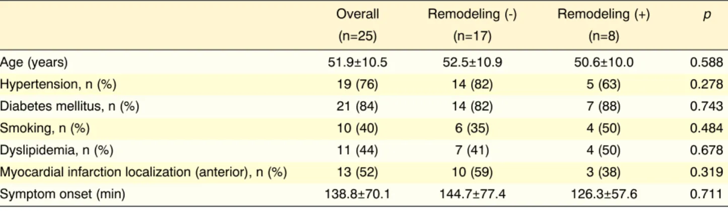
Table 1. Comparison of baseline demographic and clinical features of the study population according to the presence of left ventricular remodeling
Overall Remodeling (-) Remodeling (+) p
(n=25) (n=17) (n=8) Age (years) 51.9±10.5 52.5±10.9 50.6±10.0 0.588 Hypertension, n (%) 19 (76) 14 (82) 5 (63) 0.278 Diabetes mellitus, n (%) 21 (84) 14 (82) 7 (88) 0.743 Smoking, n (%) 10 (40) 6 (35) 4 (50) 0.484 Dyslipidemia, n (%) 11 (44) 7 (41) 4 (50) 0.678
Myocardial infarction localization (anterior), n (%) 13 (52) 10 (59) 3 (38) 0.319
Symptom onset (min) 138.8±70.1 144.7±77.4 126.3±57.6 0.711
Table 2. Comparison of revascularization parameters, serum biomarkers, and histopathological features of the aspirate according to the presence of left ventricular remodeling
Remodeling (-) Remodeling (+) p
(n=17) (n=8)
ST-segment resolution, n (%) 15 (88) 7 (87) 0.958
Myocardial blush grade 3, n (%) 13 (76) 7 (87) 0.520
Thromolysis in Myocardial Infarction frame count 22.6±6.5 21.8±5.0 0.761
High-sensitivity C-reactive protein (mg/L) 0.68±0.83 0.64±0.45 0.886
White blood cell (serum), ×1000/dL 12.7±5.7 12.7±4.9 0.994
Troponin I, ng/mL 28.2±21.7 28.3±24.3 0.989
N-terminal pro B-type natriuretic peptide (pg/mL) 192.4±141.8 180.8±122.0 0.843
Leukocyte count, per field 127.5±86.0 227.2±120.7 0.026
Significant inflammation, n (%) 6 (35) 6 (75) 0.046
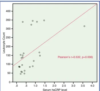
the leukocyte count in the aspirated material with the serum hsCRP level supported this association (r=0.532; p=0.006). Figure 2 illustrates the relevant results. While the values regarding the time of symp-tom onset, reperfusion determinants, and troponin I levels were comparable, the LVESV and LVEDV The second variable used to define groups was the
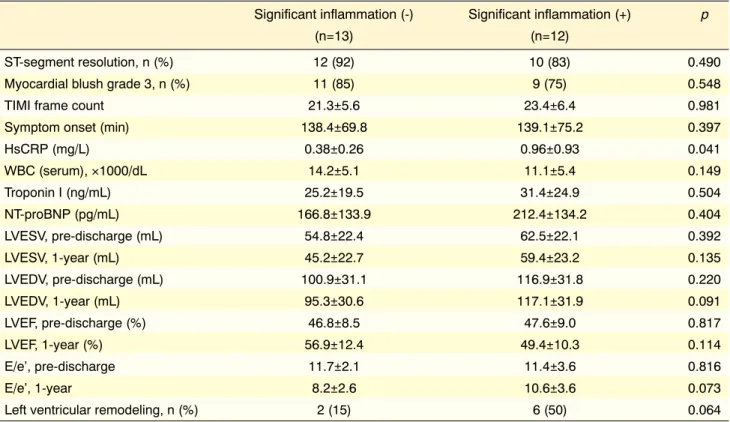
SOI (mild: ≤100 WBCs per field, significant: >100 WBCs per field) in the aspirated material (Table 4). This comparison yielded a statistically significant result in the serum hsCRP level (0.38±0.26 mg/L vs 0.96±0.93 mg/L; p=0.041). Correlation analysis of
Table 3. Comparison of pre-discharge and 1-year echocardiographic parameters of the groups with and without adverse left ventricular remodeling
Remodeling (-) Remodeling (+) p
(n=17) (n=8)
Left ventricular end-systolic volume, pre-discharge (mL) 62.4±22.2 50.5±21.2 0.262
Left ventricular end-systolic volume, 1-year (mL) 47±20.1 62.6±28.2 0.189
Left ventricular end-diastolic volume, pre-discharge (mL) 114.9±30.3 95.2±32.7 0.140
Left ventricular end-diastolic volume, 1-year (mL) 98±28.7 121.7±36.4 0.133
Left ventricular ejection fraction, pre-discharge (%) 47±8.7 47.6±8.8 0.842
Left ventricular ejection fraction, 1-year (%) 54.5±11.4 50.9±13.1 0.512
E/e’, pre-discharge 12.3±2.4 10.7±3.3 0.184
E/e’, 1-year 9.6±3.5 8.9±2.9 0.605
Significant mitral regurgitation, 1-year 11.8 (2) 25 (2) 0.570
Table 4. Comparison of revascularization parameters, serum biomarkers, and echocardiographic measures according to the severity of inflammation
Significant inflammation (-) Significant inflammation (+) p
(n=13) (n=12)
ST-segment resolution, n (%) 12 (92) 10 (83) 0.490
Myocardial blush grade 3, n (%) 11 (85) 9 (75) 0.548
TIMI frame count 21.3±5.6 23.4±6.4 0.981
Symptom onset (min) 138.4±69.8 139.1±75.2 0.397
HsCRP (mg/L) 0.38±0.26 0.96±0.93 0.041 WBC (serum), ×1000/dL 14.2±5.1 11.1±5.4 0.149 Troponin I (ng/mL) 25.2±19.5 31.4±24.9 0.504 NT-proBNP (pg/mL) 166.8±133.9 212.4±134.2 0.404 LVESV, pre-discharge (mL) 54.8±22.4 62.5±22.1 0.392 LVESV, 1-year (mL) 45.2±22.7 59.4±23.2 0.135 LVEDV, pre-discharge (mL) 100.9±31.1 116.9±31.8 0.220 LVEDV, 1-year (mL) 95.3±30.6 117.1±31.9 0.091 LVEF, pre-discharge (%) 46.8±8.5 47.6±9.0 0.817 LVEF, 1-year (%) 56.9±12.4 49.4±10.3 0.114 E/e’, pre-discharge 11.7±2.1 11.4±3.6 0.816 E/e’, 1-year 8.2±2.6 10.6±3.6 0.073
Left ventricular remodeling, n (%) 2 (15) 6 (50) 0.064
HsCRP: High-sensitivity C-reactive protein; LVEDV: Left ventricular diastolic volume; LVEF: Left ventricular ejection fraction; LVESV: Left ventricular end-systolic volume; NT-proBNP: N-terminal pro B-type natriuretic peptide; WBC: White blood cell; TIMI: Thromolysis in Myocardial Infarction.
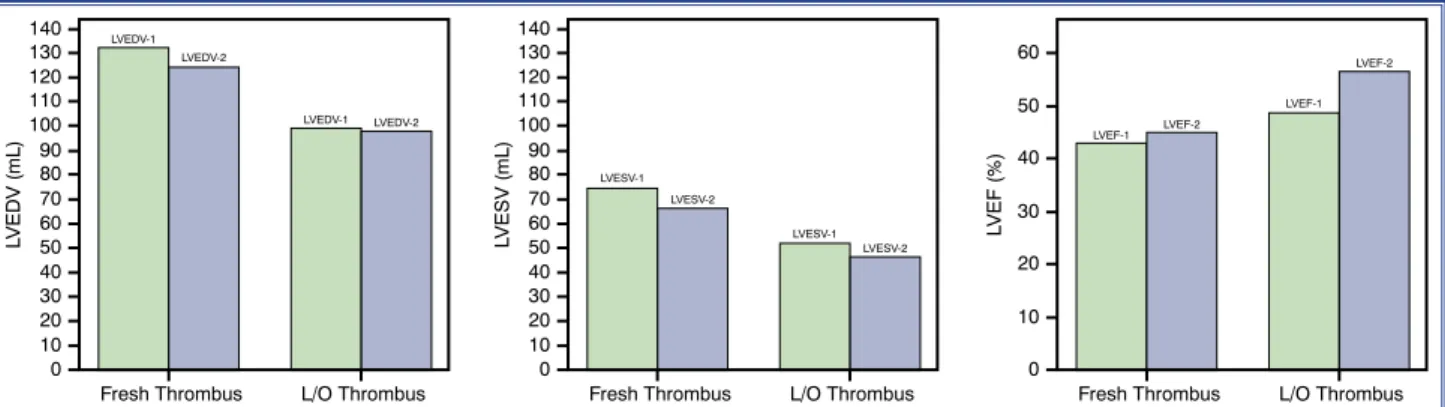
Finally, patients were grouped according to the age of the thrombus (fresh or L/O) (Table 5). The 72nd
-hour troponin I level was higher in the fresh thrombus group (fresh: 46.6±25.1 ng/mL, L/O: 21.1±16.4 ng/ mL; p=0.025). The LVEF value was lower and the ventricular volume measures were higher in the fresh thrombus group both at baseline and the follow-up echocardiographic examinations, but the results were not statistically significant (Fig. 3).
DISCUSSION
AMI is a major cause of mortality and disabil-ity worldwide. Various demographic, biochemical, imaging, and pathological parameters have been re-ported as potential determinants of prognosis in AMI.
[2,3,5,10,15,28–31] Under most circumstances, PCI is the
pri-mary treatment option for STEMI and the main goal is the immediate restoration of macro- and microvascu-lar flow.[2,16] TA was introduced as a secondary
modal-ity the 2000s, and there have been conflicting results since then regarding the impact of the method on early and long-term clinical outcomes.[2,16,18] In addition to
were higher both at baseline and the 1-year echocar-diographic assessment in individuals with significant inflammation. However, these differences did not reach the level of statistical significance.
Table 5. Comparison of revascularization parameters, serum biomarkers, and echocardiographic measures according to the age of the thrombus
Fresh thrombus L/O thrombus p
(n=7) (n=18)
ST-segment resolution, n (%) 6 (86) 16 (89) 0.826
Myocardial blush grade 3, n (%) 6 (86) 14 (78) 0.656
Thromolysis in Myocardial Infarction frame count 21.9±5.4 22.5±6.4 0.836
Symptom onset (min) 107.1±23.6 151.1±79.6 0.297
High-sensitivity C-reactive protein (mg/l) 1.09±1.15 0.50±0.39 0.110
White blood cell (serum), ×1000/dL 10.2±5.1 13.7±5.3 0.125
Troponin I (ng/mL) 46.6±25.1 21.1±16.4 0.025
N-terminal pro B-type natriuretic peptide (pg/mL) 255.7±139.9 162.6±124.9 0.125
Left ventricular end-systolic volume, pre-discharge (mL) 74.9±20.7 52.2±19.7 0.021
Left ventricular end-systolic volume, 1-year (mL) 66.7±16.2 46.3±23.8 0.055
Left ventricular end-diastolic volume, pre-discharge (mL) 132.7±30.9 99.2±27.6 0.029
Left ventricular end-diastolic volume, 1-year (mL) 124.5±25.7 98.2±32.6 0.064
Left ventricular ejection fraction, pre-discharge (%) 42.9±4.5 48.8±9.2 0.158
Left ventricular ejection fraction, 1-year (%) 44.9±8.4 56.6±11.4 0.029
E/e’, pre-discharge 11.3±2.9 11.6±2.9 0.615
E/e’, 1-year 10.2±2.4 9.0±3.5 0.198
Left ventricular remodeling, n (%) 2 (29) 6 (33) 0.819
L/O: Lytic or organized.
Figure 2. A scatter plot of the correlation between leukocyte count in the aspirate and serum high-sensitivity C-reactive protein (hsCRP) level. Leukocyte Count 100 50 0 .0 .5 1.0 1.5 2.0 2.5 3.0 3.5 4.0 200 250 400 350 300 150 Serum hsCRP level Pearson’s r=0.532, p=0.006)
The leukocyte count of the aspirated material may indicate inflammation severity from a histopatholog-ical perspective. Arakawa et al.[14] demonstrated that
the rates of incomplete STR and a low MBG were higher in the group demonstrating a neutrophil cellu-lar density of >100/0.025 mm2 in the aspirated
throm-bus sample. In this group, the LVEF was also lower at a 6-month assessment. They used immunohisto-chemical myeloperoxidase staining to quantify neu-trophils. It should be noted that the LV volume and the LVEF were evaluated with left ventriculography in this study, rather than echocardiography. Yunoki et al.[12] grouped their population as platelet-rich (low
red blood cells), mixed, or erythrocyte-rich thrombi aspirates. They found that the number of myeloperox-idase+ cells was greater in the erythrocyte-rich group. In addition, incomplete STR, low MBG, and LVR at the sixth month were observed more frequently in this group. According to our results, significant inflam-mation in the thrombus, defined as >100 WBCs per field, was associated with increased LV volume both at baseline and the 1-year assessment. Moreover, the quantity of local inflammatory cells was notably cor-related with the serum hsCRP level.
The age of the thrombus can also be a consid-erable determinant of adverse outcomes in AMI.
[4,5,10,13,15] L/O thrombi have been detected in 40% to
50% of the patients who presented for medical care within the first 12 hours of symptom onset.[5,13] The
ischemic time was proven to be longer, and the rate of incomplete STR, 30-day adverse event occurrence, and long-term mortality was higher in this group of patients in various publications.[4,5,10,11,13,15] In our
pop-ulation, the time from symptom onset to presentation the clinical benefit, histopathological analysis of these
aspirated thrombi clarified various issues about the underlying mechanisms of coronary obstruction and the thrombotic process.[5,32,33] In addition to
macro-scopic features of the thrombi, like quantity and color, pathological characteristics, such as age and content (cellular and non-cellular) were analyzed within the context of successful revascularization, adverse event occurrence, and less frequently, ventricular function.
[5,8,11,15,32,34]
A high thrombus burden is a well-documented predictor of angiographic complications, larger in-farct size, and a less favorable clinical outcome, par-ticularly due to increased rates of stent thrombosis.
[8,9,16] Moreover, it has been speculated that manual
catheters failed to guarantee a restoration of blood flow in this group.[3,35] Therefore, angiographic
indi-cators of a large thrombus were an exclusion criterion in our study.
Inflammation is recognized as a consistent compo-nent of the pathological cascade during the course of coronary occlusion in AMI.[31,36–38] C-reactive protein
(CRP), a systemic marker of inflammation, has been determined to be associated with LV function and the occurrence of clinical adverse events, including heart failure and death.[31,36,38,39] An elevated serum CRP
level has been associated with reduced LV systolic function, a rise in filling pressure, and adverse remod-eling.[36,38–40] Furthermore, a high CRP level has been
correlated with unstable plaques.[41] However, Maier
et al.[31] reported that local CRP levels were lower in
the IRA in cases of AMI. They suggested that it may have been due to local uptake and catabolism of the protein.
Figure 3. A comparison of left ventricular volume and ejection fraction at baseline and 1-year follow-up between groups of pa-tients with a fresh or lytic/organized thrombus. L/O: Lytic/organized; LVEDV: Left ventricular end-diastolic volume; LVEF: Left ventricular ejection fraction; LVESV: Left ventricular end-systolic volume.
LVEDV (mL) 140 130 120 110 100 90 80 70 60 50 40 30 20 10 0 Fresh Thrombus LVEDV-1 LVEDV-1 LVEDV-2 LVEDV-2 L/O Thrombus LVESV (mL) 140 130 120 110 100 90 80 70 60 50 40 30 20 10 0 Fresh Thrombus LVESV-1 LVESV-1 LVESV-2 LVESV-2 L/O Thrombus LVEF (%) 60 50 40 30 20 10 0 Fresh Thrombus LVEF-1 LVEF-1 LVEF-2 LVEF-2 L/O Thrombus
estimated using the 72nd-hour serum troponin I level;
cardiac magnetic resonance imaging was not used for more precise measurement of infarct size and LV vol-ume and function. Finally, newer, potent antiplatelet agents (e.g., prasugrel and ticagrelor) were not used due to local unavailability throughout the study pe-riod.
Conclusion
Adverse LVR is a well-known determinant of long-term prognosis in AMI. However, only a few stud-ies have been conducted that investigated the rela-tionship between remodeling and the content of the aspiration material. Our results demonstrated that an increased leukocyte count, indicating significant in-flammation in the thrombus material, was associated with LV remodeling and probably with increased LV volume. Another notable finding was the correlation between the leukocyte count of the aspirate and the serum hsCRP level. This may reflect the fact that not only markers of systemic inflammation, but local determinants may also alter the outcome in cases of AMI, regardless of other patient-related and proce-dure-related characteristics. Finally, a fresh thrombus was associated with a higher troponin I level and LV volume, and a lower LVEF in our study. This result, which differs from previous data, was presumably a consequence of ischemic preconditioning.
Ethics Committee Approval: The study was approved by
the local ethical committee of Kartal Kosuyolu Yuksek Ihti-sas Education and Research Hospital.
Peer-review: Externally peer-reviewed. Conflict-of-interest: None.
Authorship contributions: Concept: M.O.O., N.O.;
De-sign: M.O.O., N.O.; Supervision: C.K., N.O.; Materials: M.O.O., C.D., A.S., R.B.B., M.K.T., S.H.A., Z.B.; Data: C.D., İ.O.K., S.H.A.; Analysis: M.O.O.; İ.O.K., A.S., Z.B., Literature search: M.O.O., C.D., M.K.Y., R.B.B., N.O.; Writing M.O.O., N.O.; Critical revision: C.K., N.O.O.
REFERENCES
1. Tilsted HH, Olivecrona GK. To Aspirate or Not to Aspirate: That Is the Question. JACC Cardiovasc Interv 2015;8:585–7. 2. Vandermolen S, Marciniak M, Byrne J, De Silva K. Thrombus
aspiration in acute myocardial infarction: concepts, clinical tri-als, and current guidelines. Coron Artery Dis 2016;27:233–43. 3. Keskin M, Kaya A, Tatlısu MA, Uzman O, Börklü EB, Çinier
G, et al. Effect of Adjunctive Thrombus Aspiration on In-was lower in patients with a fresh thrombus. The 72nd
-hour troponin I level, which may reflect infarct size, and baseline and 1-year LV volumes were higher, and the LVEF was lower in this group. There was a sta-tistical significance in the comparison of the baseline LVESV and LVEDV, and the 1-year LVEF accord-ing to thrombus age. These findaccord-ings were inconsistent with previous data associating the detrimental effects of an older thrombus with episodes of flow reduction and spontaneous lysis culminating in microvascular dysfunction.[10] However, it should be emphasized that
determinants of successful restoration of microvascu-lar flow were comparable between thrombus groups in our study. We hypothesized that this contradictory relationship might be due to ischemic precondition-ing, which is not an odd phenomenon in the presence of an L/O thrombus.
Adverse remodeling has been observed in 30% to 35% of AMI patients even after a successful PCI.[44]
Remodeling is a strong predictor of the occurrence of heart failure and increased mortality.[30,42–44] The
definition of remodeling used in other studies varies substantially; however, the most frequently used is an increase in LVEDV of 20%.[42–45] Several
demo-graphic (diabetes mellitus, male gender), clinical (late revascularization, distal embolization, anterior loca-tion, larger infarct size, incomplete STR, low MBG, etc.), and echocardiographic parameters have been identified as predictors of remodeling.[16,28,29,36,42–46]
Among the histopathological features of the throm-bus, as mentioned above, Yunoki et al.[12] observed a
significant relationship to the erythrocyte count. Ad-verse remodeling was observed in 32% of our pop-ulation, which is consistent with results reported in the literature. In our study, the leukocyte count of the thrombus aspirate and the presence of significant in-flammation in the aspirate were defined as parameters solely associated with adverse remodeling.
Limitations
The main limitation of our research is the size of the sample population. The study was conducted in a sin-gle center during a specific period. The strict exclu-sion criteria used to minimize the possibility of bias, difficulties obtaining an assessable specimen and optimal echocardiographic images, and individuals lost to follow-up yielded a group of 25 participants. Therefore, the study lacks the power to estimate the clinical outcome. In addition, infarct size was only
Hospital and 3-Year Outcomes in Patients With ST-Segment Elevation Myocardial Infarction and Large Native Coronary Artery Thrombus Burden. Cardiol 2017;120:1708–14. [CrossRef]
4. Nishihira K, Hatakeyama K, Shibata Y, Kitamura K, Asada Y. Organized thrombus in aspirated coronary materials can predict in-hospital mortality of patientswith acute myocardial infarction. Circ J 2013;77:1275–80. [CrossRef]
5. Kramer MC, van der Wal AC, Koch KT, Rittersma SZ, Li X, Ploegmakers HP, et al. Histopathological features of aspirated thrombi after primary percutaneous coronary intervention in patients with ST-elevation myocardial infarction. PLoS One 2009;4:e5817. [CrossRef]
6. Steiner I, Spaček J, Matějková A, Vojáček J3 Bis J, Dušek J. Histopathology of aspirated thrombi during primary percuta-neous coronary intervention in patients with acute myocardial infarction. Cardiovasc Pathol 2014;23:267–71. [CrossRef]
7. Rittersma SZ, van der Wal AC, Koch KT, Piek JJ, Henriques JP, Mulder KJ, et al. Plaque instability frequently occurs days or weeks before occlusive coronary thrombosis: a patholog-ical thrombectomy study in primary percutaneous coronary intervention. Circulation 2005;111:1160–5. [CrossRef]
8. Martí D, Salido L, Mestre JL, Esteban MJ, Casas E, Jiménez-Mena M, et al. Impact of thrombus burden on procedural and mid-term outcomes after primary percutaneous coronary in-tervention. Coron Artery Dis 2016;27:169–75. [CrossRef]
9. Hermanides RS, van Werkum JW, Ottervanger JP, Breet NJ, Gosselink AT, van Houwelingen KG, et al. The effectof pre-hospital glycoprotein IIb-IIIa inhibitors on angiographic out-come in STEMIpatients who are candidates for primary PCI. Catheter Cardiovasc Interv 2012;79:956–64. [CrossRef]
10. Verouden NJ, Kramer MC, Li X, Meuwissen M, Koch KT, Henriques JP, et al. Histopathology of aspirated thrombus and its association with ST-segment recovery in patients undergo-ing primary percutaneous coronary intervention with routine thrombus aspiration. Catheter Cardiovasc Interv 2011;77:35– 42. [CrossRef]
11. Quadros AS, Cambruzzi E, Sebben J, David RB, Abelin A, Welter D, et al. Red versus white thrombi in patients with ST-elevation myocardial infarction undergoing primary per-cutaneous coronary intervention: clinical and angiographic outcomes. Am Heart J 2012;164:553–60. [CrossRef]
12. Yunoki K, Naruko T, Sugioka K, Inaba M, Iwasa Y, Komatsu R,et al. Erythrocyte-rich thrombus aspirated from patients with ST-elevation myocardial infarction: association with ox-idative stress and its impact on myocardial reperfusion. Eur Heart J 2012;33:1480–90. [CrossRef]
13. Li X, Kramer MC, Damman P, van der Wal AC, Grundeken MJ, van Straalen JP, et al. Older coronary thrombus is an in-dependent predictor of 1-year mortality inacute myocardial infarction. Eur J Clin Invest 2016;46:501–10. [CrossRef]
14. Arakawa K, Yasuda S, Hao H, Kataoka Y, Morii I, Kasahara Y, et al. Significant association between neutrophil aggrega-tion in aspirated thrombus and myocardial damage in patients
with ST-segment elevation acute myocardial infarction. Circ J 2009;73:139–44. [CrossRef]
15. Kramer MC, van der Wal AC, Koch KT, Ploegmakers JP, van der Schaaf RJ, Henriques JP, et al. Presence of older thrombus is an independent predictor of long-term mortality in patients with ST-elevation myocardial infarction treated with throm-bus aspiration during primary percutaneous coronary inter-vention. Circulation 2008;118:1810–6. [CrossRef]
16. Mastoris I, Giustino G, Sartori S, Baber U, Mehran R, Kini AS, et al. Efficacy and safety of routine thrombus aspiration in patients with ST-segment elevation myocardial infarction undergoing primary percutaneous coronary intervention: An updated systematic review and meta-analysis of randomized controlled trials. Catheter Cardiovasc Interv 2016;87:650–60. 17. Lipiecki J, Monzy S, Durel N, Cachin F, Chabrot P, Muliez
A, et al. Effect of thrombus aspiration on infarct size and left ventricular function in high-risk patients with acute myocar-dial infarction treated by percutaneous coronary intervention. Results of a prospective controlled pilot study. Am Heart J 2009;157:583.e1–7. [CrossRef]
18. Schiele F, Ecarnot F. Does thrombo-aspiration still have a place in the treatment of myocardial infarction? BMC Cardio-vasc Disord 2016;16:97. [CrossRef]
19. Vlaar PJ, Svilaas T, van der Horst IC, Diercks GF, Fokkema ML, de Smet BJ, et al.Cardiac death and reinfarction af-ter 1 year in the Thrombus Aspiration during Percutaneous coronary intervention in Acute myocardial infarction Study (TAPAS): a 1-year follow-up study. Lancet 2008;371:1915– 20. [CrossRef]
20. Sardella G, Mancone M, Bucciarelli-Ducci C, Agati L, Scardala R, Carbone I, et al. Thrombus aspiration during primary percutaneous coronary intervention improves my-ocardial reperfusion and reduces infarct size: the EXPIRA (thrombectomy with export catheter in infarct-related artery during primary percutaneous coronary intervention) prospec-tive, randomized trial. J Am Coll Cardiol 2009;53:309–15. 21. Fröbert O, Lagerqvist B, Olivecrona GK, Omerovic E,
Gudnason T, Maeng M, et al. Thrombus aspiration during ST-segment elevation myocardial infarction. N Engl J Med 2013;369:1587–97. [CrossRef]
22. Jolly SS, Cairns JA, Yusuf S, Meeks B, Pogue J, Rokoss MJ, et al. Randomized trial of primary PCI with or without routine manual thrombectomy. N Engl J Med 2015;372:1389–98. 23. Jones DA, Rathod KS, Gallagher S, Jain AK, Kalra SS, Lim
P, et al. Manual Thrombus Aspiration Is Not Associated With Reduced Mortality in Patients Treated With Primary Percu-taneous Coronary Intervention: An Observational Study of 10,929 Patients With ST-Segment Elevation Myocardial In-farction From the London Heart Attack Group. JACC Cardio-vasc Interv 2015;8:575–84. [CrossRef]
24. van ‘t Hof AW, Liem A, Suryapranata H, Hoorntje JC, de Boer MJ, Zijlstra F. Angiographic assessment of myocardial reper-fusion in patients treated with primary angioplasty for acute
Assa E, Keren G, et al. Association between C-reactive pro-tein level and echocardiography assessed left ventricular function in first ST-segment elevation myocardial infarction patients who underwent primary coronary intervention. J Car-diol 2014;63:402–8. [CrossRef]
37. Ross R. Atherosclerosis: an inflammatory disease. N Engl J Med 1999;340:115–26. [CrossRef]
38. Suleiman M, Khatib R, Agmon Y, Mahamid R, Boulos M, Kapeliovich M, et al. Early inflammation and risk of long-term development of heart failure and mortality in survivors of acute myocardial infarction predictive role of C-reactive protein. J Am Coll Cardiol 2006;47:962–8. [CrossRef]
39. Ørn S, Manhenke C, Ueland T, Damås JK, Mollnes TE, Ed-vardsen T, et al. C-reactive protein, infarct size, microvascular obstruction, and left-ventricular remodelling following acute myocardial infarction. Eur Heart J 2009;30:1180–6. [CrossRef]
40. Williams ES, Shah SJ, Ali S, Na BY, Schiller NB, Whooley MA. C-reactive protein, diastolic dysfunction, and risk of heart failure in patients with coronary disease: Heart and Soul Study. Eur J Heart Fail 2008;10:63–9. [CrossRef]
41. Ishikawa T, Hatakeyama K, Imamura T, Date H, Shibata Y, Hikichi Y, et al. Involvement of C-reactive protein obtained by directional coronary atherectomy in plaque instability and developing restenosis in patients with stable or unstable angina pectoris. Am J Cardiol 2003;91:287–92. [CrossRef]
42. Lacalzada J, de la Rosa A, Izquierdo MM, Jiménez JJ, Irib-arren JL, García-González MJ, et al. Left ventricular global longitudinal systolic strain predicts adverse remodeling and subsequent cardiac events in patients with acute myocardial infarction treated with primary percutaneous coronary inter-vention. Int J Cardiovasc Imaging 2015;31:575–84. [CrossRef]
43. Liszka J, Haberka M, Tabor Z, Finik M, Gąsior Z. Two-dimen-sional speckle-tracking echocardiography assessment of left ventricular remodeling in patients after myocardial infarction and primary reperfusion. Arch Med Sci 2014;10:1091–100. 44. Liistro F, Grotti S, Angioli P, Falsini G, Ducci K, Baldassarre
S, et al. Impact of thrombus aspiration on myocardial tissue reperfusion and left ventricular functional recovery and re-modeling after primary angioplasty. Circ Cardiovasc Interv 2009;2:376–83. [CrossRef]
45. Flachskampf FA, Schmid M, Rost C, Achenbach S, DeMaria AN, Daniel WG. Cardiac imaging after myocardial infarction. Eur Heart J 2011;32:272–83. [CrossRef]
46. Fan Y, Bai X, Chen Y, Shen G, Lu Q, Wan Z, et al. Late per-cutaneous coronary intervention prevents left ventricular re-modeling and improves clinical outcomes in patients with ST-elevation myocardial infarction. Clin Cardiol 2015;38:82–91. myocardial infarction: myocardial blush grade. Zwolle
My-ocardial Infarction Study Group. Circulation 1998;97:2302– 6. [CrossRef]
25. Gibson CM, Cannon CP, Daley WL, Dodge JT Jr, Alexan-der B Jr, Marble SJ, et al. TIMI frame count: a quantita-tive method of assessing coronary artery flow. Circulation 1996;93:879–88. [CrossRef]
26. Matetzky S, Novikov M, Gruberg L, Freimark D, Feinberg M, Elian D, et al. The significance of persistent ST elevation versus early resolution of ST segment elevation after primary PTCA. J Am Coll Cardiol 1999;34:1932–8. [CrossRef]
27. Lang RM, Bierig M, Devereux RB, Flachskampf FA, Foster E, Pellikka PA, et al. Recommendations for chamber quantifi-cation: a report from the American Society of Echocardiogra-phy’s Guidelines and Standards Committee and the Chamber Quantification Writing Group, developed in conjunction with the European Association of Echocardiography, a branch of the European Society of Cardiology. J Am Soc Echocardiogr 2005;18:1440–63. [CrossRef]
28. Lemesle G, Sudre A, Bouallal R, Delhaye C, Rosey G, Bauters C, et al. Impact of thrombus aspiration use and direct stenting on final myocardial blush score in patients presenting with ST-elevation myocardial infarction. Cardiovasc Revasc Med 2010;11:149–54. [CrossRef]
29. Joyce E, Hoogslag GE, Leong DP, Debonnaire P, Katsanos S, Boden H, et al. Association between left ventricular global longitudinal strain and adverse left ventricular dilatation af-terST-segment-elevation myocardial infarction. Circ Cardio-vasc Imaging 2014;7:74–81. [CrossRef]
30. van Loon RB, Veen G, Kamp O, Baur LH, van Rossum AC. Left ventricular remodeling after acute myocardial infarction: the influence of viability and revascularization - an echocar-diographic substudy of the VIAMI-trial. Trials 2014;15:329. 31. Maier W, Altwegg LA, Corti R, Gay S, Hersberger M, Maly
FE, et al. Inflammatory markers at the site of ruptured plaque in acute myocardial infarction: locally increased interleukin-6 and serum amyloid A but decreased C-reactive protein. Circu-lation 2005;111:1355–61. [CrossRef]
32. Yunoki K, Naruko T, Sugioka K, Inaba M, Itoh A, Haze K, et al. Thrombus aspiration therapy and coronary thrombus com-ponents in patients withacute ST-elevation myocardial infarc-tion. J Atheroscler Thromb 2013;20:524–37. [CrossRef]
33. Falk E, Nakano M, Bentzon JF, Finn AV, Virmani R. Update on acute coronary syndromes: the pathologists’ view. Eur Heart J 2013;34:719–28. [CrossRef]
34. Ribeiro DR, Cambruzzi E, Schmidt MM, Quadros AS. Thrombosis in ST-elevation myocardial infarction: Insights from thrombi retrieved by aspiration thrombectomy. World J Cardiol 2016;8:362–7. [CrossRef]
35. Mehta S, Oliveros E, Ishmael A, Peña C, Dahya Z. Selective strategy for thrombus management in STEMI interventions. J Invasive Cardiol 2010;22:26B–33B. [CrossRef]
36. Shacham Y, Topilsky Y, Leshem-Rubinow E, Arbel Y, Ben
Keywords: Inflammation mediators; leukocyte count; myocardial in-farction; thrombectomy; ventricular remodeling.
Anahtar sözcükler: İnflamasyon mediyatörleri; lökosit sayımı; miyo-kart infarktüsü; trombektomi; ventriküler yeniden yapılanma.