Carriage of Plasmidic AmpC Beta-Lactamase Producing Escherichia
coli in Cattle and Sheep and Characterisation of the Isolates in Terms
of Antibiogram Profiles, Phylogeny and Virulence
[1]Faruk PEHLIVANOGLU
1,a Dilek OZTURK
1,bHulya TURUTOGLU
1,c[1] The part of the data presented in this article was presented as oral presentation in Ecology and Safety 2019-28th
International Conference (28 June-02 July 2019), Burgas, Bulgaria
1 Department of Microbiology, Faculty of Veterinary Medicine, Mehmet Akif Ersoy University, TR-15030 Burdur - TURKEY
ORCIDS: a 0000-0001-9358-8007; b 0000-0002-9643-8570; c 0000-0003-0011-8597
Article ID: KVFD-2019-23541 Received: 30.10.2019 Accepted: 20.02.2020 Published Online: 01.03.2020 How to Cite This Article
Pehlivanoglu F, Ozturk D, Turutoglu H: Carriage of plasmidic AmpC beta-lactamase producing Escherichia coli in cattle and sheep and
characterisation of the isolates in terms of antibiogram profiles, phylogeny and virulence. Kafkas Univ Vet Fak Derg, 26 (4): 469-476, 2020. DOI: 10.9775/kvfd.2019.23541
Abstract
AmpC type beta-lactamase enzyme production by Escherichia coli confers resistance to penicillin and cephalosporins including oxyimino-cephalosporin, cephamycin and aztreonam (variably). Screening of AmpC beta-lactamase determinants in both commensal and pathogenic E.
coli isolates in livestock is important to reveal the resistance status of the bacteria. Therefore, we aimed to investigate the AmpC beta-lactamase
producing E. coli isolates in cattle and sheep populations in Burdur, Turkey. The fecal samples were collected from 250 Holstein cows, older than 12 months of age and apparently healthy, and from 225 sheep from different breeds, older than 6 months of age and apparently healthy. After selective isolation and identification of the agent in coliform/E. coli selective medium supplemented with cefotaxime (2 µg/mL) or ceftazidim (2 µg/mL), the cefoxitin resistant E. coli isolates were determined by agar disc diffusion test (ADDT). Then, pAmpC beta-lactamase genes were determined by multiplex polimerase chain reaction (PCR) as gold standart test for pAmpC beta-lactamase producing E. coli. Finally, the isolates were characterized by PCR for phylogeny and Enterohemorrhagic/Shiga toxin producing E. coli (EHEC/STEC) related virulence genes. Totally 17 (6.8%) cattle fecal samples were found positive for pAmpC beta-lactamase producing E. coli. None of the sheep fecal samples yielded culture positive results for the bacteria of interest. Among the E. coli isolates, only CIT (origin, Citrobacter freundii) family pAmpC gene was found. The predominant phylogenetic group was found as group A and only eae gene was detected in only one E. coli isolate. Multidrug resistance (MDR) was observed in 7 (41.2%) isolates. Consequently, the present study revealed that pAmpC beta-lactamase producing E. coli, with MDR and low phylogenetic group diversity, exists in cattle population, but not in sheep population.
Keywords: Beta-lactamase, Escherichia coli, intimin, pAmpC, Phylogeny, Ruminant
Sığır ve Koyunlarda Plasmidik AmpC Beta Laktamaz Üreten Escherichia
coli Taşıyıcılığı ve İzolatların Antibiyogram Profilleri, Filogenetik ve
Virulans Yönünden Karakterizasyonu
ÖzEscherichia coli tarafından AmpC beta laktamaz enzim üretimi penisilinlere, oksiimino sefalosporinler dahil tüm sefalosporinlere, sefamisinlere
ve değişken olmakla birlikte aztreonama direnç sağlar. Çiftlik hayvanlarında komensal ve patojenik E. coli izolatlarında AmpC beta laktamazların taranması bakterilerdeki antibiyotik dirençliliğinin durumunu göstermesi açısından önemlidir. Bu nedenle, Burdur ilindeki sığır ve koyun popülasyonunda AmpC beta laktamaz üreten E. coli yaygınlığını ortaya çıkarmayı amaçladık. Bu çalışmada, 12 aylık yaştan daha büyük ve sağlıklı görünümdeki 250 Holştayn ırkı sığır ve 6 aylıktan daha büyük ve sağlıklı görünümdeki 225 değişik ırktan koyundan dışkı örneği toplandı. Sefotaksim (2 µg/ mL) veya seftazidim (2 µg/mL) ilave edilmiş Koliform/E. coli besi yerinde selektif izolasyon ve identifikasyon gerçekleştirildikten sonra, agar disk difüzyon testi (ADDT) ile sefoksitine dirençli E. coli izolatları belirlendi. Takiben pAmpC beta laktamaz genleri, pAmpC beta laktamaz üreten E.
coli belirlenmesi için altın standart test olan multipleks polimeraz zincir reaksiyonu (PZR) ile belirlendi. Son olarak, izolatlar PZR ile filogenetik
ve enterohemorajik/Siga toksin üreten E. coli (EHEC/STEC) virulans genleri açısından karakterize edildi. Toplam 17 (%6.8) sığır dışkı örneği
pAmpC beta laktamaz üreten E. coli yönünden pozitif bulunurken koyun örneklerinin tümü negatif bulundu. Izolatlarda sadece CIT ailesi pAmpC
geni tespit edildi. İzolatlarda en yaygın filogenetik grubun grup A olduğu ve sadece 1 izolatta eae virulans geninin olduğu tespit edildi. Çoklu antibiyotik dirençliliği 7 (%41.2) izolatta tespit edildi. Sonuç olarak, bu çalışma pAmpC beta laktamaz üreten E. coli’nin koyunlarda bulunmadığı ve sığırlarda çoklu antibiyotik dirençliliğine sahip ve az sayıda filogenetik çeşitlilikte var olduğu belirlendi.
Anahtar sözcükler: Beta laktamaz, Escherichia coli, İntimin, pAmpC, Filogenetik, Ruminant
Correspondence
+90 248 2132063INTRODUCTION
Beta-lactamase enzymes that inactivate the beta-lactam antibiotics by hydrolysing the beta-lactam ring of the antibiotic [1,2] posses highly heterogenic character in nature. They were divided to several groups for classification that is updated regularly by the researchers [3,4]. Several classification schemes for bacterial beta-lactamases have been described. One of them was based on the activity of the beta-lactamases against different beta-lactam antimicrobials [5]. The other scheme developed by Ambler divides the beta- lactamase enzymes into four classes as A, B, C, and D, according to their amino acid sequence differences [6]. The group of beta-lactamases classified as Ambler Class C and named AmpC beta-lactamases can confer resistance to penicillins and cephalosporins including oxyimino-cephalosporins (e.g., cefotaxime, ceftazidime and ceftriaxone), cephamycins (e.g., cefoxitin and cefotetan), and aztreonam (variably) [5,7]. It has been reported that use of beta-lactams for treatment of several infections causes development of AmpC beta-lactamase producing E. coli isolates in animal and human intestinal microflora. This casual use also triggers an increase in AmpC beta-lactamase production in Gram-negative pathogens in humans and animals due to the horizontal transfer of resistance genes [8,9].
The genes encoding AmpC beta-lactamases can be located on a conjugative plasmid or chromosome of a Gram-negative bacterium [7].Plasmid mediated AmpC beta-lactamases (pAmpC) are composed of 6 families which were formed based on amino acid sequences. These families are named as ACC (Ambler class C), CIT (origin, Citrobacter freundii), DHA (site of discovery, Dhahran hospital in Saudi Arabia), EBC (origin, Enterobacter claocae), FOX (resistance to cefoxitin) and MOX (resistance to moxalactam). Unlike to extended-spectrum beta-lactamases (ESBL), AmpC beta-lactamases can not be inhibited by beta-lactamase inhibitors (clavulanic acid and tazobactam) [7,8].
Escherichia coli strains causing diarrhea in human have been classified into several pathotypes based on virulence characteristics and infection mechanisms. There have been described 5 main intestinal pathogenic E. coli strains named as enteropathogenic E. coli (EPEC), enterohemorrhagic E. coli (EHEC), enteroaggregative E. coli (EAEC), enterotoxigenic E. coli (ETEC) and enteroinvasive E. coli (EIEC). EHEC strains are responsible from bloody diarrhoea, haemorrhagic colitis (HC) and the Haemolytic Uraemic Syndrome (HUS) in human and can be transmitted to human by consumed food [10]. All are life threating infections for human. On the other hands, animals especially cattle are known the reservoir of EHEC/Shiga toxin producing strains (STEC) and especially carriage status of O157:H7 strains have been revealed by several researchers in cattle [11]. Therefore, it is important to investigate the multidrug resistant E. coli isolates, such as ampC beta-lactamase producing E. coli, for EHEC/STEC virulence determinants to reduce the fecal
shedding of such E. coli isolates in animals that prevents fecal contamination of food of animal origin.
The studies conducted in several countries in the world report the existence and prevalence of AmpC beta-lactamase producing Gram-negative bacteria isolated from live- stock [2,12-15]. In Turkey, Pehlivanoglu [16] reported the presence of pAmpC producing E. coli in laying hens, Aslantaş et al.[17] reported in cattle, Gumus et al.[18] reported in dogs and cats and Aslantas and Yilmaz [19] reported in dogs, but more studies are needed to reveal the true prevelance of AmpC producing E. coli in Turkey. Hence, the present study was carried out to investigate the existence of pAmpC beta-lactamase producing E. coli isolates in healthy cattle and sheep in Burdur, Turkey and to characterize the isolates for antibiotic susceptibility pattern, phylogeny and virulence.
MATERIAL and METHODS
Sampling
Approximate sample size of the study was determined to be 138 using a 10% expected field prevalence [16,17] at the 95% confidence level and the desired absolute precision of 5% [20], but more animals were included to the present study for higher precise results. Twenty dairy cattle and 12 sheep farms with no close contact to cattle farms were selected randomly from different locations of Burdur. Approximately 50% of animals were selected randomly from each cattle and sheep farms. Total 250 Holstein cows, older than 12 months of age and apparently healthy, and 225 sheep from different breeds, older than 6 months of age and apparently healthy, were included in the study. Fecal samples (at least 5 grams) were collected from rectum of the animals by using separate disposable examination gloves for each animals. The fecal samples were put into sterile screw-top vials, transported to the laboratory on ice in a cooler within 2 h and kept at 4ºC until processing within 24 h. The protocol for fecal collection from animals in the present study was approved by Burdur Mehmet Akif Ersoy University (Turkey) Animal Care and Use Committee (approval number: 07.09.2012/05).
Selective Isolation
Isolation was initiated with preparation of a 10% suspension of each fecal sample in buffered peptone water (Lab M, UK) and incubation at 37ºC for 24 h under aerobic conditions. Fifty microliters from each suspension was plated onto Brilliance E. coli/coliform Selective Agar (Oxoid, UK) supplemented with cefotaxime (CTX, 2 µg/mL) (Sigma Aldrich, Germany) or ceftazidime (CAZ, 2 µg/mL) (Sigma Aldrich, Germany) and the plates were incubated at 37ºC for 24 h under aerobic conditions.
Presumptive E. coli colonies (purple or blue colour) from each plate (one colony from the selective agar with CTX and one colony from the selective agar with CAZ) per culture
positive fecal sample were selected randomly. Identification of the suspicious colonies were carried out by the bio-chemical tests [21]. Finally, molecular confirmation of the E.
coli isolates were performed by PCR [22] after DNA extraction yielded by boiling method [23] (Table 1).
Determination of the Presumptive AmpC Beta-lactamase Producing E. coli
Firstly, the E. coli isolates were tested in terms of ESBL production by agar disc diffusion test (ADDT) [24]. In this test, aztreonam (ATM, 30 µg), cefotaxime (CTX, 30 µg), cefpodoxime (CPD, 10 µg), ceftazidime (CAZ, 30 µg) and ceftriaxone (CRO, 30 µg) discs were used [24]. The isolate resistant to at least one of them were further tested with ESBL confirmatory test [24] and 34 isolates found positive
for ESBL production were excluded from the study. Then, non-ESBL-producing isolates were tested for cefoxitin resistance by ADDT for phenotypic determination of AmpC beta-lactamase producers [24,25]. ADDT was performed by plating of each E. coli isolates with an inoculum (McFarland turbidity 0.5) on Mueller Hinton Agar (MHA) (Oxoid, UK) plates followed by the disc placement of cefoxitin (FOX, 30 μg) (Oxoid, UK). The plates were incubated at 37ºC for 24 h. Inhibition zone diameter lower than 18 mm was accepted for the evidence of FOX resistance [24].
PCR Analysis of Plasmid Mediated AmpC Beta-lactamase Genes (pAmpC)
As the gold standard test for detemination of pAmpC producing E. coli, PCR was performed for pAmpC genes
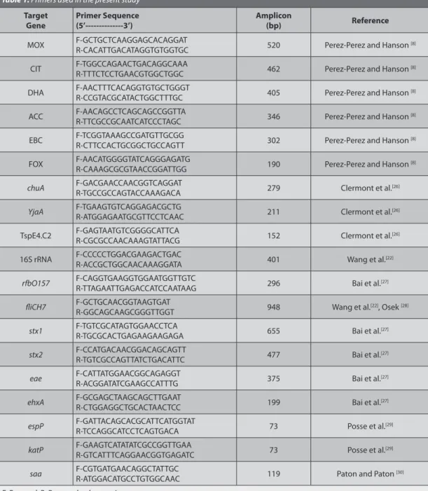
Table 1. Primers used in the present study
Target
Gene Primer Sequence(5’---3’) Amplicon(bp) Reference
MOX F-GCTGCTCAAGGAGCACAGGATR-CACATTGACATAGGTGTGGTGC 520 Perez-Perez and Hanson [8]
CIT F-TGGCCAGAACTGACAGGCAAAR-TTTCTCCTGAACGTGGCTGGC 462 Perez-Perez and Hanson [8]
DHA F-AACTTTCACAGGTGTGCTGGGTR-CCGTACGCATACTGGCTTTGC 405 Perez-Perez and Hanson [8]
ACC F-AACAGCCTCAGCAGCCGGTTAR-TTCGCCGCAATCATCCCTAGC 346 Perez-Perez and Hanson [8]
EBC F-TCGGTAAAGCCGATGTTGCGGR-CTTCCACTGCGGCTGCCAGTT 302 Perez-Perez and Hanson [8]
FOX F-AACATGGGGTATCAGGGAGATGR-CAAAGCGCGTAACCGGATTGG 190 Perez-Perez and Hanson [8]
chuA F-GACGAACCAACGGTCAGGATR-TGCCGCCAGTACCAAAGACA 279 Clermont et al.[26]
YjaA F-TGAAGTGTCAGGAGACGCTGR-ATGGAGAATGCGTTCCTCAAC 211 Clermont et al.[26]
TspE4.C2 F-GAGTAATGTCGGGGCATTCAR-CGCGCCAACAAAGTATTACG 152 Clermont et al.[26]
16S rRNA F-CCCCCTGGACGAAGACTGACR-ACCGCTGGCAACAAAGGATA 401 Wang et al.[22]
rfbO157 F-CAGGTGAAGGTGGAATGGTTGTCR-TTAGAATTGAGACCATCCAATAAG 296 Bai et al.[27]
fliCH7 F-GCTGCAACGGTAAGTGATR-GGCAGCAAGCGGGTTGGT 948 Wang et al.[22], Osek [28]
stx1 F-TGTCGCATAGTGGAACCTCAR-TGCGCACTGAGAAGAAGAGA 655 Bai et al.[27]
stx2 F-CCATGACAACGGACAGCAGTTR-TGTCGCCAGTTATCTGACATTC 477 Bai et al.[27]
eae F-CATTATGGAACGGCAGAGGTR-ACGGATATCGAAGCCATTTG 375 Bai et al.[27]
ehxA F-GCGAGCTAAGCAGCTTGAATR-CTGGAGGCTGCACTAACTCC 199 Bai et al.[27]
espP F-GATTACAGCACGCATTCATGGTATR-TCCAGGCATCCTCAGTGACA 73 Posse et al.[29]
katP F-GAAGTCATATATCGCCGGTTGAAR-GTCATTTCAGGAACGGTGAGATC 73 Posse et al.[29]
saa F-CGTGATGAACAGGCTATTGCR-ATGGACATGCCTGTGGCAAC 119 Paton and Paton [30]
according to the method developed by Perez-Perez and Hanson [8]. The PCR protocol was modified slightly in our laboratory as follow: Two sets of triplex PCR (set 1: ACC, CIT, FOX and set 2: DHA, EBC, MOX) were established for detection of pAmpC genes. The first triplex PCR was adjusted as 25 µL consisted of; 50 mM KCl, 0.2 mM each dNTP, 1.5 mM MgCl2, 0.4-0.6 µM primer sets (specific for FOX, ACC and CIT, respectively), 1.25 U of Taq DNA polymerase (Thermo Scientific) and 2 µL template DNA. The second triplex PCR was adjusted as 25 µL consisted of; 50 mM KCl, 0.2 mM each dNTP, 1.5 mM MgCl2, 0.5-0.6 µM primer sets (specific for EBC, DHA and MOX, respectively), 1.25 U of Taq DNA polymerase (Thermo Scientific) and 2 µL template DNA. Thermal cycling conditions for both of triplex PCRs were 5 min at 94ºC for initial denaturation, followed by 35 cycles of 45 sec at 94ºC, 45 sec at 64ºC and 1 min at 72ºC, and a final elongation step of 7 min at 72ºC. The primer sequences used were presented in Table 1.
Antibiotic Susceptibility Profiles, Phylogroups and Virulence Genes of the Isolates
Susceptibility of pAmpC beta-lactamase producing E. coli isolates to beta-lactam antibiotics and to other classes of antibiotics were determined by ADDT [24,31,32]. The beta-lactams antibiotic discs (Oxoid, UK) tested were ampicillin (AMP, 10 μg), cefepime (FEP, 30 μg), cefuroxime sodium (CXM, 30 μg), cephalothin (CEF, 30 μg) and imipenem (IPM, 10 μg). The antibiotics (Oxoid, UK) from other classes tested were chloramphenicol (C, 30 μg), ciprofloxacin (CIP, 5 μg), enrofloxacin (ENR, 5 μg), florfenicol (FFC, 30 μg), gentamicin (CN, 10 μg), kanamycin (K, 30 μg), nalidixic acid (NA, 30 μg), streptomycin (S, 10 μg), sulfamethoxazole-trimethoprim (SXT, 25 μg) and tetracycline (TE, 30 μg). E. coli ATCC 25922 strain was used as the control strain in ADDT. The inhibition zone diameters were evaluated according to CLSI critical zone diameters in CLSI document M10 0-S26 [24], M31-A3 [31] and VET01-S2 [32]. Based on the results, the isolates were classified as resistant, intermediate or susceptible. In the present study, a pAmpC beta-lactamase producing E. coli isolate that was resistant to at least 3 different classes of antibiotics excluding beta-lactams was accepted as multidrug-resistant (MDR) strain.
The phylogroups (groups A, B1, B2 and D) of the E. coli isolates were determined according to a triplex PCR protocol as described elsewhere [26], with the modified PCR conditions by Higgins et al.[33]. The triplex PCR is based on the amplification of a 279 bp fragment of the chuA gene, 211 bp fragment of the yjaA gene and 152 bp fragment of TspE4.C2 (a noncoding DNA region of E. coli genome). The phylogenetic groups of the isolates were assigned according to following criteria: the phylogenetic group A (chuA-, TspE4.C2-), B1 (chuA-, TspE4.C2+), B2 (chuA+, yjaA+), or D (chuA+, yjaA-). Additionally, phylogenetic subgroups (A: A0 and A1; B2: B22 and B23; D: D1 and D2) were investigated as described by Escobar-Páramo et al.[34].
E. coli ATCC 25922 was used as positive control strain
(chuA+, yjaA+ and TspE4.C2+) in the triplex PCR.
The pAmpC beta-lactamase producing E. coli isolates were screened by PCR for serotype O157:H7 (rfbO157 and fliCH7 genes) [27,28] and eae (intimin, attaching and effacing protein), ehxA (enterohemolysin), espP (extra-cellular serine protease), katP (catalase-peroxidase), saa (autoagglutinating adhesin), stx1 (Shiga toxin 1) and stx2 (Shiga toxin 2) to determine if the isolates were Enterohemorrhagic E. coli (EHEC) [27,29,30].
RESULTS
As the results of culture of cattle fecal samples, presumptive E. coli colonies were observed on at least one of both media (supplemented with CTX or CAZ) from 51 fecal samples. All colonies were identified as E. coli by phenotypic tests and PCR. After ESBL confirmatory test, E. coli isolates from 34 fecal samples were separated as ESBL-producing isolates. The remaining E. coli isolates (non-ESBL producers) were from 17 fecal samples and growth of E. coli colonies on these fecal samples (n=17) were observed on both medium (supplemented with CTX or CAZ) and therefore total 34 non-ESBL-producing E. coli were obtained. All of 34 non-ESBL- producing E. coli isolates were found to be resistant to cefoxitin by ADDT and therefore they were accepted as potential AmpC producers. In the present study, both potential AmpC beta-lactamse producing E. coli isolates (one from selective agar with CTX and the other one from the selective agar with CAZ) from a single fecal sample showed the same antibiotic susceptibility profile and the same phylogenetic group in all of the fecal samples. Therefore, the prevalence was estimated as 6.8% (17/250) for cattle in the study. E. coli was not isolated from sheep fecal samples.
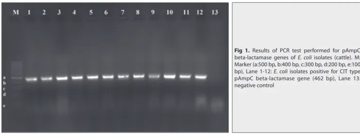
All E. coli isolates from cattle harbored only CIT family pAmpC gene (Fig. 1). In total, 5 (25%, 5/20) cattle herds were found positive in terms of carrying pAmpC beta-lactamase producing E. coli isolates (Table 2).
According to phylogenetic analysis by PCR, 11 (11/17, 64.7%) E. coli isolates were found belong to group A (subgroup A1), 3 (3/17, 17.6%) isolates to group D (subgroup D1), 2 (2/17, 11.8%) isolates to group B2 (subgroup B22) and 1 (1/17, 5.9%) isolate to group B1 (Table 2).
In total, 7 of 17 (41.2%) pAmpC beta-lactamase producing E. coli isolates were found MDR. Among the MDR isolates, 3 isolates belonged to phylogoup A(subgroup A1), one isolate to phylogoup B1 and one isolate to D (subgroup D1) (Table 2). The highest resistance was found against TET (9/17, 52.9%) whereas the lowest resistance was against CIP, ENR and NA (1/17, 5.9%). All isolates were susceptible to FEP and IPM (Table 3).
Among the virulence genes screened among the isolates, only eae (intimin) gene was detected in only one isolate
Table 2. Distrubition of the pAmpC beta-lactamase producing E. coli isolates (cattle) according to herds, phylogenetic group and antibiogram profiles Farm No E. coli Isolates (n) E. coli Isolates (n):
Phylogenetic Group Resistance Profile
blapAmpC Gene Family 3 8 7:A (A1) - CIT 1:B2 (B22) - CIT 6 1 A (A1) CN, S, K, FFC, C, TE* CIT 7 6 1:A (A1) TE CIT
1:B1 CN, S, K, SXT, NA, ENR, CIP, FFC, C, TE* CIT 1:B2 (B22) S, K, TE CIT 3:D (D1) S, SXT, TE* CIT 8 1 A (A1) CN, S, K, SXT, FFC C, TE* CIT 9 1 A (A1) CN, S, K, FFC, C, TE* CIT
* Multidrug resistant isolate (MDR, resistant to at least 3 classes of antibiotics except beta-lactam antibiotics). C: chloramphenicol, CIP: ciprofloxacin,
ENR: enrofloxacin, FFC: florfenicol, CN: gentamicin, K: kanamycin, NA: nalidixic acid, S: streptomycin, SXT: sulfamethoxazole-trimethoprim, TE: tetracycline
Table 3. Antibiotic susceptibilities of pAmpC beta-lactamase producing E. coli isolates (cattle)
Beta- Lactams
pAmpC Producing
E. coli (n=17) Other Antibiotics pAmpC ProducingE. coli (n=17)
R (n) I (n) R(n) I (n) AMP 17 0 CN 4 0 ATM 17 n/a K 5 0 FEP 0 0 S 8 1 CTX 17 n/a CIP 1 0 CPD 17 n / a ENR 1 0 CAZ 17 n/a NA 1 0 CRO 17 n /a TE 9 0 CXM 13 0 SXT 5 0 CEF 17 0 FFC 4 0 IPM 0 0 C 4 0
n/a: not applicable, R: resistant, I: intermediate, AMP: ampicillin, ATM: aztreonam, CAZ: ceftazidime, FEP: cefepime, CPD: cefpodoxime, CRO: ceftriaxone, CXM: cefuroxime, CEF: cephalothin, CTX: cefotaxime, FOX: cefoxitin, IPM: imipenem, C: chloramphenicol, CIP: ciprofl oxacin, ENR: enrofl oxacin, FFC: fl orfenicol, CN: gentamicin, K: kanamycin, NA: nalidixic acid, S: streptomycin, SXT: sulfamethoxazole-trimethoprim, TE: tetracycline
Fig 1. Results of PCR test performed for pAmpC
beta-lactamase genes of E. coli isolates (cattle). M: Marker (a:500 bp, b:400 bp, c:300 bp, d:200 bp, e:100 bp), Lane 1-12: E. coli isolates positive for CIT type pAmpC beta-lactamase gene (462 bp), Lane 13: negative control
from Farm 3. This E. coli isolate belonged to B22 phylo- genetic group and was not resistant to any of the antibiotics (except beta lactams) tested. None of the pAmpC beta-lactamase producing E. coli isolates was O157:H7 serotype of E. coli.
DISCUSSION
The real prevalence of pAmpC beta-lactamase producing E. coli is still unknown in healthy cattle and sheep in Turkey. There is only one local study conducted on this topic in healthy cattle in Turkey and it was found only one E. coli isolate carrying ampC (cmy) gene out of 312 cattle rectal swab samples [17]. On the other hand, there is no report for pAmpC beta-lactamase producing E. coli in healthy sheep in Turkey. Even though some studies have shown the existence of AmpC beta-lactamase producing E. coli and AmpC genes on cattle origin food products (cheese, meat, and milk) in different parts of Turkey [35-37], these studies do not reflect exactly the extent of these isolates in live animals in Turkey due to possible contamination of the animal origin food during processing. Therefore, the present study conducted have given additional information about the presence of AmpC beta-lactamase producing E. coli in cattle and sheep in Turkey.
Although we detected AmpC beta-lactamase producing E. coli in 17 cattle, no sheep was detected positive for AmpC beta-lactamase producing E. coli. This can be attributed to use of the beta-lactams and other classes of antibiotics (aminoglycosides, beta-lactams, phenicols, quinolones, sulfamethoxazole-trimethoprim and tetracycline) more widely in the prevention and treatment of wide variety of infections (mastitis, lameness, calf diarrhea, metritis, arthritis, pneumoniae, salmonellosis, urinary tract infections, septisemia, etc.) in cattle population than sheep population in Turkey and some of them may cause co-selection of AmpC beta-lactamase producing E. coli isolates in gut microflora. It has been known that ampC genes are located on a large plasmid together with other antimicrobial resistance genes such as the genes responsible for aminoglycosides, phenicols, quinolones, sulfamethoxazole-trimethoprim and tetracycline resistances and frequent use of these anti- biotics in livestock for several purposes leads the selection pressure for AmpC beta-lactamase producing Gram-negative bacteria in gut microflora [2]. In line with this it was detected co-resistance to at least one of these antibiotics in 9 (9/17, 52.9%) isolates and MDR in 7 (41.2%) isolates in this study.
Escherichia coli isolates obtained in this study were found fall into four main phylogenetic groups (A, B1, B2 and D). It is known that E. coli strains belonging to group A and B1 are primarily found in the commensal microflora [26,34]. The pathogen E. coli strains associated with extra- intestinal infections and diarrhea mainly belong to B2 and D groups [26,34]. Later, Escobar-Paramo et al.[34] stated
phylogenetic subgroups (A: A0 and A1; B2: B22 and B23; D: D1 and D2). Likewise, in the present study, phylogentic analysis of E. coli isolates showed that group A (subgroup A1) (n=11) is the predominant group, followed by group D (subgroup D1) (n=3), group B2 (subgroup B22) (n=2) and group B1 (n=1). On the other hand, the reports indicate that E. coli strains from B2 and D phylogroups possess more virulence factors but less MDR pattern than A and B1 phylogroups [38,39]. Similarly, in the present study the E. coli isolates from A and B1 phylogroups showed resistance to more antibiotics than the B2 and D phylogroup isolates and EHEC/STEC related virulence gene (eae, intimin) detected was found in the E. coli isolate belonging to B22 phylogroup with no co-resistance to other classes of antibiotics. The eae gene encodes the intimin protein on the surface of EPEC/STEC/EHEC isolates and it is located on the locus of enterocyte effacement (LEE) pathogenicity island. The intimin protein is important for intimate attachment to the intestinal mucosa and the formation of the attaching and effacing lesions in EPEC and STEC/EHEC infections [40]. Therefore, it is possible to state that the eae gene positive and pAmpC beta-lactamase producing E. coli isolate from B22 phylogenetic group determined in cattle in this study can be pathogenic for both human and calf even thogh this isolate is not a STEC/EHEC isolate.
Overall, based on the similarity of phylogenetic analysis results and antibiogram profiles of the isolates, three farms (Farm 6, 8 and 9) had only one isolate, one farm (Farm 3) had 2 different isolates and one farm (Farm 7) had 4 different isolates. Additionally, all isolates had the same blapAmpC gene family (CIT). Hence, it can be stated that few parent E. coli strains with the same pAmpC gene (CIT) were circulating in the cattle farms in Burdur, Turkey. It is generally accepted that food producing animals serve as reservoir for MDR E. coli strains and they can be transmitted to human by direct contact and/or via food chains. This issue has also been considered for AmpC beta-lactamase producing E. coli since there are many studies showing the similar pAmpC genes and plasmids in both animal and animal owners or farm workers [41-43]. Therefore, we can mention the possible health risk for people close contact to the cattle population in the present study. In conclusion, the present study showed the absence of pAmpC beta-lactamase producing E. coli in sheep population and presence of few multi-drug resistant pAmpC beta-lactamase producing E. coli strains with only one type pAmpC gene family (CIT) in cattle population in Burdur, Turkey. However, more studies are needed to reveal and understand the course of prevalence of pAmpC beta-lactamase producing E. coli (pathogen and commensal) and diversity in pAmpC genes in livestock populations in Turkey. The other point that should be considered, as known, emergence of E. coli isolates possesing pAmpC genes may also bring the increase in production of the AmpC beta-lactamases in other Gram-negative bacteria due to horizontal
transfer of plasmids between Gram-negative bacteria species. Hence, the necessary preventive measurements should be taken, for examples, livestock sector workers and veterinarians should be informed regularly about increase in antimicrobial-resistant strains and proper selection of antimicrobials should be provided in the treatment of the infections in animals. Also, monitoring should be performed for antimicrobial resistance levels to limit the escalating trend in antimicrobial resistance and the emergence of resistance traits in genetic material of Gram-negatives.
D
eclarationofc
onflictingi
nterests The authors declare no conflict of interest. REFERENCES1. Frere JM: Beta-lactamases and bacterial resistance to antibiotics. Mol
Microbiol, 16, 385-395, 1995. DOI: 10.1111/j.1365-2958.1995.tb02404.x
2. Seiffert SN, Hilty M, Perreten V, Endimiani A: Extended-spectrum
cephalosporin-resistant gram-negative organisms in livestock: An emerging problem for human health? Drug Resist Updat, 16, 22-45, 2013. DOI: 10.1016/j.drup.2012.12.001
3. Bush K, Jacoby GA: Updated functional classification of
beta-lactamases. Antimicrob Agents Chemother 54 (3): 969-976, 2010. DOI: 10.1128/AAC.01009-09
4. Vardanyan R, Hruby V: Antibiotics. In, Sysnthesis of Best Seller Drugs.
573-643, Elsevier B.V., 2016.
5. Bush K, Jacoby GA, Medeiros AA: Functional classification scheme
for β-lactamases and its correlation with molecular structure. Antimicrob
Agents Chemother, 39 (6): 1211-1233, 1995. DOI: 10.1128/AAC.39.6.1211
6. Ambler RP: The structure of β-lactamases. Philos Trans R Soc Lond B Biol
Sci, 289 (1036): 321-331, 1980. DOI: 10.1098/rstb.1980.0049
7. Jacoby GA: AmpC β-lactamases. Clin Microbiol Rev, 22, 161-182, 2009.
DOI: 10.1128/CMR.00036-08
8. Pérez-Pérez FJ, Hanson ND: Detection of plasmid-mediated AmpC
β-lactamase genes in clinical isolates by using multiplex PCR. J Clin
Microbiol, 40, 2153-2162, 2002. DOI: 10.1128/JCM.40.6.2153-2162.2002
9. Lerner A, Matthias T, Aminov R: Potential effects of horizontal gene
exchange in the human gut. Front Immunol, 8:1630, 2017. DOI: 10.3389/ fimmu.2017.01630
10. Bugarel M, Martin A, Fach P, Beutin L:Virulence gene profiling of
enterohemorrhagic (EHEC) and enteropathogenic (EPEC) Escherichia
coli strains: A basis for molecular risk assessment of typical and atypical
EPEC strains. BMC Microbiol, 11:142, 2011. DOI: 10.1186/1471-2180-11-142
11. Venegas-Vargas C, Henderson S, Khare A, Mosci RE, Lehnert JD, Singh P, Ouellette LM, Norby B, Funk JA, Rust S, Bartlett PC, Grooms D, Manning SD: Factors associated with Shiga toxin-producing
Escherichia coli shedding by dairy and beef cattle. Appl Environ Microbiol,
82, 5049-5056, 2016. DOI: 10.1128/AEM.00829-16
12. Asai T, Masani K, Sato C, Hiki M, Usui U, Baba K, Ozawa M, Harada K, Aoki H, Sawada T: Phylogenetic groups and cephalosporin resistance
genes of Escherichia coli from diseased food-producing animals in Japan.
Acta Vet Scand, 53:52, 2011. DOI: 10.1186/1751-0147-53-52
13. Hille K, Fischer J, Falgenhauer L, Sharp H, Brenner GM, Kadlec K, Friese A, Schwarz S, Imirzalioglu C, Kietzmann M, Von Münchhausen C, Kreienbrock L: On the occurence of extended-spectrum-and
AmpC-beta-lactamase-producing Escherichia coli in livestock: Results of selected European studies. Berl Munch Tierarztl Wochenschr, 127 (9-10): 403-411, 2014.
14. EFSA (European Food Safety Authority) and ECDC (European Centre for Disease Prevention and Control): The European Union
summary report on antimicrobial resistance in zoonotic and indicator bacteria from humans, animals and food in 2017. EFSA J, 17 (2):e05598,
2019. DOI: 10.2903/j.efsa.2019.5598
15. Tewari R, Mitra S, Ganaie F, Das S, Chakraborty A, Venugopal N, Shome R, Rahman H, Shome BR: Dissemination and characterisation
of Escherichia coli producing extended-spectrum β-lactamases, AmpC β-lactamases and metallo-β-lactamases from livestock and poultry in Northeastern India: A molecular surveillance approach. J Glob Antimicrob
Resist, 17, 209-215, 2019. DOI: 10.1016/j.jgar.2018.12.025
16. Pehlivanoglu F: Existence of plasmid mediated AmpC
beta-lactamase-producing Escherichia coli isolates in healthy laying hens. Van
Vet J, 28 (2): 63-67, 2017.
17. Aslantaş Ö, Elmacıoğlu S, Yılmaz EŞ: Prevalence and characterization
of ESBL-and AmpC-producing Escherichia coli from cattle. Kafkas Univ Vet
Fak Derg, 23, 63-67, 2017. DOI: 10.9775/kvfd.2016.15832
18. Gumus B, Celik B, Kahraman BB, Siğirci BD, Ak S: Determination
of extended spectrum beta-lactamase (ESBL) and AmpC beta-lactamase producing Escherchia coli prevalence in faecal samples of healthy dogs and cats. Revue Med Vet, 168, 46-52, 2017
19. Aslantas O, Yilmaz ES: Prevalence and molecular characterization
of extended-spectrum lactamase (ESBL) and plasmidic AmpC beta-lactamase (pAmpC) producing Escherichia coli in dogs. J Vet Med Sci, 79, 1024-1030, 2017. DOI: 10.1292/jvms.16-0432
20. Thrusfield M: Surveys. In, Veterinary Epidemiology. 3rd ed., 228-242, Blackwell Publishing, Ames, Iowa, 2007.
21. Winn W, Allen S, Janda W, Koneman E, Procop G, Schreckenberger P, Woods G: The Enterobactericeae. In, Koneman EW (Ed): Koneman’s
Color Atlas and Textbook of Diagnostic Microbiology. 6th ed., 211-302, Lippincott Williams and Wilkins, Philadelphia, 2006.
22. Wang G, Clark CG, Rodgers FG: Detection in Escherichia coli of the
genes encoding the major virulence factors, the genes defining the O157:H7 serotype, and components of the type 2 shiga toxin family by multiplex PCR. J Clin Microbiol, 40, 3613-3619, 2002. DOI: 10.1128/ JCM.40.10.3613-3619.2002
23. Pehlivanoglu F, Turutoglu H, Ozturk D: CTX-M-15-type
extended-spectrum beta-lactamase-producing Escherichia coli as causative agent of bovine mastitis. Foodborne Pathog Dis, 13 (9): 477-482, 2016. DOI: 10.1089/fpd.2015.2114
24. CLSI (Clinical and Laboratory Standards Institute): Performance
standards for antimicrobial susceptibility testing. 26th Informational Supplement, CLSI Document M100-S26, Pennsylvania, USA, 2016.
25. Polsfuss S, Bloemberg GV, Giger J, Meyer V, Böttger EC, Hombach M: Practical approach for reliable detection of AmpC beta-lactamase
producing Enterobactericeae. J Clin Microbiol, 49, 2798-2803, 2011. DOI: 10.1128/JCM.00404-11
26. Clermont O, Bonacorsi S, Bingen E: Rapid and simple determination
of the Escherichia coli phylogenetic group. Appl Environ Microbiol, 66, 4555-4558, 2000. DOI: 10.1128/aem.66.10.4555-4558.2000
27. Bai J, Paddock ZD, Shi X, Li S, An B, Nagaraja TG: Application of a
multiplex PCR to detect the seven major shiga toxin-producing Escherichia
coli based on genes that code for serogroup-specific O-antigens and
major virulence factors in cattle feces. Foodborne Pathog Dis, 9, 541-548, 2012. DOI: 10.1089/fpd.2011.1082
28. Osek J: Development of a multiplex PCR approach for the
identification of shiga toxin-producing Escherichia coli strains and their major virulence factor genes. J Appl Microbiol, 95, 1217-1225, 2003. DOI: 10.1046/j.1365-2672.2003.02091.x
29. Posse B, Zutter LD, Heyndrickx M, Herman L: Metabolic and genetic
profiling of clinical O157 and non-O157 shiga-toxin-producing Escherichia
coli. Res Microbiol, 158, 591-599, 2007. DOI: 10.1016/j.resmic.2007.06.001
30. Paton AW, Paton JC: Direct detection and characterization of Shiga
toxigenic Escherichia coli by multiplex PCR for stx1, stx2, eae, ehxA and saa. J Clin Microbiol, 40, 271-274, 2002. DOI: 10.1128/JCM.40.1.271-274.2002
31. CLSI (Clinical and Laboratory Standards Institute): Performance
standards for antimicrobial disk and dilution susceptibility tests for bacteria isolated from animals, Approved standard, 3rd Ed., CLSI document M31-A3, Pennsylvania, USA, 2010.
standards for antimicrobial disk and dilution susceptibility tests for bacteria isolated from animals, 2nd informational supplement, CLSI document VET01-S2, Pennsylvania, USA, 2013.
33. Higgins J, Hohn C, Hornor S, Frana M, Denver M, Joerger R:
Genotyping of Escherichia coli from environmental and animal samples. J
Microbiol Methods, 70, 227-235, 2007. DOI: 10.1016/j.mimet. 2007.04.009
34. Escobar-Páramo P, Le Menac’h A, Le Gall T, Amorin C, Gouriou S, Picard B, Skurnik D, Denamur E: Identification of forces shaping the
commensal Escherichia coli genetic structure by comparing animal and human isolates. Environ Microbiol, 8, 1975-1984, 2006. DOI: 10.1111/ j.1462-2920.2006.01077.x
35. Pehlivanlar Onen S, Aslantas O, Yılmaz ES, Kurekci C: Prevalence of
β-lactamase -producing Escherichia coli from retail meat in Turkey. J Food
Sci, 80 (9): M2023-M2029, 2015. DOI: 10.1111/1750-3841.12984
36. Ozpinar H, Tekiner IH, Sarici B, Cakmak B, Gokalp F, Ozadam A:
Phenotypic characterization of ESBL- and AmpC- type beta- lactamases in Enterobacteriaceae from chicken meat and dairy products. Ankara Univ
Vet Fak Derg, 64 (4): 267-272, 2017.
37. Ozdikmenli Tepeli S, Demirel Zorba NN: Frequency of
extended-spectrum β-lactamase (ESBL)- and AmpC β-lactamase-producing
Enterobacteriaceae in a cheese production process. J Dairy Sci, 101,
2906-2914, 2018. DOI: 10.3168/jds.2017-13878
38. Smith JL, Fratamico PM, Gunther NW: Extraintestinal pathogenic
Escherichia coli. Foodborne Pathog Dis, 4 (2): 134-163, 2007. DOI: 10.1089/
fpd.2007.0087
39. Chakraborty A, Saralaya V, Adhikari P, Shenoy S, Baliga S, Hegde A: Characterization of Escherichia coli phylogenetic groups associated
with extraintestinal infections in South Indian population. Ann Med
Health Sci Res, 5 (4): 241-246, 2015.
40. McDaniel TK, Jarvis KG, Donnenberg MS, Kaper JB: A genetic locus
of enterocyte effacement conserved among diverse enterobacterial pathogens. Proc Natl Acad Sci USA, 92 (5): 1664-1668, 1995. DOI: 10.1073/ pnas.92.5.1664
41. Voets GM, Fluit AC, Scharringa J, Schapendonk C, Van Den Munckhof T, Leverstein-Van Hall MA, Stuart JC: Identical plasmid
AmpC beta-lactamase genes and plasmid types in E. coli isolates from patients and poultry meat in the Netherlands. Int J Food Microbiol, 167, 359-362, 2013. DOI: 10.1016/j.ijfoodmicro.2013.10.001
42. Huijbers PMC, Graat EAM, Haenen APJ, Van Santen MG, Van Essen-Zandbergen A, Mevius DJ, Van Duijkeren E, Van Hoek AHAM:
Extended-spectrum and AmpC β-lactamase-producing Escherichia coli in broilers and people living and/or working on broiler farms: Prevalence, risk factors and molecular characteristics. J Antimicrob Chemother, 69 (10): 2669-2675, 2014. DOI: 10.1093/jac/dku178
43. Ljungquist O, Ljungquist D, Myrenås M, Ryden C, Finn M, Bengtsson B: Evidence of household transfer of ESBL-/pAmpCproducing
Enterobacteriaceae between humans and dogs - A pilot study. Infect Ecol Epidemiol, 6:31514, 2016. DOI: 10.3402/iee.v6.31514