МЕДИЧНІ ПЕРСПЕКТИВИ / MEDICNI PERSPEKTIVI
UDC 616.379-008.64:616.8-009.7]-085-092.9:634.8 https://doi.org/10.26641/2307-0404.2019.4.189197A. Yurt
1,
B. Köksal
2,
P. Gürbüz
3,
A. Yıldız
3,
N. Vardı
3,
E. Alçin
3I
I
N
N
V
V
E
E
S
S
T
T
I
I
G
G
A
A
T
T
I
I
N
N
G
G
E
E
F
F
F
F
E
E
C
C
T
T
S
S
O
O
F
F
G
G
R
R
A
A
P
P
E
E
S
S
E
E
E
E
D
D
E
E
X
X
T
T
R
R
A
A
C
C
T
T
O
O
N
N
N
N
E
E
U
U
R
R
O
O
P
P
A
A
T
T
H
H
I
I
C
C
P
P
A
A
I
I
N
N
I
I
N
N
T
T
H
H
E
E
S
S
T
T
R
R
E
E
P
P
T
T
O
O
Z
Z
O
O
T
T
O
O
C
C
I
I
N
N
-
-
I
I
N
N
D
D
U
U
C
C
E
E
D
D
D
D
I
I
A
A
B
B
E
E
T
T
I
I
C
C
M
M
I
I
C
C
E
E
Igdır University, Vocational School of Health Services 1
Igdır, Turkey
Lokman Hekim University, Faculty of Medicine 2
Ankara, Turkey
İnönü University, Faculty of Medicine 3
Malatya, Turkey Університет Ігдір, Професійне училище охорони здоров'я 1 Ігдір, Туреччина Університет Локмана Хекіма, медичний факультет 2 Анкара, Туреччина Університет Іненю, медичний факультет 3 Малатья, Туреччина
Corresponding author Burcu Köksal, e-mail: [email protected] Цитування: Медичні перспективи. 2019. Т. 24, № 4. С. 51-58
Cited: Medicni perspektivi. 2019;24(4):51-58
Key words: diabetes mellitus, neuropathic pain, grape seed extraction, hot plate, streptozocin (STZ)
Ключові слова: цукровий діабет, невропатичний біль, екстракція виноградних кісточок, гаряча пластина,
стрептозоцин
Ключевые слова: сахарный диабет, невропатическая боль, экстракция виноградных косточек, горячая
пластина, стрептозоцин
Abstract. Investigating effects of grape seed extract on neuropathic pain in the streptozotocin-induced diabetic mice. Yurt A., Köksal B., Gürbüz P., Yıldız A., Vardı N., Alçin E. Diabetes mellitus is a complicated and serious
health problem involving peripheral neuropathy. This situation causes to loss of senses, tingle and pain. Diabetic peripheral neuropathy (DPN) affects 236 million people around the World. Hence there is a need to investigate alternative ways of cure focusing on DPN. Flavonoids have potential on pain due to their permeability characteristic in the capillary microcirculation system and in lowering blood pressure. Flavonoids are common in grape seed. The main flavonoids of grape seed involve proanthocyanidins which might be an effective agent in cure of pain in DPN. The purpose of this study is to investigate effects of grape seed extract on neuropathic pain in the Streptozotocin-induced diabetic mice. In the study, 50 eight-week old BALB-C strain mice in five different groups (Control, Diabetic Control, Control+25 mg/kg, Diabetes+25 mg/kg and Diabetes+50 mg/kg) were used. To induce diabetes in thirty of these animals, single dose Streptozotocin (180 mg/kg) was administered intraperitoneally. After diabetes was observed, grape seed extract (25 mg/kg and 50 mg/kg body weight) was administered by oral gavage in three groups (Control+25 mg/kg, Diabetes+25 mg/kg and Diabetes+50 mg/kg) during six weeks. At the end of the second and sixth weeks, pain threshold measurements in hot plate test were performed in line with a predetermined thermal pain model. Also the tissues of the sciatic nerve and abdominal aorta from the animals were histologically investigated. As a result hot plate measurements, pain threshold values of the animals in Diabetes+50mg/kg group significantly differed from the measurements of the animals in control group in the first measurements and from the animals in Diabetes+25 mg/kg group in the second measurements (p<0.05). However pain threshold values of the animals in Diabetes+25 mg/kg group differed significantly from the values of the animals in control+25 mg/kg group and control group. It means pain threshold values of the animals in Diabetes+50 mg/kg and Diabetes+25 mg/kg groups were significantly lower than the values of the animals in the other groups. The results of histological investigations showed that degenerations of myelin sheet and axons in diabetic control group were decreased significantly in Diabetes+50 mg/kg and Diabetes+25 mg/kg groups. Moreover degenerations of aorta tissues of animals in diabetic group were not seen in the animals of Diabetes+50 mg/kg except for tunica adventitia inflammation. It can be said that grape seed extract decreased threshold of neuropathic pain in the Streptozotocin-induced diabetic mice and prevented degenerations of myelin sheet and axons, and aorta tissues.
КЛІНІЧНА МЕДИЦИНА
Реферат. Вивчення впливу екстракту виноградних кісточок на біль при невропатії в мишей з діабетом, який викликаний стрептозотоцином. Юрт А., Кексал Б., Гюрбюз П., Йлдіз А., Варді Н., Алчін Е. Цукровий діабет – складна й серйозна проблема для здоров'я, яка пов'язана з периферичною невропатією. Вона викликає втрату чуттів, поколювання і біль. Діабетична периферична невропатія (ДПН) уражає 236 мільйонів осіб у всьому світі. Отже, існує необхідність досліджувати альтернативні способи лікування з акцентом на ДПН. Флавоноїди мають потенціал щодо больового синдрому через їх проникність, характерну для капілярної системи мікроциркуляції, зниження артеріального тиску. Флавоноїди є у виноградних кісточках. Основні флавоноїди виноградних кісточок включають проантоціанідини, які можуть бути ефективним засобом при лікуванні болю при ДПН. Метою цього дослідження є вивчення впливу екстракту виноградних кісточок на біль при невропатії в мишей з діабетом, викликаним стрептозотоцином. У дослідженні використовували 50 мишей віком 8 тижнів штаму BALB-C у п'яти різних групах (контроль, діабетичний контроль, контроль +25 мг/кг, діабет + 25 мг/кг і діабет +50 мг/кг). Щоб викликати діабет у тридцяти з цих тварин, внутрішньочеревинно вводили одноразову дозу стрептозотоцину (180 мг/кг). Після того, як спостерігався діабет, екстракт виноградних кісточок (25 мг/кг і 50 мг/кг маси тіла) вводили пероральним зондом у трьох групах (контроль + 25 мг/кг, діабет +25 мг/кг і діабет +50 мг/кг ) протягом шести тижнів. На прикінці другого та шостого тижнів виконували вимірювання порога болю в тесті гарячою пластиною відповідно до заздалегідь визначеної моделі термічного болю. Також були досліджені тканини сідничного нерва і черевної аорти від тварин. У результаті вимірювань на гарячій пластині порогові значення болю в тварин у групі діабету +50 мг/кг значно відрізнялися від вимірювань у тварин у контрольній групі в перших вимірах і від тварин у групі діабету +25 мг/кг у групі при других вимірах (р<0.05). Однак порогові значення болю у тварин у групі діабету +25 мг/кг значно відрізнялися від значень у тварин у контрольній групі +25 мг/кг і контрольній групі. Це означає, що порогові значення болю у тварин у групах діабету +50 мг/кг і діабету +25 мг/кг були значно нижче, ніж значення у тварин в інших групах. Результати гістологічних досліджень показали, що дегенерації мієлінового шару й аксонів у контрольній групі з діабетом значно зменшилися в групах діабету +50 мг/кг і діабету +25 мг/кг. Більше того, дегенерації тканин аорти тварин у групі діабету не спостерігалося у тварин з діабетом +50 мг/кг, за винятком запалення адвентиціальної оболонки. Можна сказати, що екстракт вино-градних кісточок знижував поріг невропатичного болю в мишей з діабетом, який викликаний стрепто-зотоцином, і запобігав дегенерації мієлінового шару й аксонів, а також тканин аорти.Diabetes mellitus is a metabolic disease affecting 382 million people around the World [1, 2]. Dia-betes involves either insufficient insulin secretion or deficiency in tissue response to insulin [3]. Based on the deficiency of insulin metabolism, diabetes causes different complications involving hypoglycemia [4, 5], ketoacidosis [6] and peripheral neuropathy [7]. Neuropathy is characterized by loss of sense, tingle and pain [7]. In neuropathy deficiency of distal axons, loss of axons and loss of myelin in neuron fibers are observed [8]. Moreover the neuropathic patients feel depression and pain [9]. Neuropathic pain affects 6-8% of the World population and its prevalence increases with diabetes mellitus [10]. Neuropathic pain frequently causes to burning sensations, to sleep disturbances and to tiredness [11, 12]. These symptoms prevent the patients from working and social life.
In neuropathy, production of free oxygen radicals and increased load of mitochondrial electron transfer cause damage to cells. Since accumulation of free oxygen radicals lead to increased levels of lipid, DNA and protein peroxidation which induce cell death and decrease blood flow in nervous system [13, 14]. Hence prevention of peroxidation caused by free oxygen radicals might be a beginning point in decreasing neuropathic pain in diabetic patients. Flavonoids provide a way to investigate this claim,
since content of flavonoids involves antioxidant activity [15], especially proantocyanidin which is a solvable short chain polymer found in grape seed is a strong anti-oxidant (free radical scavenger) [16]. Proantocyanidin’s entioxidant activity is predicted by its phenolic hydrogen content [17, 18]. However for defining a proanthocyanidin as an antioxidant two basic conditions must be provided: the first is that when its concentrations is low relative to the substrate, it must prevent oxidative injury while the second is that the product following the scavenging must be stable [19].
Proantocyanidin scavenges peroxyl and hydroxyl radicals and prevents lipid peroxidation [20, 21]. Moreover a recent study showed preventive effect of grape seed extract on DNA damage [22]. Grape seed extract also contributes to prevention of capillary blood vessels by decreasing the effect of free radical harms on the walls of the vessels; hence it decreases the risk of platelet aggregations and arteriosclerosis [23, 24]. Therefore the purpose of this study to investigate effects of grape seed extract on neuropathic pain in the Streptozotocin-induced diabetic mice
MATERIALS AND METHODS OF RESEARCH
Experimental Animals: In the study 50
eight-week BALB-C strain female mice were used (30±5 g). The mice were hold in a room at 22-25°C
МЕДИЧНІ ПЕРСПЕКТИВИ / MEDICNI PERSPEKTIVI
and they were exposured to 12 dark and 12 light cycle. The animals were in cages for five mice and they were fed with tap water and standard pellet food for mice. Then, the animals were divided into five different groups (10 mice per group):
I. Group: Control (n=10)
II. Group: Diabetic Control (n=10)
III. Group: Control + 25 mg/kg grape seed extract (n=10)
IV. Group: Diabetes+25 mg/kg grape seed extract (n=10)
V. Group: Diabetes +50 mg/kg grape seed extract (n=10)
Inducing Diabetes: In three groups of the study,
Streptozotocin (STZ) (Sigma-Aldrich) (180 mg/kg) solved in 0.4 ml (0.1 M) sodium citrate (pH=4.5) were intraperitonally injected to the animals as a single dose. Blood glucose levels were measured by taking blood from tails of the animals and using glucose meter. Animals having satiety blood glucose levels over 200 mg/dl were accepted as diabetics.
Application of Grape Seed Extract: The extracts
(25 ve 50 mg/kg) were prepared daily and fresh by solving them in distillated water. The extracts were given by oral gavage way provided by plastic part of the grey vascular access tool (16G). During the gavage process, the animals were anesthetized by ether and the extracts were given at the same time of days.
Hot Plate Test: In the test the animals were put
on a hot platform and time of their reactions were recorded for determining pain threshold value. For the mice appropriate Hot Plate Analgesiameter (Harvard, Edenbridge, UK) was used. Before the experiments all of the animals were tested in hot plate test during 5 days to provide accommodation to the system (147). Then the animals were rested for one day and they were tested two times in sixth day. During the experimental Hot Plate test the temperature of the platform was adjusted to 50±0.5 °C, the mice could not go out of the system due to its barriers and they were tested for pain threshold. After the animals were put into the platform, a chronometer was started and their retract times of extremities and licking them were recorded in a silent environment. If the animals did not retract the extremities in 60 sec., they were taken from the system to prevent tissue harms and they were not included in the study. The test was applied to the animals after the oral gavage process.
Histological Applications
Sciatic Nerve Tissue: Sciatic nerve tissues of the
animals were fixed in form aldehyde (%10). Then the tissues were embedded into the paraffin after washing them in tap water, dehydration and
poli-shing. Samples in 4 µm thickness were taken from the paraffin blocks and, deparafinisation and rehy-dration processes were applied to the samples. Then the samples were stained by hematoksilen-eozin (H-E) method and they were observed by Leica DFC-280 research microscope.
Sciatic Nerve: Changes in nerve cells (axonal
changes) were analyzed by Leica Q Win image ana-lysis system. During the anaana-lysis each slice of the samples stained by H-E method were observed by using 100:1 (100X) magnifying power. Axonal changes were scored as none (0), slight (1), mild (2) and strong (3).
Aorta Tissue: Aorta tissues of the animals were
fixed in form aldehyde (%10). Similar to sciatic nerve tissues, aorta tissues were embedded into the paraffin after washing them in tap water, dehy-dration and polishing. Then slices from the samples in 4 µm thickness were taken from the paraffin blocks. The samples were stained by hematoksilen-eozin (H-E) and orsein methods, and then Leica DFC-280 research microscope was used to observe stained samples.
Abdominal Aorta: For the analysis of abdominal
aorta, tunica media thicknesses of randomly selected six samples stained by hematoksilen-eozin (H-E) method were measured by using 40X magnifying power and Leica Q Win image analysis system. Elastic fibers stained by orsein were analyzed by considering loss of elastic fibers and defects in undu-lated-waved structure. Defects in elastic fibers were scored as strong (0), slight (1), mild (2) and severe (3) by observing them in 40X magnifying power.
Statistical Analysis: For analysis of quantitative
data, descriptive (mean±standart deviation) values were represented. In following inferential statistics Wilcoxon Signed Ranks Test was used to compare first and second scores on Hot Plate Test while paired samples t-test was used to compare total first and second scores on Hot Plate Test. Between-group comparisons of Hot Plate scores and histological data were made by Mann Whitney U test. For all of the analyses, 0.05 was accepted as significance threshold.
Findings
Findings on Blood Glucose Levels and Hot Plate Test
Blood glucose levels of the animals before and after grape seed extract application showed that all of the diabetes groups showed significant rise in their blood glucose levels relative to control group animals in both before and after the application. Table 1 represents results of analysis on blood glu-cose levels of the animals before and after grape seed extract application.
КЛІНІЧНА МЕДИЦИНА
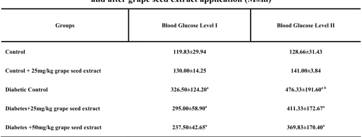
T a b l e 1
Analysis of blood glucose levels of the animals before
and after grape seed extract application (M±m)
Groups Blood Glucose Level I Blood Glucose Level II
Control 119.83±29.94 128.66±31.43
Control + 25mg/kg grape seed extract 130.00±14.25 141.00±3.84
Diabetic Control 326.50±124.20a 476.33±191.60a b
Diabetes+25mg/kg grape seed extract 295.00±58.90a 411.33±172.67a
Diabetes +50mg/kg grape seed extract 237.50±42.65a 369.83±170.40a
Note. a – Significant difference with control group at 0.05, b – Significant difference between first and second levels at 0.05.
In terms of blood glucose levels, just diabetic control animals represented a significant increase in their blood glucose levels between two measu-rements. After determining blood glucose levels, pain threshold data were analyzed and the results
showed that pain threshold values of the all groups were significantly increased except for the control group. Table 2 represents results of statistical analysis of hot plate test data.
T a b l e 2
Results of statistical analysis of hot plate test data of the groups (M±m)
Groups Hot Plate Test I Hot Plate Test II p
Control 17.09±2.94 sec. 20.02±5.03 sec. 0.16
Control + 25mg/kg grape seed extract 14.36±1.55 sec. 19.38±3.46 sec. 0.01b
Diabetic Control 15.71±2.10 sec. 22.67±5.23 sec. 0.02b
Diabetes+25mg/kg grape seed extract 15.88±3.07 sec. 24.35±4.48 sec. 0.00b
Diabetes +50mg/kg grape seed extract 14.33±1.71 sec. 19.17±4.36 sec. 0.02b
Note. b – Significant difference between first and second levels at 0.05
When the groups were compared in terms of their scores on two hot plate test applications, it was seen that there were significant differences in hot plate scores of the experimental groups and control groups. Results on comparisons of the groups in terms of hot plate scores are seen in table 3.
Table 3 showed that there was statistically sig-nificant difference between hot plate test I scores of the animals in Control and Diabetes+50 mg/kg
groups. Similarly hot plate test I scores of the animals in Control group were significantly higher than the scores of the animals in Control+25 mg/kg group. When looked at the hot plate test II scores, it was seen that the scores of the animals in Diabetes+25 mg/kg were significantly higher than the scores of the control group animals, animals in Control+25 mg/kg group and the animals in Diabetes+50 mg/kg group.
МЕДИЧНІ ПЕРСПЕКТИВИ / MEDICNI PERSPEKTIVI
T a b l e 3
Analysis results on comparisons of the groups in terms of hot plate scores
Groups Hot Plate Test I (p-value) Hot Plate Test II (p-value)
Control- Diabetic Control 0.36 0.21
Control- Diabetes+25mg/kg 0.41 0.03*
Control- Diabetes+50mg/kg 0.02* 0.93
Control- Control+25mg/kg 0.02* 0.65
Diabetic Control- Diabetes+25mg/kg 0.82 0.60
Diabetic Control- Diabetes+50mg/kg 0.17 0.21
Diabetes +25mg/kg- Diabetes+50mg/kg 0.35 0.03*
Control + 25mg/kg- Diabetic Control 0.28 0.11
Control + 25mg/kg- Diabetes +25mg/kg 0.36 0.03*
Control + 25mg/kg- Diabetes+50mg/kg 0.93 0.74
Note. * – Significant difference at 0.05
Findings Histolopathological Parameters
Histological analyses were done in two ways as statistical comparison and microscope observation. Statistical analyses of two tissues showed that histopathological scores of the animal tissues in
diabetes control group were significantly higher than control group animals in terms of sciatic nerve damage and elastic fiber defects. The results are represented in table 4.
T a b l e 4
Statistical analyses of two tissues in terms of histopathological scores (M±m)
Groups Histopathological score (Sciatic Nerve Tissue)Histopathological score (Aorta tunica Media
Thickness)
Histopathological score (Defects of Elastic
Fibers og Aorta)
Control 0.48±0.65 35.44±4.07 0.50±0.55
Control + 25mg/kg grape seed extract 0.16±0.37 35.04±2.61 0.67±0.52
Diabetic Control 2.12±0.83a 37.14±4.94 1.67±0.82a
Diabetes+25mg/kg grape seed extract 1,68±0.69 37.35±3.35 1.33±0.52
Diabetes +50mg/kg grape seed extract 1.64±0.91 33.96±8.37 1.00±0.89
Note. a – Significant difference with control group at 0.05.
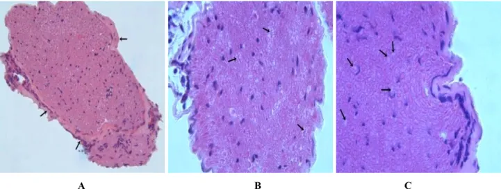
Sciatic nerve tissue damages of the animals in diabetic control group involved thick perineuriums, shrinkage in axons and degenerations in myelin
sheet structure. Figure 1 represents the damages of sciatic nerve tissue.
КЛІНІЧНА МЕДИЦИНА
A B C Fig. 1. The damages of sciatic nerve tissue of the animals in diabetic control group. A. Thick perineuriums (Arrows), H-E, X40. B. Shrinkage in axons (Arrows), H-E, X100.
C. Degenerations in myeline sheet structure (Arrows), H-E, X100 Moreover aorta tissue damages of the animals in
diabetic control group involved loss of elastic fibers and defects in undulated-waved structure. Figure 2 represents the damages of aorta tissue.
As seen in the figures, the animals in diabetic control group had damages and deficits in their tissues of aorta and sciatic nerve. However there were no significant damages and deficits in the tissues of the animals in the other groups.
D
Fig. 2. The damages of aorta tissue of the animals in diabetic control group. D. Loss of elastic fibers and defects in
undulated-waved structure (Stars), Orsein, X100 RESULTS AND DISCUSSION
Findings of the study showed that content of the grape seed extract prevented significant increases in
blood glucose levels of diabetic animals. This situation might be related to effect of flavonoids in the extract on increasing plasma insulin levels in rats. One of the study reported that narigine flavo-noid induced increase of plasma insulin levels in STZ-induced diabetic rats [25]. In another study, the grape seed extract showed antihyperglisemic feature and it prevented increase in blood glucose levels of the animals [26]. Similarly Wu et al. applied the grape seed extract to the rats and their findings showed that the grape seed extract prevented abnor-mal elevation of blood glucose in diabetic rats [27]. In following analyses, pain thresholds were also in focus and the findings showed that the thresholds increased significantly except for control group animals. Also the threshold value of the diabetes group taking the grape seed extract (50 mg/kg) differed significantly in the first measurement while the other diabetes group taking the grape seed extracts (25 mg/kg) represented significantly higher threshold value than the control group animals in the second measurement. Actually the threshold values of the animals in diabetic control in two measu-rements were not different from the values of control group animals. This means change in pain threshold occurs during diabetes because diabetic neuropathy involves thermal hipo and hiperalgesia, allodini and degenerations in nerve fibers [28]. However contri-bution of the grape seed extract depends on dose and time. Hence similarity of pain threshold values of group taking the grape seed extract with those for control group animals might be related to dose and time factors [29].
When looked at the histopathological findings, it is seen that the animals in diabetic control group
МЕДИЧНІ ПЕРСПЕКТИВИ / MEDICNI PERSPEKTIVI
represent thick perineuriums in sciatic nerve tissue, shrinkage in axons, degenerations in myelin sheet structure, loss of elastic fibers and defects in undu-lated-waved structure in aorta tissue. The results led us to think that applying grape seed extract contri-buted to prevent histological damages and defects of diabetes, since the animal tissues in the groups taking grape seed extract did not significantly differ from control group. In a previous study, it was shown that the grape seed extract contributed to healing of Schwan cell damage, decrease of de-myelinisations, and prevention of structural abnor-malities in peripheral neurons [30]. Also the extract was shown to be having protective feature on nerve tissue function [31]. For vessel function it was also shown that the grape seed extract contributed to microcirculation of blood and to prevention of endo-thelial dysfunction [32]. These contributions of the grape seed extract are thought to be related to its anti-oxidant [33], anti-proliferative [34] and neuro-protective [35] features.
CONCLUSION
In sum the findings of this study indicate that the grape seed extract is an effective agent in healing of damage in sciatic nerve tissues in diabetic neuro-pathy. Also it is partially effective on healing of damage in vascular tissues. In spite of these contri-butions, dose and application time are the determi-nants of the effectiveness. In following studies, dose and application time should be studied to see the pic-ture clearly. As another point, analgesic effects of the extract was not determined in this study, this parameter should be studied by considering dose and application time.
Acknowledgement. This study was supported by Inonu University Scientific Research Project Fund with the project number; 2013/178. Portions of this work were presented at the TFBD 41. National Phy-siology Congress (Çanakkale, Turkey, 2015). This research did not receive any specific funding.
Conflict Statement. No potential conflict of inte-rest was reported by the authors.
R
R
E
E
F
F
E
E
R
R
E
E
N
N
C
C
E
E
S
S
1. International Diabetes Federation: Diabetes Atlas, 6th Edition; 2014. Available from: http://www.idf.org/Diabetesatlas.org.
2. Tapp R, Shaw J. Epidemiology of diabetic neuro-pathy,” in Diabetic Neuropathy. In: Tesfaye S, Boulton A editors. Oxford University Press: Oxford, UK; 2009. doi: https://doi.org/10.1093/med/9780199551064.003.0001
3. Barrett AM, Lucero MA, Le T, Robinson RL, Dworkin RH, Chappell AS. Epidemiology, public health burden, and treatment of diabetic peripheral neuropathic pain: a review. Pain Medicine. 2007;8(2):50-62. doi: https://doi.org/10.1111/j.1526-4637.2006.00179.x
4. Altuntaş Y. Acute metabolic complications of diabetes. Turkey Clinics Journal of Endocrinology. 2003;1(3):214-8.
5. Williams G, John C Pickup. Handbook of Dia-betes. Blackwell Publishing: Oxford. UK; 2004.
6. Kitabchi AE, Umpierrez GE, Murphy MB, Bar-rett EJ, Kreisberg RA, Malone JI, Wall BM. Management of hyperglycemic crises in patients with Diabetes. Diabetes Care. 2001;24(1):131-53. doi: https://doi.org/10.2337/diacare.24.1.131
7. Tesfaye S. Recent advances in the management of diabetic distal symmetrical polyneuropathy. Journal of Diabetes Investigation. 2011;2(1):33-42. doi: https://doi.org/10.1111/j.2040-1124.2010.00083.x
8. Yagihashi S, Matsunaga M. Ultrastructural patho-logy of peripheral nerves in patients with diabetic neuro-pathy. The Tohoku Journal of Experimental Medicine, 1979;129(4):357-66.
doi: https://doi.org/10.1620/tjem.129.357
9. Quattrini C, Jeziorska M, Boulton AJ, Malik RA. Reduced vascular endothelial growth factor expression
and intra-epidermal nerve fiber loss in human diabetic neuropathy. Diabetes Care. 2008;31(1):140-5. doi: https://doi.org/10.2337/dc07-1556
10. Bouhassira D, Lantéri-Minet M., Attal N., Lau-rent B., Touboul C. Prevalence of chronic pain with neu-ropathic characteristics in the general population. Pain. 2008;136(3):380-7.
doi: https://doi.org/10.1016/j.pain.2007.08.013
11. Williams R, Larsen PR., Kronenberg H. Williams textbook of endocrinology Saunders. Philadelphia (PA), USA; 2003.
12. Quattrini C, Tesfaye S. Understanding the impact of painful diabetic neuropathy. Dia-betes/Metabolism Research and Reviews. 2003;19(1):2-8. doi: https://doi.org/10.1002/dmrr.360
13. Vincent AM, Callaghan BC, Smith AL, Feld-man EL. Diabetic neuropathy: cellular mechanisms as thera-peutic targets. Nature Reviews Neurology. 2011;7(10):573-83. doi: https://doi.org/10.1038/nrneurol.2011.137
14. Leinninger GM., Vincent AM, Feldman EL. The role of growth factors in diabetic peripheral neuropathy. Journal of the Peripheral Nervous System. 2004;9(1):26-53. doi: https://doi.org/10.1111/j.1085-9489.2004.09105.x
15. Forgacs E, Cserhati T. Thin-layer chromatogra-phy of natural pigments: new advances. Journal of Liquid Chromatography & Related technologies. 2002;25(10-11):1521-41. doi: https://doi.org/10.1081/JLC-120005702 16. Bagchi D, Sen CK, Ray SD, Das DK, Bagchi M, Preuss HG, Vinson JA. Molecular mechanisms of cardio-protection by a novel grape seed proanthocyanidin extract. Mutation Research/Fundamental and Molecular Mechanisms of Mutagenesis. 2003;523:87-97. doi: https://doi.org/10.1016/S0027-5107(02)00324-X
КЛІНІЧНА МЕДИЦИНА
17. Chen ZY, Chan PT, Ho KY, Fung KP, Wang J. Antioxidative activity of natural flavonoids is governed by number and location of their aromatic hydroxyl groups, Chem. Phys. Lipids. 1996;79:157-63. doi: https://doi.org/10.1016/0009-3084(96)02523-6
18. Rice-Evans CA, Miller NJ, Paganda G. Structure-antioxidant activity relationships of flavonoids and phenolic acids, Free Rad. Biol. Med. 1996;20:933-56. doi: https://doi.org/10.1016/0891-5849(95)02227-9
19. Shahidi F, Wanasundara PKJ. Phenolic antioxi-dants, Crit. Rev. Food Sci. Nutr. 1992;32:67-103. doi: https://doi.org/10.1080/10408399209527581
20. Da Silva J M R, Darmon N, Fernandez Y, Mitja-vila S. Oxygen free radical scavenger capacity in aqueous models of different procyanidins from grape seeds. Journal of Agricultural and Food Chemistry. 1991;39(9):1549-52. doi: https://doi.org/10.1021/jf00009a002
21. Çölkesen A, Aydın A, Işımer A, Orhan G, Gener B. Comparative Free Radical Scavenging Capacity of the Seed Extracts Obtained From The White and red Grape Berries Used For Wine-making in Turkey. Turkish J. Pharm. Sci. 2006;3(3):177-85.
22. Morin B, Narbonne JF, Ribera D, Badouard C, Ravanat JL. Effect of dietary fat-soluble vitamins A and E and proanthocyanidin-rich extract from grape seeds on oxidative DNA damage in rats. Food and Chemical Toxicology. 2008;46(2):787-96.
doi: https://doi.org/10.1016/j.fct.2007.10.011
23. Başer KHC. Fonksiyonel gıdalar ve nutrasötikler, 14. Bitkisel İlaç Hammaddeleri Toplantısı, Bildiriler, 29-31 Mayıs 2002, Eskişehir. 2002. ISBN 975-94077-2-8.
24. Yıldız SD. Enoant ve sağlık üzerine etkileri. Gıda Teknolojileri Elektronik Dergisi. 2007;1:65-70.
25. Ali MM, El Kader MA. The influence of naringin on the oxidative state of rats with streptozotocin-induced acute hyperglycaemia. Zeitschrift fur Naturforschung C. 2004;59:726-33.
doi: https://doi.org/10.1515/znc-2004-9-1018
26. Pinent M, Blay M, Blade MC, Salvado MJ, Arola L, Ardevol A. Grape seed-derived procyanidins have an antihyperglycemic effect in streptozotocin-induced diabe-tic rats and insulinomimediabe-tic activity in insulinsensitive cell lines. Endocrinology. 2004;145(11):4985-90. doi: https://doi.org/10.1210/en.2004-0764
27. Wu Z, Shen S, Jiang J, Tan D, Jiang D, Bai B, Sun X, Fu S. Protective effects of grape seed extract
frac-tions with different degrees of polymerisation on blood glucose, lipids and hepatic oxidative stress in diabetic rats, Natural Product Researchю (2015) 29(10), 988-992, doi: https://doi.org/10.1080/14786419.2014.965165
28. Vareniuk I, Pacher P, Pavlov IA, Drel VR, Obro-sova IG. Peripheral neuropathy in mice with neuronal nitric oxide synthase gene deficiency. International Journal of Molecular Medicine. 2009;23(5):571-80. doi: https://doi.org/10.3892/ijmm_00000166
29. Tesfaye S, Chaturvedi N, Eaton SE, Ward JD, Manes C. Ionescu Tirgoviste C, Fuller JH. Vascular risk factors and diabetic neuropathy. New England Journal of Medicine. 2005;352(4):341-50.
doi: https://doi.org/10.1056/NEJMoa032782
30. Cui XP, Li BY, Gao HQ, Wei N, Wang WL, Lu M. Effects of grape seed proanthocyanidin extracts on peripheral nerves in streptozocin-induced diabetic rats. Journal of Nutritional Science and Vitaminology. 2008;54(4):321-8.
doi: https://doi.org/10.3177/jnsv.54.321
31. Ding Y, Dai X, Zhang Z, Jiang Y, Ma X, Cai X, Li Y. Proanthocyanidins protect against early diabetic peripheral neuropathy by modulating endoplasmic reticulum stress. The Journal of Nutritional Biochemistry. 2014;25(7):765-72.
doi: https://doi.org/10.1016/j.jnutbio.2014.03.007 32. Salmasi A-M, Belcaro G, Nicolaides AN. Impai-red venoarteriolar reflex as a possible cause for nife-dipine-induced ankle oedema. International Journal of Cardiology. 1991;30(3):303-7.
doi: https://doi.org/10.1016/0167-5273(91)90007-C 33. Li X, Xu L, Gao H, Li B, Cheng M. Effects of grape seed proanthocyanidins extracts on AGEs and expression of bone morphogeneticprotein-7 in diabetic rats. Journal of Nephrology. 2007;21(5):722-33.
34. Netticadan T, Temsah RM, Kent A, Elimban V, Dhalla NS. Depressed levels of Ca2+-cycling proteins may underlie sarcoplasmic reticulum dysfunction in the diabetic heart. Diabetes. 2001;50(9):2133-8. doi: https://doi.org/10.2337/diabetes.50.9.2133
35. Kandhare AD, Raygude KS, Ghosh P, Ghule AE, Bodhankar SL. Neuroprotective effect of naringin by modu-lation of endogenous biomarkers in streptozotocin induced painful diabetic neuropathy. Fitoterapia. 2012;83(4):650-9. doi: https://doi.org/10.1016/j.fitote.2012.01.010
Стаття надійшла до редакції 31.07.2019