http://journals.tubitak.gov.tr/botany/ © TÜBİTAK
doi:10.3906/ bot-1505-23
Ameliorative role of β-estradiol against lead-induced oxidative stress
and genotoxic damage in germinating wheat seedlings
Mucip GENİŞEL1,*, Hülya TÜRK2,3, Serkan ERDAL2, Yavuz DEMİR4, Ebru GENÇ2, İrfan TERZİ5
1Department of Crop and Animal Production, Vocational High School, Ağrı İbrahim Çeçen University, Ağrı, Turkey
2Department of Biology, Faculty of Science, Atatürk University, Erzurum, Turkey
3East Anatolian High Technology Research and Application Center, Atatürk University, Erzurum, Turkey
4Department of Biology, Kazım Karabekir Faculty of Education, Atatürk University, Erzurum, Turkey
5Department of Biology, Faculty of Education, Dumlupınar University, Kütahya, Turkey
1. Introduction
Plants, which cannot change their locations, are inevitably affected by environmental factors (Krasensky and Jonak, 2012). One of the major environmental factors is heavy metal toxicity. Heavy metals usually cause phytotoxicity and decrease of growth, yield, and quality in agricultural crops, even leading to deterioration of human health by entering the food chain (Yang et al., 2011).
Due to its widespread distribution and high toxic potential for all living organisms, lead has been accepted as one of the most dangerous substances by the Agency for Toxic Substances and Disease (Fahr et al., 2013). Today, smelting of lead ores, mining, burning of fossil fuels, fertilizers, pesticides, and metal plating plants are the most important sources of lead pollution (Basharat et al., 2014). Although lead is not essential for plant nutrition, it is accumulated in plant organs by being taken up by the roots. It negatively affects plant metabolic processes such as seed germination, photosynthesis, and carbohydrate
metabolism, and it can cause many physiological, biochemical, and structural disorders such as declines in chlorophyll contents, photosynthetic rate, and biomass as well as inhibition of the root and shoot growth (Verma and Dubey, 2003; Lamhamdi et al., 2011). Lead and other heavy metals also lead to the deterioration of nutrient balance in plants. Lead can inhibit the exchange of cations such as potassium, calcium, magnesium, iron,and zinc in roots (Fahr et al., 2013; Basharat et al., 2014).
Unlike redox-active metals such as iron and copper, lead does not cause direct generation of reactive oxygen species (ROS) like superoxide anion (O2-), hydroxyl radical
(∙OH), and hydrogen peroxide (H2O2) by contributing to an increase in biological redox reactions such as Haber–Weiss and Fenton reactions. However, it leads to indirect increase in ROS formation by enhancing the activity of NADPH oxidases, displacing essential cations from specific binding sites of enzymes, and inhibiting the enzymatic activities due to the lead affinity of SH groups Abstract: In the present study, to determine the effects of β-estradiol on the ability of plants to tolerate lead toxicity, β-estradiol (10 µM) and lead (1.75 mM), singly or in combination, were exogenously applied to wheat seeds. Although lead resulted in a marked increase in the activities of antioxidant enzymes, including superoxide dismutase, guaiacol peroxidase, ascorbate peroxidase, and glutathione reductase (but not catalase), as well as an increase in the level of antioxidant compounds such as ascorbic acid and glutathione, this was insufficient to ameliorate the lead-induced oxidative injury or the superoxide anion, hydrogen peroxide, and malondialdehyde levels. However, β-estradiol was able to reduce the lead-induced oxidative damage and improved the antioxidant system. Similarly, β-estradiol reduced lead-induced α-amylase activity. The effects of lead toxicity on genetic material were also determined using the randomly amplified polymorphic DNA technique. While lead led to DNA damage in wheat seedlings, β-estradiol significantly mitigated this damage. Our element analysis results show that β-estradiol did not prevent lead uptake by roots, even it did stimulate the accumulation there. Taken together, our data demonstrate for the first time that β-estradiol-induced lead tolerance is associated with many biochemical and molecular mechanisms, including the antioxidant system, detoxification of reactive oxygen species, modulation of uptake and accumulation of lead, and protection of genetic material.
Key words: Wheat, lead stress, oxidative stress, antioxidant enzymes, DNA mutations
Received: 14.05.2015 Accepted/Published Online: 27.10.2015 Printed: 21.12.2015 Research Article
on the enzyme (Shahid et al., 2013). The overproduction of ROS is the best indicator of secondary stress, which results from electron transport activities on the membrane as well as a number of metabolic pathways (Verma and Dubey, 2003). ROS are highly reactive and alter normal cellular metabolism by triggering lipid peroxidation, protein denaturing, and DNA mutation (Wang et al., 2012). To cope with the oxidative damage, plants have antioxidants such as glutathione and ascorbic acid and antioxidant enzymes such as superoxide dismutase (SOD), guaiacol peroxidase (GPX), catalase (CAT), ascorbate peroxidase (APX), and glutathione reductase (GR) (Zhou et al., 2014).
The toxic level of lead for plants varies among varieties within plant species. Lead toxicity also depends on several factors including dose and duration of exposure. Today, many researchers are focused on reducing the toxic effect of lead with combined applications of lead and various compounds such as nitric oxide, polyamines, sulfur, and plant growth regulators (Chaoui and El Ferjani, 2013). These studies showed that plant tolerance capability against heavy metals could be enhanced with some of these substances (Yu et al., 2010). Among the substances being used for this purpose are mammalian sex hormones (MSHs), including progesterone, estrone, estriol, estradiol, and androsterone.
MSHs are members of the group of steroids derived from isoprenoids, which are lipophilic and low-molecular-weight compounds. They are naturally present in organs such as the roots, leaves, and flowers of plant, and they have decisive effects on differentiation, development, and homeostasis of higher eukaryotes. They also have bioregulatory roles for various metabolic pathways (Erdal and Dumlupinar, 2010); however, they have not been accepted as plant hormones, yet.
Prior studies reported that exogenously applied MSHs affect plant growth and development; stimulate seed germination, flowering, pollination, and fertilization; and enhance root meristem activity (Erdal and Dumlupinar, 2011; Janeczko et al., 2015). At the biochemical level, MSHs increase the content of inorganic elements, chlorophyll, carotenoid, sugar, and protein (Dumlupinar et al., 2011; Chaoui and El Ferjani, 2013). In recent times, the specific binding sites and receptors of MSHs in plants have been detected in cytosol, nuclei, and cell membranes (Yang et al., 2005), and it was declared that these binding sites and receptors are similar to those in mammals (Janeczko et al., 2015). In addition to effects of MSHs on growth and development of plants under unstressed conditions, many studies have been carried out to demonstrate their effects on plant resistance and defense mechanisms. In these studies, it was determined that MSHs improved plant tolerance against various environmental stresses like cold, salinity, heat, and high light (Erdal, 2012a, 2012b; Genisel
et al., 2013; Su et al., 2014). There are also a few studies on effects of MSHs on metal toxicity in plants. They showed that β-estradiol, a member of the MSHs, is an effective compound on the defense system of seeds and seedlings of lentil against copper- and cadmium-induced oxidative stress (Chaoui and El Ferjani, 2013, 2014). However, in the literature, there is no study exhibiting the effects of MSHs on plants exposed to lead toxicity. Thus, to elucidate the possible protective roles of β-estradiol against lead toxicity, we investigated for the first time the changes in morphological, biochemical, and molecular responses in germinating wheat seeds exposed to lead stress in the absence and presence of β-estradiol.
2. Materials and methods
2.1. Treatments and germination conditions
Wheat seeds (Triticum aestivum L.) were surface-sterilized with 5% sodium hypochlorite for 10 min, followed by thorough rinsing in distilled water a few times. The imbibition operations of seeds were carried out by immersing them in lead (1.75 mM (Pb(NO3)2)) alone and lead supplemented with 10 µM β-estradiol solution (a preliminary study showed that 10 µM β-estradiol solution was optimum for enhancing lead tolerance in wheat seeds) in plant growth cabinets in the dark at 25 °C for 6 h. Control seeds were soaked in distilled water. Swollen seeds were then sown on petri dishes (10 cm in diameter, 15 seeds per dish) with a double layer of filter paper (Whatman No. 1) moistened with 10 mL of these solutions in which seeds were immersed. The covers of the petri dishes were closed and they were kept at 25 ± 1 °C in the dark in growth cabinets for 5 days. The root and coleoptile lengths of germinated seeds were measured at day 5 of incubation. Our preliminary studies showed that this duration was required to determine the distinctive ameliorative effect against lead toxicity of β-estradiol. Lead and other chemical substances used in this work were obtained from the Sigma-Aldrich Co. (St. Louis, MO, USA).
2.2. Determination of root and coleoptile lengths
The growth values of 5-day-old wheat seedlings were assessed by recording root and coleoptile lengths.
2.3. Determination of amylase activity
For determination of amylase activity, crude enzyme extracts (CEs) were prepared by the method described by Juliano and Varner (1969). The endosperms (0.5 g) were ground in liquid nitrogen and homogenized in 0.05 M cold Tris-maleate buffer (pH 7.0). The homogenates were centrifuged in 2400 × g for 20 min. The supernatants were used as CE. The α-amylase activity in endosperms was determined according to Chrispeels and Varner (1967).
2.4. Determination of lead content
Root and coleoptile samples were oven-dried at 68 °C for 48 h, and then 0.25 g of dried samples were ground with liquid N2 in a mortar to pass a 1-mm sieve. The concentration of lead was determined with inductively coupled plasma mass spectrophotometry (ICP/OES) (Horwitz and Latimer, 2005).
2.5. Determination of total soluble protein content and antioxidant enzyme activities
Total soluble protein content was determined by the method of Smith et al. (1985), in which bovine serum albumin was used as standard. Samples of 0.5 g were homogenized with 0.1 M phosphate buffer (pH 6.75). After centrifugation, about 5 µL of supernatant was transferred to wells of the plate and BCA (bicinchoninic acid + FeCl3) reagents were added to the samples. The plate was incubated at 65 °C for 15 min. Absorbance values at 562 nm corresponding to protein values were obtained utilizing the standard chart.
For the determination of activities of antioxidant enzymes (SOD, GPX, CAT, APX, and GR), leaf tissue (0.5 g) was homogenized in 5 mL of 0.1 M phosphate buffer (pH 7) containing 4% (w/v) polyvinylpyrrolidone and 1 mM ethylenediaminetetraacetic acid (EDTA). The homogenate was centrifuged at 12,000 × g for 15 min at 4 °C and then the supernatant was used as the source of enzymes.
SOD activity was determined according to method of Agarwal and Pandey (2004) following the inhibition of photochemical reduction due to nitroblue tetrazolium chloride (NBT). GPX activity was measured based on the rate of decomposition of H2O2 by peroxidase, with guaiacol as a hydrogen donor, by measuring the color development at 470 nm (Yee et al., 2002). CAT activity was determined using the method described Havir and McHale (1987). APX was determined according to Nakano and Asada (1981). GR activity was determined using the method described by Foyer and Halliwell (1976).
2.6. Determination of contents of antioxidant compounds
The contents of total ascorbate (AsA) and total glutathione (GSH) were determined according to the method of Wu et al. (2009), modified by the method of Hodges et al. (1996).
2.7. Determination of oxidative stress parameters
The method described by Elstner and Heupel (1976) with a slight modification was applied to quantify superoxide production. Sodium nitrite was used as a standard solution to calculate the production rate of superoxide anion.
H2O2 content was determined as described by Velikova et al. (2000). The content of H2O2 was calculated by comparison with a standard calibration curve, previously plotted by using different concentrations of H2O2.
The level of lipid peroxidation in the leaf tissue was measured in terms of malondialdehyde (MDA) content,
a product of lipid peroxidation, determined according to Velikova et al. (2000). The MDA content (ng g–1 FW) was
calculated according to the molar extinction coefficient of 155 (mM cm–1).
2.8. DNA isolation and RAPD-PCR amplification
The roots of 5-day-old seedlings (1.5 mg) were collected and snap-frozen in liquid nitrogen. DNA isolation of samples was carried out according to the method described by Liu and Quiros (2001) with some modifications.
In this study, the random amplified polymorphic DNA (RAPD) technique was used to determine modifications in the structure of DNA. A total of 28 RAPD primers were tested and 17 primers were selected and used for RAPD-polymerase chain reaction (PCR) reactions. After the PCR reactions were carried out, the electrophoresis of the PCR products was performed according to the method described by Bozari et al. (2013). The amplified DNA bands were detected by the Vilber Lourmat-Fusion System and evaluated using computer software (Total Lab TL120 v.2009). Genomic template stability (GTS) was calculated for each primer by using the following formula (GTS%): GTS = (1 – a / n) × 100. The average number of polymorphic bands detected in each sample (a) and the number of total bands in the control (n) were used for calculation.
2.9. Statistical analysis
The experiment was carried out with a randomized design. Analysis of variance (ANOVA) was computed for statistically significant differences determined based on the appropriate one-way variance analysis. The results are the mean of three independent replicates with two parallels. The mean differences were compared utilizing Duncan’s multiple range tests with SPSS 20.0.
3. Results and discussion
3.1. Effects of β-estradiol on root and coleoptile lengths of germinating wheat seedlings exposed to lead stress
As a toxic substance for all organisms, lead can induce various types of toxicity, including genotoxicity and immunotoxicity, and can inhibit the growth of organisms. The earliest symptom of lead toxicity in germinating wheat seedlings was the inhibition of growth in roots and coleoptiles compared with control seedlings (Figure 1). It has already been reported that lead negatively affects plant growth and development even at low concentrations (Nguyen et al., 2012). The decreased root and coleoptile lengths can be explained by the phytotoxic effects of lead, including the deformation of the root systems, and alterations in water and nutritional status of plants. Lead is moved through water from soil to plant roots and is quickly transported to various sites in the shoots, and
thus it inhibits the metabolic and biochemical processes in many parts of plants (Verma and Dubey, 2003). Prior reports confirmed that lead has marked inhibitor effects on plant growth, and this inhibition might be the result of disturbances either in cell division and/or cell elongation within the root meristem (Wang et al., 2010; Yang et al., 2010). Although lead toxicity alone significantly reduced root and coleoptile growth as compared to the control group, β-estradiol addition conspicuously lessened its inhibitory effect on postgermination events. The protective effect of the combination of lead and β-estradiol on root and coleoptile growth is corroborated by the data in Figure 1. Increment in growth rate of β-estradiol-applied seedlings might be associated with high antioxidant activity and increase in biosynthesis reactions including protein synthesis.
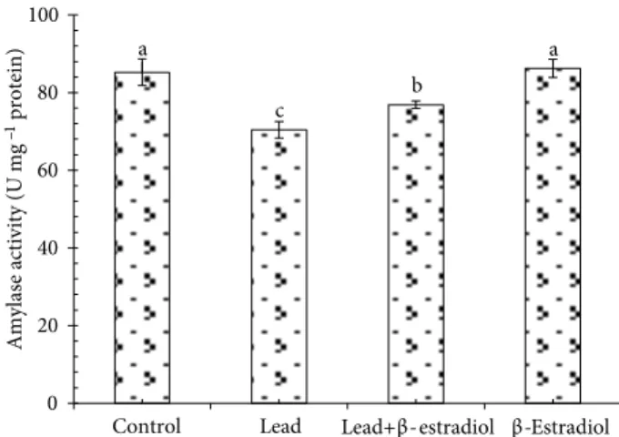
3.2. Effect of β-estradiol on amylase activity in roots of germinating wheat seedlings exposed to lead stress
Heavy metal treatments seriously affect the activities of many enzymes, which are effective in different metabolic processes of plants (Lamhamdi et al., 2011). Changes in activity of amylase, one of the key indicators of reserve mobilization, were examined to determine possible protective mechanisms of β-estradiol in the response of germinating wheat seedlings to lead stress. Amylase is active during germination and allows the mobilization of starch reserves, an essential process for providing metabolic substrates to growing cells (Lamhamdi et al., 2011). In this study, lead reduced amylase activity in germinating wheat seeds (Figure 2). However, the combination of lead and β-estradiol reversed this inhibitory effect on the enzymatic activity. Prior researchers reported that amylase activity was stimulated by β-estradiol and progesterone in germinating maize and chickpea seeds (Erdal and
Dumlupinar, 2010; Erdal et al., 2010). On the other hand, there are many studies reporting inhibitory effects of metals on amylase activity. Karmous et al. (2012) found that cadmium and copper inhibited amylase activity in cotyledons of germinating seeds of numerous legumes. The inhibiting effect of lead on amylase activity might have resulted from the directly toxic effect of lead ions by displacing Ca2+ ions. Because α-amylases are calcium
metalloenzymes, they are completely unable to function in the absence of calcium (Lamhamdi et al., 2011).
3.3. Effect of β-estradiol on lead content in roots and coleoptiles of germinating wheat seedlings exposed to lead stress
The roots and coleoptiles of seedlings grown in unstressed conditions had very low values of lead. In low quantities, the presence of lead in control seedlings might result from contamination caused by agricultural chemicals and the original presence of lead in seeds. When exposed to lead toxicity, roots quickly took up lead, as expected (Figure 3), and lead content highly increased. In the roots of β-estradiol-applied seedlings, lead content was higher than that of seedlings treated with lead alone. Unlike the roots, in coleoptiles of lead-applied seedlings, lead accumulation was not different from that of control seedlings (Figure 3). These data clearly showed that the lead taken up by roots was not transported to coleoptiles to cope with the excessive lead toxicity in the surroundings. Conversely, it accumulated in the roots. The β-estradiol provided further increase in the lead accumulation in the roots. Similar to our data, Verma and Dubey (2003) found that lead was accumulated in roots rather than shoots in rice when plants were exposed to lead stress. These data show that the counteraction strategy of germinating wheat seedlings against lead toxicity was to accumulate the lead in their roots.
b d c a b d c a 0 1 2 3 4 5 6 7 8 0 2 4 6 8 10 12 14 16 18
Control Lead Lead+β-estradiol β-Estradiol
Coleoptile length (cm)
Root length (cm)
root coleoptile
Figure 1. Effects of β-estradiol on root and coleoptile lengths of 5-day-old wheat seedlings exposed to lead toxicity. Different letters in the same group indicate statistically significant differences (P < 0.05). Error bars mean ±SE.
a c b a 0 20 40 60 80 100
Control Lead Lead+β-estradiol β-Estradiol
Amylase activity (U mg
–1protein)
Figure 2. Effects of β-estradiol on α-amylase activity of 5-day-old wheat seedlings exposed to lead toxicity. Different letters indicate statistically significant differences (P < 0.05). Error bars mean ±SE.
3.4. Effect of β-estradiol on soluble protein in roots of germinating wheat seedlings exposed to lead stress
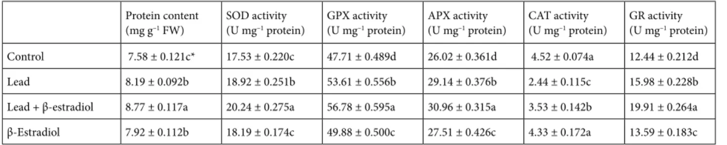
The effects of lead and β-estradiol treatments on soluble protein contents in roots of wheat seedlings are shown in Table 1. Soluble protein contents were significantly in-creased by lead application. These increases might have re-sulted from de novo synthesis of stress proteins provoked by metal exposure (Biteur et al., 2011). In the absence of lead, exogenous β-estradiol application provided a marked increase in the soluble protein contents compared with the control seedlings. Enhanced protein content under unstressed conditions could be linked to acceleration of synthesis reactions needed for growth and development. Similarly, previous studies showed that MSH application increased soluble protein contents of plants under stressed and unstressed conditions. Erdal and Dumlupinar (2011) reported that exogenous MSH application increased the protein content in chickpea. Similarly, Dogra and Kaur
(1994) demonstrated that protein contents of wheat seeds at the germination stage were significantly increased by es-trone, testosterone, and progesterone. In the present study, lead-induced elevation in protein content was significantly increased by the exogenous application of β-estradiol. This increase could be explained by the fact that β-estradiol might have resulted in an increase in both the rate of syn-thesis reactions related to development and synsyn-thesis of stress proteins related to osmoregulation.
3.5. Effects of β-estradiol on antioxidant system and oxidative stress in roots of germinating wheat seedlings exposed to lead stress
To demonstrate the ameliorative role of β-estradiol on lead-induced oxidative damage, changes in enzymatic and nonenzymatic antioxidant systems, membrane damage, and ROS content were investigated under stressed and unstressed conditions. The changes in the capacity of the antioxidant system are the most important responses against oxidative stress in plants (Choudhary et al., 2012). The high capacity of the antioxidant system enhances plant resistance against environmental stresses by protecting the plant cell structures from oxidative damage (Yang et al., 2011). To deal with lead stress, antioxidant enzyme activities may be differentially regulated in plants. The overall effect of the antioxidant system depends on the intracellular balance among antioxidant enzymes rather than a single component (Yang et al., 2010). Recently, it has also been reported that the activities of antioxidant enzymes increased in several higher plants under heavy metal stresses (Biteur et al., 2011; Yang et al., 2011). The data from the present study showed that activities of SOD, GPX, APX, and GR increased markedly in the roots of wheat under lead stress alone compared to control plants, whereas CAT activity was decreased severely by lead application. Moreover, β-estradiol application under lead stress significantly improved activities of SOD (indicating a better O2-∙ scavenging ability), GPX, APX, and GR, and
it considerably reversed the lead-induced reduction in the CAT activity in the roots. Furthermore, β-estradiol
a c a a a b a c 0 10 20 30 40 50 60 70 80
Control Lead Lead+β-estradiol β-Estradiol
Lead content
(ppb
)
coleoptile root
Figure 3. Effects of β-estradiol on lead content in roots and coleoptiles of 5-day-old wheat seedlings exposed to lead toxicity. Different letters in the same group indicate statistically significant differences (P < 0.05). Error bars mean ±SE.
Table 1. Effects of β-estradiol on protein content and activities of SOD, GPX, APX, CAT, and GR in roots of 5-day-old wheat seedlings exposed to lead toxicity.
Protein content
(mg g–1 FW) SOD activity(U mg–1 protein) GPX activity(U mg–1 protein) APX activity(U mg–1 protein) CAT activity(U mg–1 protein) GR activity(U mg–1 protein)
Control 7.58 ± 0.121c* 17.53 ± 0.220c 47.71 ± 0.489d 26.02 ± 0.361d 4.52 ± 0.074a 12.44 ± 0.212d Lead 8.19 ± 0.092b 18.92 ± 0.251b 53.61 ± 0.556b 29.14 ± 0.376b 2.44 ± 0.115c 15.98 ± 0.228b Lead + β-estradiol 8.77 ± 0.117a 20.24 ± 0.275a 56.78 ± 0.595a 30.96 ± 0.315a 3.53 ± 0.142b 19.91 ± 0.264a β-Estradiol 7.92 ± 0.112b 18.19 ± 0.174c 49.88 ± 0.500c 27.51 ± 0.426c 4.33 ± 0.172a 13.59 ± 0.183c * Different letters in the same column indicate statistically significant differences (mean ± SE) (P < 0.05).
application alone as compared to the control was effective on the increase of antioxidant enzyme activities, except that of CAT (Table 1). It is known that CAT, GPX, and APX scavenge excess H2O2 (Racchi, 2013). The high increases in the enzyme activities in response to stress can be explained by possible de novo synthesis of the enzymatic proteins by β-estradiol (Sheng et al., 2007). Increased GPX activity in lead-stressed roots might be due to the increase in the release of peroxidases localized in the cell walls (Lamhamdi et al., 2011) and can serve as an intrinsic defense tool to resist lead-induced oxidative damage (Yang et al., 2011). Enhancements in antioxidant enzyme activities indicate a more powerful defense capacity of plants against lead stress (Biteur et al., 2011). Our results also demonstrated that the antioxidative enzymes can play an important role against oxidative injury induced by lead toxicity. The protective role of β-estradiol against lead stress might be due to the fact that β-estradiol has a signal molecule role upregulating the antioxidant activities and thus it helps indirectly to the scavenge ROS. The high antioxidant activity induced by β-estradiol might provide better oxidative stress management in plants (Choudhary et al., 2012). In parallel to our findings, several previous works reported that MSHs have stimulatory effects on the antioxidant systems of plants. Chaoui and El Ferjani (2014) found that β-estradiol application enhanced antioxidant defense capacity of lentil seedlings. Similarly, Su et al. (2014) reported that progesterone improved antioxidant capacity of wheat under heat and high light stress.
In plants exposed to stress conditions, activities of APX and GR are usually regarded as an indicator of the tolerance of the plant against stress conditions (Bowler et al., 1992). The present study showed that lead stress promoted both total AsA and GSH contents and the APX and GR activities. AsA and GSH also have an important role as substrates in the ascorbate-glutathione cycle (Racchi, 2013). APX needs AsA as a substrate in order to detoxify H2O2 and AsA can be regenerated by using GSH as an electron donor (Carvalho et al., 2015). The other
important enzyme of the ascorbate-glutathione cycle, GR is effective in the reduction of oxidized glutathione to glutathione. Further increases in activities of APX and GR and levels of AsA and GSH were observed in β-estradiol-applied seedlings. Taken together, these findings clearly signify that β-estradiol application activates the ascorbate-glutathione cycle by upregulating the activities of APX and GR and thereby causes the regeneration of total AsA and GSH. The marked activation of the ascorbate-glutathione cycle can be attributed to detoxification of H2O2 caused by high SOD activity (Shahid et al., 2013). AsA and GSH can also directly inhibit the ROS (Agati et al., 2007). Therefore, these substances can contribute to a reduction of the oxidants by directly inhibiting the ROS such as superoxide and hydroxyl.
The O2-∙, H
2O2, and MDA amounts are frequently used
as indicators of oxidative stress degree. Table 2 highlights the changes in these parameters in the roots of wheat affected by treatments of β-estradiol and lead stress. It was determined that lead stress caused a serious elevation in these parameters up to approximately two-fold that of the control groups in radish seedlings (Biteur et al., 2011). The elevation in these parameters clearly showed lead-induced oxidative damage. The elevated levels of O2-∙, H
2O2, and
MDA indicated that antioxidative systems were not able to scavenge the ROS and sustain them at a constant and safe level. Exogenous application of β-estradiol significantly lowered O2-∙, H
2O2, and MDA contents under lead stress.
A lower level of MDA means a lower degree of membrane damage (Wang and Song, 2009). The reductions in O2-∙,
H2O2, and MDA levels could be attributed to β-estradiol-induced high antioxidant capacity. Therefore, it is highly possible that antioxidant systems activated by β-estradiol may be profitable for plants to remove excess ROS and thus inhibit lipid peroxidation.
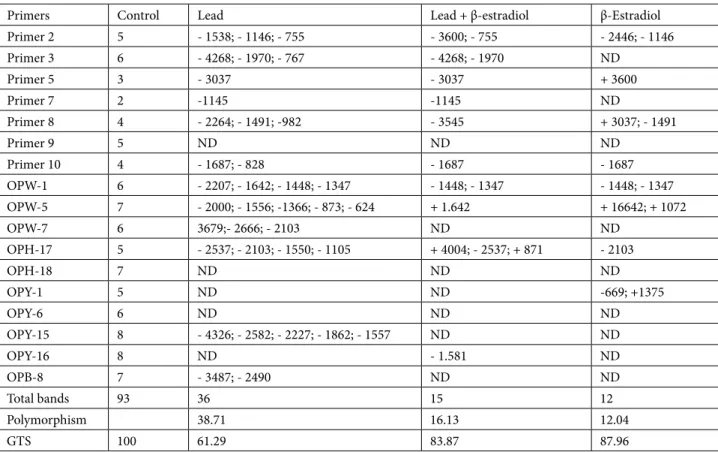
3.6. Effect of lead stress on the DNA profile in roots of germinating wheat seedlings exposed to lead stress
As seen from Table 3, RAPD profiles of the control and experimental groups showed significant differences. Lead Table 2. Effects of β-estradiol on contents of superoxide anion, hydrogen peroxide, malondialdehyde, glutathione, and ascorbate in roots of 5-day-old wheat seedlings exposed to lead toxicity.
Superoxide anion
content (µg g–1 FW) Hydrogen peroxide content (µg g–1 FW) Malondialdehyde content (ng g–1 FW) Total GSHcontent (ng g–1 FW) Total AsA content(ng g–1 FW)
Control 4.22 ± 0.118c* 19.54 ± 0.314c 2.95 ± 0.218c 2.35 ± 0.069c 94.68 ± 1.108d
Lead 5.35 ± 0.128a 42.99 ± 0.490a 6.11 ± 0.205a 2.64 ± 0.032b 116.14 ± 1.360b
Lead + β-estradiol 4.69 ± 0.094b 36.21 ± 0.391b 4.75 ± 0.253b 2.91 ± 0.071a 131.11 ± 1.414a β-Estradiol 4.30 ± 0.135c 19.13 ± 0.291c 2.86 ± 0.197c 2.41 ± 0.066c 99.89 ± 1.275c * Different letters in the same column indicate statistically significant differences (mean ± SE) (P < 0.05).
stress-induced changes were reflected in the disappearance of 36 existing bands as compared to the control. However, β-estradiol application significantly lessened lead-induced changes; β-estradiol-induced changes were the appearance of only 3 new and disappearance of 12 existing bands
compared to the control. The lead + β-estradiol treatment with respect to the control led to the appearance of 4 new and disappearance of 8 existing bands. The polymorphic bands showed variability in each application group in response to the different primers (Figure 4).
Table 3. Effects of β-estradiol on DNA toxicity in roots of 5-day-old wheat seedlings exposed to lead toxicity (disappearance (-), appearance (+) of the bands with molecular sizes (base pairs)).
Primers Control Lead Lead + β-estradiol β-Estradiol
Primer 2 5 - 1538; - 1146; - 755 - 3600; - 755 - 2446; - 1146 Primer 3 6 - 4268; - 1970; - 767 - 4268; - 1970 ND Primer 5 3 - 3037 - 3037 + 3600 Primer 7 2 -1145 -1145 ND Primer 8 4 - 2264; - 1491; -982 - 3545 + 3037; - 1491 Primer 9 5 ND ND ND Primer 10 4 - 1687; - 828 - 1687 - 1687 OPW-1 6 - 2207; - 1642; - 1448; - 1347 - 1448; - 1347 - 1448; - 1347 OPW-5 7 - 2000; - 1556; -1366; - 873; - 624 + 1.642 + 16642; + 1072 OPW-7 6 3679;- 2666; - 2103 ND ND OPH-17 5 - 2537; - 2103; - 1550; - 1105 + 4004; - 2537; + 871 - 2103 OPH-18 7 ND ND ND OPY-1 5 ND ND -669; +1375 OPY-6 6 ND ND ND OPY-15 8 - 4326; - 2582; - 2227; - 1862; - 1557 ND ND OPY-16 8 ND - 1.581 ND OPB-8 7 - 3487; - 2490 ND ND Total bands 93 36 15 12 Polymorphism 38.71 16.13 12.04 GTS 100 61.29 83.87 87.96
Figure 4. RAPD profiles of selected four primers of genomic DNA extracted from roots of germinating wheat seedlings under lead toxicity with and without β-estradiol for 5 days. C: Control; L: lead-applied plants; L + E: lead + β-estradiol-applied plants; E: β-estradiol-applied plants.
The value of genomic template stability (GTS%), which indicates the quantitative changes in RAPD pattern, was 61.29%, 83.87%, and 87.96% for treatment groups with lead stress, β-estradiol, and lead + β-estradiol, respectively. The calculation of GTS% was performed for 17 primers and the results of GTS% are given in Table 3. As indicated by the data, the highest GTS% value was obtained with β-estradiol treatment and the changes in polymorphism rate were small (12.04%). Treatment with β-estradiol against lead stress increased the value of GTS% and led to the approach of this value to the value of the control. This means that β-estradiol reduced DNA damage caused by lead stress in wheat. The results obtained from this study indicate that there was a positive correlation between genomic template stability and other parameters (root and coleoptile growth, ROS contents, and oxidative damage) under lead stress.
Taken together, the results of our study confirmed the positive effects of β-estradiol on mitigation of lead toxicity, as reflected by higher SOD, GPX, APX, and GR activities; total AsA and GSH contents; and lower amounts of oxidative damage indicators (O2-∙, H
2O2, and MDA)
in germinating wheat seedlings. In addition, β-estradiol reduced lead-induced DNA damages. It can be expected that β-estradiol may be used as a useful substance for the reduction of lead toxicity in plants.
Acknowledgment
This work was supported by a grant from the research funds appropriated to Atatürk University, Erzurum, Turkey (2011-107).
References
Agarwal S, Pandey V (2004). Antioxidant enzyme responses to NaCl stress in Cassia angustifoli. Biol Plantarum 48: 555–560. Agati G, Matteini P, Goti A, Tattini M (2007). Chloroplast-located
flavonoids can scavenge singlet oxygen. New Phytol 174: 77– 89.
Ahmad MSA, Ashraf M, Tabassam Q, Hussain M, Firdous H (2011). Lead (Pb)-induced regulation of growth, photosynthesis, and mineral nutrition in maize (Zea mays L.) plants at early growth stages. Biol Trace Elem Res 144: 1229–1239.
Basharat A, Theodore MM, Rafaqat AG, Chong Y, Shafaqat A, Muhammad KD, Yueyan W, Weijun Z (2014). Improvement of element uptake and antioxidative defense in Brassica napus under lead stress by application of hydrogen sulfide. Plant Growth Regul 74: 261–273.
Biteur N, Aoues A, Kharoubi O, Slimani M (2011). Oxidative stress induction by lead in leaves of radish (Raphanus sativus) Seedlings. Not Sci Biol 3: 93–99.
Bowler C, Montagu MV, Inze D (1992). Superoxide dismutase and stress tolerance. Annu Rev Plant Physiol Plant Mol Biol 43: 83–116.
Bozari S, Agar G, Aksakal O, Erturk FA, Yanmis D (2013). Determination of chemical composition and genotoxic effects of essential oil obtained from Nepeta nuda on Zea mays seedlings. Toxicol Ind Health 29: 339–348.
Carvalho LC, Vidigal P, Amâncio S (2015). Oxidative stress homeostasis in grapevine (Vitis vinifera L.). Front Environ Sci 3: 1–15.
Chaoui A, El Ferjani EE (2013). β-estradiol protects embryo growth from heavy-metal toxicity in germinating lentil seeds. J Plant Growth Regul 32: 636–645.
Chaoui A, El Ferjani EE (2014). Heavy metal-induced oxidative damage is reduced by β-estradiol application in lentil seedlings. Plant Growth Regul 74: 1–9.
Choudhary SP, Oral HV, Bhardwaj R, Yu JQ, Tran LSP (2012). Interaction of brassinosteroids and polyamines enhances copper stress tolerance in Raphanus sativus. J Exp Bot 63: 5659–5675.
Chrispeels MJ, Varner JE (1967). Gibberellic acid enhanced synthesis and release of α-amylase and ribonuclease by isolated barley aleurone layers. Plant Physiol 42: 398–406.
Dogra R, Kaur A (1994). Effect of steroids on some growth and biochemical parameters of Triticum aestivum L. during germination. Crop Res 8: 611–620.
Dumlupinar R, Genisel M, Erdal S, Korkut T, Taspinar MS, Taskin M (2011). Effects of progesterone, β-estradiol, and androsterone on the changes of inorganic element content in barley leaves. Biol Trace Elem Res 143: 1740–1745.
Elstner E, Heupel A (1976). Inhibition of nitrite formation from hydroxylammoniumchloride: a simple assay for superoxide dismutase. Anal Biochem 70: 616–620.
Erdal S (2012a). Exogenous mammalian sex hormones mitigate inhibition in growth by enhancing antioxidant activity and synthesis reactions in germinating maize seeds under salt stress. J Sci Food Agr 92: 839–843.
Erdal S (2012b). Alleviation of salt stress in wheat seedlings by mammalian sex hormones. J Sci Food Agric 92: 1411–1416.
Erdal S, Dumlupinar R (2010). Progesterone and β-estradiol stimulate the seed germination in chickpea by causing important changes in biochemical parameters. Z Naturforsch C 65: 239–244.
Erdal S, Dumlupinar R (2011). Mammalian sex hormones stimulate antioxidant system and enhance growth of chickpea plants. Acta Physiol Plant 33: 1011–1017.
Erdal S, Dumlupinar R, Cakmak T, Genisel M (2010). Mammalian sex hormones influence germination velocity and enzyme activities in germinating maize seeds. Fresen Environ Bull 19: 1458–1465.
Fahr M, Laplaze L, Bendaou N, Hocher V, Mzibri ME, Bogusz D, Smouni A (2013). Effect of lead on root growth. Front Plant Sci 4: 175.
Foyer CH, Halliwell B (1976). The presence of glutathione and glutathione reductase in chloroplasts: a proposed role in ascorbic acid metabolism. Planta 133: 21–25.
Genisel M, Turk H, Erdal S (2013). Exogenous progesterone application protects chickpea seedlings against chilling-induced oxidative stress. Acta Physiol Plant 35: 241–251. Havir EA, McHale NA (1987). Biochemical and development
characterization of multiple forms of catalase in tobacco leaves. Plant Physiol 84: 450–455.
Hodges DM, Andrews CJ, Johnson DA, Hamilton RI (1996). Antioxidant compound responses to chilling stress in differentially sensitive inbred maize lines. Plant Physiol 98: 685–692.
Horwitz W, Latimer GW (2005). Official Methods of Analysis of AOAC International. Gaithersburg, MD, USA: AOAC.
Janeczko A, Oklestkova J, Novak O, Sniegowska-Swierk K, Snaczke Z, Pociecha E (2015). Disturbances in production of progesterone and their implications in plant studies. Steroids 96: 153–163. Juliano BO, Varner JE (1969). Enzymatic degradation of starch
granules in the cotyledons of germinating peas. Plant Physiol 44: 886–892.
Karmous I, Jaouani K, Chaoui A, Ferjani EE (2012). Proteolytic activities in Phaseolus vulgaris cotyledons under copper stress. Physiol Mol Biol Plants 18: 337–343.
Krasensky J, Jonak C (2012). Drought, salt, and temperature stress-induced metabolic rearrangements and regulatory networks. J Exp Bot 3: 1593–1608.
Lamhamdi M, Bakrim A, Aarab A, Lafont R, Sayah F (2011). Lead phytotoxicity on wheat (Triticum aestivum L.) seed germination and seedlings growth. C R Biol 334: 118–126.
Li G, Quiros CF (2001). Sequence-related amplified polymorphism (SRAP), a new marker system based on a simple PCR reaction: its application to mapping and gene tagging in Brassica. Theor Appl Genet 103: 455–461.
Nakano Y, Asada K (1981). Hydrogen peroxide is scavenged by ascorbate-specific peroxidase in spinach chloroplasts. Plant Cell Physiol 22: 867–880.
Nguyen LTH, Vandegehuchte MB, van der Geest HG, Janssen CR (2012). Evaluation of the mayfly Ephoron virgo for European sediment toxicity assessment. J Soils Sediments 12: 749–757. Racchi ML (2013). Antioxidant defenses in plants with attention to
Prunus and Citrus spp. Antioxidants 2: 340–369.
Shahid M, Dumat C, Pourrut B, Silvestre J, Laplanche C, Pinelli E (2013). Influence of EDTA and citric acid on lead-induced oxidative stress to Vicia faba roots. J Soils Sediment 14: 835– 843.
Sheng J, Liu K, Fan B, Yuan Y, Shen L, Ru B (2007). Improving zinc content and antioxidant activity in transgenic tomato plants with expression of mouse metallothionein-I by mt-I gene. J Agr Food Chem 55: 9846–9849.
Smith P, Krohn R, Hermanson G, Mallia A, Gartner F, Provenzano M, Fujimoto E, Goeke N, Olson B, Klenk D (1985). Measurement of protein using bicinchoninic acid. Anal Biochem 150: 76–85. Su X, Wu S, Yang L, Xue R, Li H, Wang Y, Zhao H (2014). Exogenous progesterone alleviates heat and high light stress-induced inactivation of photosystem II in wheat by enhancing antioxidant defense and D1 protein stability. Plant Growth Regul 74: 311–318.
Velikova V, Yordanov I, Edreva A (2000). Oxidative stress and some antioxidant systems in acid rain-treated bean plants protective role of exogenous polyamines. Plant Sci 151: 59–66.
Verma S, Dubey RS (2003). Lead toxicity induces lipid peroxidation and alters the activities of antioxidant enzymes in growing rice plants. Plant Sci 164: 645–655.
Wang CQ, Song H (2009). Calcium protects Trifolium repens L. seedlings against cadmium stress. Plant Cell Rep 28: 1341– 1349.
Wang P, Zhangn S, Wang C, Lu J (2012). Effects of Pb on the oxidative stress and antioxidant response in a Pb bio accumulator plant
Vallisneria natans. Ecotox Environ Safe 78: 28–34.
Wu T, Hsu Y, Lee T (2009). Effects of cadmium on the regulation of antioxidant enzyme activity, gene expression, and antioxidant defenses in the marine macroalga Ulva fasciata. Bot Stud 50: 25–34.
Yang XH, Xu ZH, Xue HW (2005). Arabidopsis Membrane Steroid Binding Protein 1 is involved in inhibition of cell elongation. Plant Cell 17: 116–131.
Yang Y, Wei X, Lu J, You J, Wang W, Shi R (2010). Lead-induced phytotoxicity mechanism involved in seed germination and seedling growth of wheat (Triticum aestivum L.). Ecotox Environ Safe 73: 1982–1987.
Yang Y, Zhang Y, Wei X, You J, Wang W, Lu J, Shi R (2011). Comparative antioxidative responses and proline metabolism in two wheat cultivars under short term lead stress. Ecotox Environ Safe 74: 733–740.
Yee Y, Tam N, Wong Y, Lu C (2002). Growth and physiological responses of two mangrove species (Bruguira gymnorrhiza and Kandelia candel) to water logging. Environ Exp Bot 49: 209–221.
Yu C, Xie F, Ma L (2014). Effects of exogenous application of ascorbic acid on genotoxicity of Pb in Vicia faba roots. Int J Agric Biol 16: 831–835.
Zhou F, Wang J, Yang N (2014). Growth responses, antioxidant enzyme activities and lead accumulation of Sophora japonica and Platycladus orientalis seedlings under Pb and water stress. Plant Growth Regul 75: 383–389.