Contribution of Polymorphism in PPARα Untranslated Region to The
Development of Axial Spondyloarthritis
Ekrem AKBULUT1,2,*, Metin ÖZGEN3,4
1Munzur University, Faculty of Health Sciences, Department of Physiotherapy and Rehabilitation, Tunceli, Turkey
2Malatya Turgut Özal University, Faculty of Engineering and Natural Sciences, Department of Bioengineering, Malatya, Turkey
[email protected], ORCID: https: 0000-0002-7526-9835
3İnönü University, Faculty of Medicine, Department of Rheumatology, Malatya, Turkey 4Ondokuz Mayıs University, Faculty of Medicine, Department of Rheumatology, Samsun, Turkey
[email protected], ORCID: 0000-0002-6842-2918
Received: 25.11.2019 Accepted: 18.04.2020 Published: 25.06.2020
Abstract
The study aimed to contribute to the overall understanding of axial spondyloarthritis by investigating the polymorphism in 5’ untranslated region of peroxisome proliferator-activated receptor alpha gene in patients. The study included 194 patients and 197 controls recruited. The DNA obtained from the samples was genotyped by multiplex Polymerase Chain Reaction and then Matrix-Assisted Laser Desorption Ionization- Time of Flight Mass Spectrometry. Data were analyzed by logistic regression. Five polymorphic regions analyzed in this study, three of the sites were associated with disease risk. An allele in the rs1800204 polymorphic region (p < 0.001), C allele in the rs4253657 polymorphic region (p = 0.040) and C allele in the rs13909022 polymorphic region (p = 0.005) were associated with disease risk. The association of disease risk could not be detected with G and A alleles in the rs881740 (p = 0.456) and rs115640476 (p = 0.674) polymorphic regions, respectively. PPARα 5' untranslated region polymorphism, which is
shown to be associated with disease risk, is thought to contribute to the elucidation of the molecular mechanism of the disease. In axial spondyloarthritis, studying the effects of genetic changes in PPARα on other genes with which it interacts will contribute to the full understanding of the molecular mechanism of the disease.
Keywords: Axial spondyloarthritis; Peroxisome proliferator activated receptor alpha;
Genetic predisposition; Disease risk.
PPAR
α Kodlanmayan Bölge Polimorfizminin Aksiyal Spondiloartrit Gelişimine
Katkısı
Öz
Bu çalışma peroksizom çoğaltıcılar tarafından aktive edilen reseptör alfa geninin 5’ ucu kodlanmayan bölge polimorfizminin aksiyel spondiloartrit hastalığının gelişimine olan katkısını araştırmayı ve hastalığın gelişim mekanizmasının aydınlatılmasına katkı sunmayı amaçlamaktadır. Çalışma 194 hasta ve 197 kontrol ile gerçekleştirildi. Örneklerden elde edilen DNA multipleks PCR sonrası matris aracılı lazer dezorpsiyon iyonizasyon uçuş zamanı kütle spektrometresi yöntemi kullanılarak genotipleme işlemi yapıldı. Elde edilen veriler lojistik regresyon ile analiz edildi. Bu çalışma ile analiz edilen beş polimorfik bölgenin üçü hastalık riski ile ilişkilendirildi. rs1800204 polimorfik bölgesinde A alelinin (p < 0.001), rs4253657 için C alelinin (p = 0.040) ve rs13909022 için C alelinin (p = 0.005) hastalık riski ile ilişkili olduğu tespit edildi. rs881740 polimorfik bölgesi için G aleli (p = 0.456) ve rs115640476 için A alelinin (p = 0.674) hastalık riski ile ilişkisi tespit edilemedi. Hastalık riski ile ilişkisi gösterilen PPARα 5’ kodlanmayan bölge polimorfizminin, hastalığın moleküler mekanizmanın aydınlatılmasına katkı sunacağı düşünülmektedir. Poligenik bir hastalık olan ax-SpA’de, PPARα’daki genetik değişikliklerin etkileştiği diğer genler üzerinde ortaya çıkaracağı etkilerin çalışılması hastalığın moleküler mekanizmasının tam olarak aydınlatılmasını sağlayacaktır.
Anahtar Kelimeler: Aksiyal spondiloartrit; Peroksizom çoğaltıcılar tarafından aktive
edilen reseptör alfa; Genetik yatkınlık; Hastalık riski.
1. Introduction
Axial spondyloarthritis (axSpA), a polygenic and chronic inflammatory disease with an unknown pathophysiology, affects the axial skeleton and spine. The most common form of axSpA is ankylosing spondylitis (AS) [1]. More than 7 million people worldwide are estimated to be affected by AS [2]. Peroxisome proliferator-activated receptors (PPARs), known as transcription
factors, are members of the nuclear receptor superfamily involved in the regulation of various essential biological processes. PPAR-alpha (PPARα), a type of PPAR, is a transcription factor that is activated by a fatty acid belonging to a nuclear hormone receptor family [3]. Due to its collective functions in different tissues, PPARα is an important focus of research. Although it is known to play a role in the development of several chronic diseases and in inflammatory processes, their role in the mechanism of disease development has not yet been fully identified. PPARs function as physiological sensors under different stress conditions and are valuable targets for innovative therapies [4]. Single-nucleotide polymorphisms (SNP) that have a high frequency in the human genome are known to provide important data regarding disease susceptibility, risk, etiopathogenesis and prognosis [5-7]. In addition, the gene expression of the sequences expressed as untranslated regions (UTRs) at the 5’ and 3’ ends of the gene regions on the genome reportedly determine the affinity of the mRNA to the ribosome, post-translational modifications, stability of the protein molecule and the stability of the three-dimensional structure [8].
In this study, we evaluated the contribution of polymorphisms in PPARα 5’untranslated region to the development of axSpA.
2. Materials and Methods 2.1. Study groups
This study included 194 ax-SpA patients (141 male and 53 female; mean age 38.10 ± 11.04; range 18 to 65 years) and 197 controls (99 male and 98 female; mean age 39.61 ± 10.34; range 20 to 69 years) recruited from the department of Rheumatology, İnönü University in Malatya, Turkey. The study protocol was approved by the ethics committee of Malatya Clinical Investigations (2016/44). A complete clinical evaluation was performed for all patients. Healthy individuals were selected for control group. Ethical rules were followed in this study and written consent of volunteers was obtained. Data collection form included information such as gender, age, disease duration, familial disease story and several clinical characteristics (Table 1).
2.2. Genomic DNA isolation
DNA isolation from peripheral blood samples was performed using Invisorb Spin Blood Mini Kit™ (Catalog Number: 1031100300). DNA concentration measurement before multiplex PCR was done using Nanodrop™. To ensure successful multiplex PCR, it was ensured that DNA concentrations were at least 20 ng/µl for each sample.
Table 1: Clinical and demographic data
Demographic Data Case
(n=194) Control (n=197)
p-Value Odds Ratio (%95CI) Age (Mean ± SD) 38.10 ± 11.04 39.61 ± 10.34 -
Sex (Male) %72.7 %50.25 2.634(1.728-4.014) <0.001
Disease duration (Year ± SD) 8.57 ± 4.72 - -
Diagnosis delay (Year ± SD) 2.82 ± 1.41 - -
Diagnosis of lumbar hernia before
Ax-SpA (%) 45.36 - -
Familial Ax-SpA
disease story %28.87 -
<0.001 3.266 (1.888-5.649) Surgical intervention before
symptoms occur %10.82 %7.11
0.381 1.393 (0.664-2.922) Suffered severe infectious disease %11.86 %3.05 4.434(1.740-11.300) 0.002
2.3. Genotyping
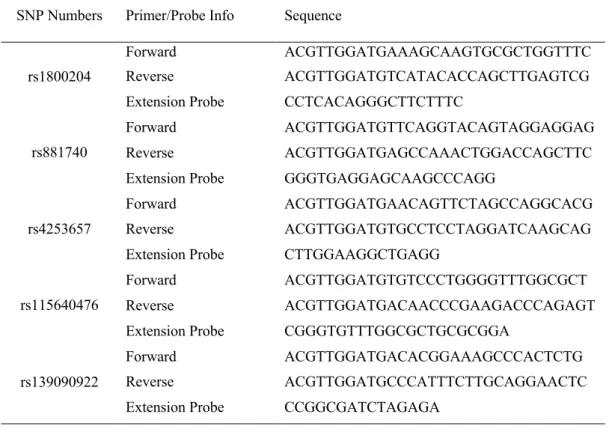
Polymorphic regions were amplified by multiplex PCR using designed primer pairs. The reaction was performed as 45 cycles with pre-denaturation at 95°C for 3 min, denaturation at 95°C for 30 sec, binding at 56°C for 30 sec, elongation at 72°C for 1 min and final elongation at 72°C for 5 min. The total volume of reaction was 5 μl with the 2.5 mM of MgCl2, 2 ng of sample DNA, 1 enzyme unit, 0.1 μM of primer and 500 μM of dNTP as the final concentration in PCR. Next, 0.5 ml of Shrimp alkaline phosphatase was added to the samples for dNTP neutralisation after multiplex PCR, and the samples were then incubated at 37°C for 40 min. The samples were further incubated at 85°C for 5 min for enzyme inactivation. Next, 0.041 μl 1X enzyme, 0.940 μl elongation primer and 0.2 μl termination mixture were added to the samples for the identification of the polymorphic base, and PCR was performed as 42 cycles with pre-denaturation at 95°C for 30 sec, denaturation at 95°C for 5 sec, binding at 52°C for 5 sec, elongation at 80°C for 5 sec and final elongation at 75°C for 2 min. After the single-base elongation reaction, 16 µl of ddH2O was added to each sample. The samples were resin-coated before MALDI TOF analysis to optimise background noise. The samples were transferred to 384-element SpectroCHIP® II, and genotype analysis was performed by MALDI TOF. Primer pairs and probe information used in the single-base elongation reaction and multiplex PCR are presented in Table 2.
Table 2: Sequences of Multiplex PCR primers and single base extension probes
SNP Numbers Primer/Probe Info Sequence
rs1800204
Forward ACGTTGGATGAAAGCAAGTGCGCTGGTTTC Reverse ACGTTGGATGTCATACACCAGCTTGAGTCG Extension Probe CCTCACAGGGCTTCTTTC
rs881740
Forward ACGTTGGATGTTCAGGTACAGTAGGAGGAG
Reverse ACGTTGGATGAGCCAAACTGGACCAGCTTC Extension Probe GGGTGAGGAGCAAGCCCAGG
rs4253657
Forward ACGTTGGATGAACAGTTCTAGCCAGGCACG
Reverse ACGTTGGATGTGCCTCCTAGGATCAAGCAG Extension Probe CTTGGAAGGCTGAGG
rs115640476
Forward ACGTTGGATGTGTCCCTGGGGTTTGGCGCT Reverse ACGTTGGATGACAACCCGAAGACCCAGAGT Extension Probe CGGGTGTTTGGCGCTGCGCGGA
rs139090922
Forward ACGTTGGATGACACGGAAAGCCCACTCTG
Reverse ACGTTGGATGCCCATTTCTTGCAGGAACTC Extension Probe CCGGCGATCTAGAGA
2.4. Statistical analysis
The association of SNP alleles and genotypes with disease predisposition was analysed by logistic regression test (SPSS 24.0, Chicago). Power analysis was performed by using PS Power and Sample Size Program 3.1.2. Hardy-Weinberg equilibrium was evaluated by χ2 analysis. Demographic data were given as mean ± standard deviation and min/max values. “p” value smaller than 0.05 was regarded as statistically significant.
3. Results
Clinical and demographic data of patients with axSpA and healthy subjects were presented in Table 1. Disease diagnosis was delayed by 2.82 years. In this study, 28% of the patients had a family history of similar disease (p < 0.001), 45% of the patients had been diagnosed with lumbar disc herniation before being diagnosed with axSpA. Also 10% of patients had undergone surgery before disease symptoms manifested and 11% of patients had an important infectious disease before disease symptoms were observed (p = 0.002). Disease prevalence was 2.6-fold higher in males than in females [p < 0.001; OR 95% CI: 2.634 (1.728–4.014)] (Table 1).
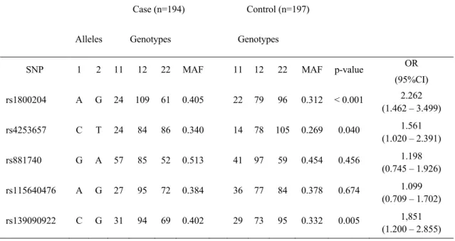
analyzed. Three of them were associated with disease risk. A allele in the rs1800204 polymorphic region was found to be associated with disease risk (p < 0.001) and a 2-fold increase in disease risk was noted [OR 95% CI: 2.262 (1.462–3.499)]; C allele in the rs4253657 polymorphic region was found to be associated with disease risk (p = 0.040) and a 1.5-fold increase in disease risk was noted [OR 95% CI: 1.561 (1.020–2.391)] and C allele in the rs13909022 polymorphic region was found to be associated with disease risk and a 1.8-fold increase in disease risk was noted [OR 95% CI: 1.851 (1.200–2.855)]. The relationship G and A alleles in the rs881740 and rs115640476 polymorphic regions, were not found to be associated with disease risk (p = 0.456 and p = 0.674, respectively).
Table 3: PPAR-α 5’untranslated region genotyping results
Case (n=194) Control (n=197) Alleles Genotypes Genotypes
SNP 1 2 11 12 22 MAF 11 12 22 MAF p-value OR (95%CI) rs1800204 A G 24 109 61 0.405 22 79 96 0.312 < 0.001 2.262 (1.462 – 3.499) rs4253657 C T 24 84 86 0.340 14 78 105 0.269 0.040 1.561 (1.020 – 2.391) rs881740 G A 57 85 52 0.513 41 97 59 0.454 0.456 1.198 (0.745 – 1.926) rs115640476 A G 27 95 72 0.384 36 77 84 0.378 0.674 1.099 (0.709 – 1.702) rs139090922 C G 31 94 69 0.402 29 73 95 0.332 0.005 1,851 (1.200 – 2.855) 4. Discussion
axSpA is an inflammatory disease which caused deformation of axial skeleton and consequent movement restriction and labor loss [9-11]. One of the major problems associated with axSpA is the 2 to 5year delay in its diagnosis [12, 13]. Our study is the first in literature to associate polymorphism in PPARα with disease risk and to identify the polymorphism as an alternative genetic target for early diagnosis, elucidation of molecular mechanisms of the disease and disease risk in patients with axSpA. This study showed that prevalence of disease in males was 2.6-fold higher than in females [p < 0.001; OR 95% CI: 2.634 (1.728–4.014)]. This result was consistent with the data obtained in a prevalence study conducted by Esther [14]. The mean delay of 2.82 ± 1.41 years in disease diagnosis observed in our study was showed similarity with
infectious diseases before disease symptoms had manifested [p = 0.002; OR 95% CI: 4.434 (1.740–11.300)]. This finding supports the previously reported statement that microorganism activity may have a role in disease development [16, 17]. 28.87% of the patients had a family history of axSpA [p < 0.001; OR 95% CI: 3.266 (1.888–5.649)] and the results were consistent with the data obtained by Serrano (38.4%) and Wang (36%) regarding the ethnic and community parameters [18, 19].
Although no study has yet elucidated the role of PPARα in axSpA development, several scientific studies have reported that PPARα is associated with the development of different diseases [20-22]. Dhaini et al. state that PPAR polymorphism may be related to the risk of developing cancer [23]. Dong et al. reported that PPAR polymorphism may have a role in the development of diseases such as Type 2 diabetes and metabolic syndrome [24]. Similarly, Andrulionytė et al. reported that the rs1800206 polymorphism increased the risk of developing diabetes [25]. In another study, Qian et al. analyzed the effect of PPARα and other members of the PPAR superfamily on the risk of developing coronary heart disease. In one of the studies, PPARα intron polymorphism (7G/C polymorphism [CC+GC vs GG; p = 0.003; OR 95% CI: 1.42 (1.13–1.78)] and L162V polymorphism [VV+LV vs LL; p = 0.031; OR 95% CI: 0.74 (0.56– 0.97)] were found to be associated with the risk of developing coronary heart disease [26]. Fan et al. found that PPARα polymorphism increases the risk of developing atherosclerosis and cardiovascular disease [27].
The human PPARα gene is a 93.2-Kb gene located on chromosome 22 at position 22q12-q13.1 encoding a protein of 468 amino acids [28]. The gene contains a total of 8 exons. The 5' UTR, including the polymorphic regions that are the focus of our study, is encoded by exons 1 and 2 and partially by exon 3 [28, 29]. PPARα has an important role in reducing inflammation. PPARα activity has an effect on neutrophils and macrophages in both acute and chronic inflammatory diseases. Leukotriene B4 (LTB4), a potent chemotactic inflammatory eicosanoid, is an endogenous PPARα ligand. Like other PPARα ligands, it induces the transcription of the genes of β- and PP-oxidation pathways that neutralize and reduce LTB4, to regulate inflammatory response [30]. PPARα regulates the duration of the inflammatory response by limiting cytokine expression and inducing genes that metabolize LTB4 [30].
5. Conclusions
The data obtained from the present study showed that the rs1800204, rs115640476 and rs139090922 polymorphic regions located in 5’ UTR of PPARα, which are responsible for suppressing inflammatory response, are increased axSpA risk. Evaluation of results suggests that
changes in 5’ UTR can downregulate PPARα expression. This may suppress LTB4-metabolising genes leading to increased LTB4 activity, which ultimately contributes to the prolonged duration of the inflammatory response. The genes that are involved in the inflammatory response and interact with PPARα may offer significant opportunities to completely understand the disease mechanisms and to determine appropriate treatment strategies.
Acknowledgement
This study was supported financially by Munzur University Health Faculty Research Budget (2017-DT).
Ethical Considerations
Ethical rules were followed in this study and written consent of volunteers was obtained. The study protocol was approved by the ethics committee of Malatya Clinical Investigations (2016/44).
Conflict of Interest
There is no conflict of interest among the authors.
References
[1] Sharma, S.M., Choi, D., Planck, S.R., Harrington, C.A., Austin, C.R., Lewis, J.A., et al., Insights in to the pathogenesis of axial spondyloarthropathy based on gene expression
profiles, Arthritis Research & Therapy, 11(6), R168, 1-9, 2009.
[2] Dean, L.E., Jones, G.T., MacDonald, A.G., Downham, C., Sturrock, R.D., Macfarlane, G. J., Global prevalence of ankylosing spondylitis, Rheumatology, 53, 650-657, 2014.
[3] Dubrac, S., Schmuth, M., PPAR-alpha in cutaneous inflammation, Dermato-Endocrinology, 3, 23-26, 2011.
[4] Yessoufou, A., Wahli, W., Multifaceted roles of peroxisome proliferator-activated
receptors (PPARs) at the cellular and whole organism levels, Swiss Med Wkly, 140, w13071,
2010.
[5] Ruan, W.F., Xie, J.T., Jin, Q., Wang, W.D., Ping, A.S., The diagnostic and prognostic
role of interleukin 12B and Interleukin 6R gene polymorphism in patients with ankylosing spondylitis, JCR: Journal of Clinical Rheumatology, 24,18-24, 2018.
[6] Wordsworth, B.P., Cohen, C.J., Vecellio, M., Quantifying the genetic risk for the
development of axial spondyloarthropathy: could this become a diagnostic tool?, Current Opinion
[7] Fabris, M., Quartuccio, L., Fabro, C., Sacco, S., Lombardi, S., Ramonda, R., et al.,
The-308 TNFα and the-174 IL-6 promoter polymorphisms associate with effective anti-TNFα treatment in seronegative spondyloarthritis, The Pharmacogenomics Journal, 16, 238, 2016.
[8] Chatterjee, S., Pal, J.K., Role of 5′‐and 3′‐untranslated regions of mRNAs in human
diseases, Biology of The Cell, 101, 251-262, 2009.
[9] Sieper, J., Poddubnyy, D., Axial spondyloarthritis, The Lancet, 390, 73-84, 2017.
[10] Barlow, J., Wright, C., Williams, B., Keat, A., Work disability among people with
ankylosing spondylitis, Arthritis Care & Research: Official Journal of The American College of
Rheumatology, 45, 424-429, 2001.
[11] Ramonda, R., Marchesoni, A., Carletto, A., Bianchi, G., Cutolo, M., Ferraccioli, G., et al., Patient-reported impact of spondyloarthritis on work disability and working life: the
ATLANTIS survey, Arthritis Research & Therapy, 18, 78, 2016.
[12] Behar, V.M., Dougados, M., Etcheto, A., Kreis, S., Fabre, S., Hudry, C., et al.,
Diagnostic delay in axial spondyloarthritis: A cross-sectional study of 432 patients, Joint Bone
Spine, 84, 467-471, 2017.
[13] Redeker, I., Callhoff, J., Hoffmann, F., Haibel, H., Sieper, J., Zink, A., et al., Determinants of diagnostic delay in axial spondyloarthritis: an analysis based on linked claims and patient-reported survey data, Rheumatology, 58(9), 1634-1638, 2019.
[14] Yi, E., Ahuja, A., Rajput, T., George, A.T., Park, Y., Clinical economic and
humanistic burden associated with delayed diagnosis of Axial Spondyloarthritis: A Systematic Review, Rheumatology Therapy, 7, 65–87, 2020.
[15] Nie, A., Wang, C., Song, Y., Xie, X., Yang, H., Chen, H., Prevalence and factors
associated with disturbed sleep in outpatients with ankylosing spondylitis, Clinical
Rheumatology, 37, 2161-2168, 2018.
[16] Zhang, L., Zhang, Y.J., Chen, J., Huang, X.L., Fang, G.S., Yang, L.J., et al., The
association of HLA-B27 and Klebsiella pneumoniae in ankylosing spondylitis: A systematic review, Microbial Pathogenesis, 117, 49-54, 2018.
[17] Braun, J., Sieper, J., Ankylosing spondylitis, The Lancet, 369, 1379-90, 2007.
[18] Serrano, P., Navarro-Compán, V., Rodríguez, S., Fernández, M., Zarco, P., de Miguel, E., SAT0419 Similarities and differences between HLA B27 positive and HLA B27 negative
spondyloarthritis: results from the esperanza cohort, Annals of The Rheumatic Disease, 76(2),
930, 2017.
[19] Wang, R., Gabriel, S.E., Ward, M.M., Progression of nonradiographic axial
spondyloarthritis to ankylosing spondylitis: a population‐based cohort study, Arthritis &
Rheumatology, 68, 1415-1421, 2016.
[20] Ruscica, M., Busnelli, M., Runfola, E., Corsini, A., Sirtori, C.R., Impact of
PPAR-alpha polymorphisms-the case of metabolic disorders and atherosclerosis, Int. J. Mol. Sci.,
[21] Sergeeva, E.G., Berkovich, O.A., Ionova, Z.I., Zaraisky, M.I., Baranova, E.I., L162v
Polymorphism of Par-Α gene, A603g polymorphism of tissue factor gene and risk of coronary heart disease in Russian population, Journal of Bioinformatics and Diabetes, 1(4), 1-11, 2019.
[22] Rashid, A., Jafar, S., Yaqub, R.K., Role of peroxisome proliferator-activated receptor
(PPAR)-α gene in Dyslipidemia, Rawal Medical Journal, 45(1), 54-57, 2020.
[23] Dhaini, H.R., Daher, Z., Genetic polymorphisms of PPAR genes and human cancers:
evidence for gene–environment interactions, Journal of Environmental Science and Health, 37(3),
146-179, 2019.
[24] Dong, C., Zhou, H., Shen, C., Yu, L.G., Ding, Y., Zhang, Y.H., et al., Role of
peroxisome proliferator-activated receptors gene polymorphisms in type 2 diabetes and metabolic syndrome, World Journal of Diabetes, 6(4), 654-661, 2015.
[25] Andrulionytė, L., Kuulasmaa, T., Chiasson, J.L., Laakso, M., Single Nucleotide
Polymorphisms of the Peroxisome Proliferator–Activated Receptor-α Gene (PPARA) Influence the Conversion from Impaired Glucose Tolerance to Type 2 Diabetes: The STOP-NIDDM Trial,
Diabetes, 56, 1181-1186, 2007.
[26] Qian, Y., Li, P., Zhang, J., Shi, Y., Chen, K., Yang, J., et al., Association between
peroxisome proliferator-activated receptor-alpha, delta, and gamma polymorphisms and risk of coronary heart disease: A case–control study and meta-analysis, Medicine, 95(32), e4299, 1-9,
2016.
[27] Fan, W., Shen, C., Wu, M., Zhou, Z.Y., Guo, Z.R., Association and interaction of
PPARα, δ, and γ gene polymorphisms with low-density lipoprotein-cholesterol in a Chinese Han population, Genetic Testing and Molecular Biomarkers, 19, 379-386, 2015.
[28] Sher, T., Yi, H.F., McBride, O.W., Gonzalez, F.J., cDNA cloning chromosomal
mapping and functional characterization of the human peroxisome proliferator activated receptor, Biochemistry, 32, 5598-5604, 1993.
[29] Desvergne, B., Wahli, W., Peroxisome proliferator-activated receptors: nuclear
control of metabolism, Endocrine Reviews, 20, 649-688, 1999.
[30] Devchand, P.R., Keller, H., Peters, J.M., Vazquez, M., Gonzalez, F.J., Wahli, W., The