CLINICAL STUDY
Mesenchymal stem cells treatment in COVID-19 patient with
multi-organ involvement
Yilmaz R
1, Adas G
2,3, Cukurova Z
1, Kart Yasar K
4, Isiksacan N
5, Oztel ON
6, Karaoz E
6,7,8Department of Biochemistry and Immunology, Bakirkoy Dr. Sadi Konuk Education and Research Hospital, Health Science University, Istanbul, Turkey. [email protected]
ABSTRACT
The aim of this study is to evaluate the therapeutic effect of mesenchymal stem cells (MSCs) in a severe case of brain and multiple organ involvement in a patient with COVID-19. Here, a 51-year-old male patient with multi-organ involvement due to COVID-19 infection and developing cardiac arrest is presented. MSCs were transplanted to the patient four times systematically and once intrathecally. As a result, the application of MSCs has been found to have a healing effect on organs in this patient with severe COVID-19 infection. In addition, transplantation of MSCs both systematically and intrathecally is considered to be effective in the treatment of the central nervous system (Tab. 2, Fig. 2, Ref. 24). Text in PDF www.elis.sk
KEY WORDS: mesenchymal stem cell, COVID-19, organ involvement.
1Department of Anesthesia and Intensive Care, Bakirkoy Dr. Sadi Konuk Edu-cation and Research Hospital, Health Science University, Istanbul, Turkey, 2Department of Surgery, Bakirkoy Dr. Sadi Konuk Education and Research Hospital, Health Science University, Istanbul, Turkey, 3Stem Cell and Gene Therapies Center, Health Science University, Istanbul, Turkey, 4Department of Infectious Diseases, Bakirkoy Dr. Sadi Konuk Education and Research Hospital, Health Science University, Istanbul, Turkey, 5Department of Bio-chemistry and Immunology, Bakirkoy Dr. Sadi Konuk Education and Re-search Hospital, Health Science University, Istanbul, Turkey, 6Liv Hospital, Center for Regenerative Medicine and Stem Cell Manufacturing (LivMed-Cell), İstanbul, Turkey, 7Faculty of Medicine, Department of Histology & Em-bryology, Istinye University, Istanbul, Turkey, and 8Center for Stem Cell and Tissue Engineering Research & Practice, Istinye University, Istanbul, Turkey
Address for correspondence: N. Isiksacan, Department of Biochemistry
and Immunology, Bakirkoy Dr. Sadi Konuk Education and Research Hospital, Zuhuratbaba Mahallesi, Tevfi k Saglam Caddesi, No: 11, 34000, Istanbul, Turkey.
Introduction
Clinical symptoms of COVID-19 infection include fever, cough, and myalgia or fatigue with pneumonia demonstrated on chest CT scan imaging (1). The clinical situation of patients varies from mild fever to acute respiratory distress syndrome (ARDS) and death (2, 3). Patients with severe disease were likely to dis-play neurologic symptoms such as acute cerebrovascular diseases, impaired consciousness, loss of smell and taste, and skeletal mus-cle injury (2, 4).
Mesenchymal stem cells (MSCs) have been widely used in cell-based therapy, from basic research to clinical trials (5, 6, 7). Wharton’s jelly mesenchymal stem cells (WJ-MSCs) have dis-tinct advantages of being abundant, easy to obtain with minimal invasiveness, and readily cultured in a suffi cient number for
trans-plantation without ethical issues of allografting (8). The aim of the case report is to determine WJ-MSCs treatment’s effi cacy in a severe case of multiple organ failure and brain involvement in a patient with COVID-19 infection. In addition, another aim of the study is to get to understand the way in which MSCs are given, especially in patients with brain involvement.
Case report
This study was undertaken in the Bakirkoy Dr. Sadi Konuk Education and Training Hospital at Health Science University and Istinye University with the permission of the Ministry of Health. The study protocol was approved by the Ethical Committee. The informed consent was obtained from the patient’s relatives.
A 51-year-old male patient started to complain of cough, myalgia, high fever (39.5 °C), and diarrhea on March 11, 2020. Upon the progression of symptoms, the patient was hospitalized in the infection service on March 16. Thorax tomography was performed and throat swab was taken. Radiological involvement related to bilateral COVID-19 infection was observed in both lungs (Fig. 1A). The patient had no previous chronic illnesses or addi-tional pathologies in his medical records. A swab was taken, and the diagnosis of COVID-19 was made using the PCR method. A supportive therapy was initiated. Upon progression of respiratory distress during his daily follow-up, the patient was transferred to the intensive care unit (ICU).
During the ICU follow-up, the patient was intubated oro-tra-cheally due to high fever; DSS: 33-40 breaths per minute (bpm), low O2 saturation, and gradually increasing respiratory distress. Due to hypoxemia in arterial blood gas (ABG) values and dete-rioration in P/F (Horowitz) values (< 150), the patient was placed in a prone position immediately after intubation.
Methylpredniso-lone was added to the treatment of the patient for 5 days. In the prone position, CPR was applied to the patient who developed sudden cardiac arrest for 10 minutes. Two hours after the arrest, a targeted temperature management was started. Body temperature was adjusted to 33 degrees. It was sedated to be between 20 and 40 according to patient state index. The antiviral treatment of the
patient was changed to Avigan. The patient was thought to have a cytokine storm. On March 25, the patient was treated with tocili-zumab (IL-6 antagonist) for 2 days.
Echocardiography performed after cardiac arrest revealed a global dyskinesia compatible with myocarditis, and ejection frac-tion (EF) of 25 % with severe apical akinesia. The patient
deve-B1 B2 B3
A1 A2 A3
C1 C2 C3
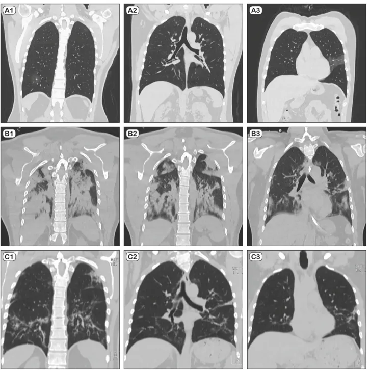
Fig. 1. Radiological evaluation of patient lungs. A1‒3: focal ground-glass opacity in the lower part of the lung observed on day 5 after fi rst symptom onset. B 1‒3:on day 10 day after fi rst symptom onset, it is observed that lesions increase in both upper and lower lung lobes. It is seen that the lesions are converted into consolidation form and merge. C 1‒3: on day 25 after fi rst symptom onset, interlobular and intralobular septal thickening is observed in parenchyma of both lungs. In his previous fi lm, the common consolidation areas and patched ground-glass densities are observed to be resorbed.
loped a severe bilateral pneumonia (Fig. 1B), ARDS, and multiple organ failure, while no response was received.
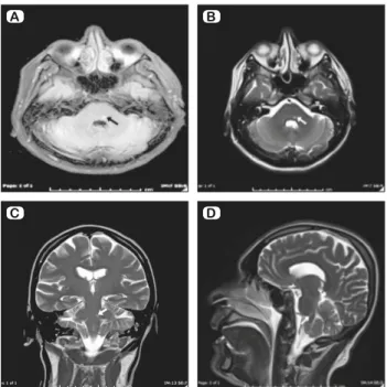
The patient did not wake up and did not show any clinical im-provement, even though his medical sedations were closed after the entire treatment period. On monitorization, delta waves were observed, and contrast enhanced cranial MR and CT were taken. Hyperintense signal change in T2A-FLAIR sequence in pons was observed in cranial MR (Fig. 2). After the general condition of the patient had not improved as a result of treatment, it was decided by the medical committee to perform MSC transplantation. WJ-MSCs were transplanted to the patient four times intravenously (IV). No side effects were observed in the patient. After stem cell transplantation, the patient underwent control echocardiography. Cardiac activity and myocardial structure were found to be nor-mal. EF was evaluated as 60 %.
On April 07, 2020, the patient was extubated with high-fl ow nasal cannula. Upon neurological examination, the patient was observed to be orientated and cooperative. After 5 days of fol-low-up, the patient was taken to medical service. The patient was diagnosed with upper gastrointestinal bleeding. After the patient’s anti-thromboembolic treatment had been discontinued, his vital signs were stable, and it was decided to follow him up at the clinic. The patient was discharged from hospital on April 20, 2020 as symptom-free and with no complaints. Information about the patient’s laboratory examination results are given in Table 1. Drug treatment of the patient was made on the basis of inter-national literature and guideline prepared by the scientifi c com-mittee related to this issue and affi liated to the ministry of health. The MSCs were slowly drawn into the syringe without pressure,
suspended in 250 ml of 0.9 % NaCl, and then given intravenously over 1 hour (Tab. 2).
The levels of alanine transaminase, aspartate transaminase, total protein, albumin, total bilirubin, direct bilirubin, ferritin, triglycerides, D-dimer, troponin I, myoglobin, procalcitonin, am-monia, c-reactive protein, pro B-type natriuretic peptide, creatine kinase, and alkaline phosphatase were determined in venous blood samples using Beckman Coulter AU5800 analyzer (Beckman Coulter, Brea, CA, USA). The complete blood count was analyzed with ADVIA 2120i autoanalyzer (Siemens Healthcare Diagnostics, Erlangen, Germany).
For coagulation assay, all analytical procedures were carried out on a random-access coagulation analyzer (Beijing Succeeder Technology Inc. China).
Flow cytometric analyses were performed with Navios cyto-meter (BECLS)-Kaluza Software. Whole blood was stained with anti-human FITC-CD45, PE-CD4, ECD-CD8, PE-CY5.5-CD3 (Beckman Coulter, Brea, California) antibodies.
All samples of WJ-MSCs as cell therapy medicinal products were isolated, expanded, and analyzed in cGMP-certifi ed facility at Liv Hospital Center for Regenerative Medicine and Stem Cell Manufacturing (LivMedCell). Human WJ-MSCs were prepared and tested as described in our previous clinical trials (8, 9, 10).
Cryopreserved vials from each donor were thawed and mixed in the same tubes before seeded at a cell density of 4,000 cells/cm2.
After harvest at the fourth passage, quality control tests were per-formed, such as fl ow cytometry analysis, endotoxin, rapid microbio-logical and sterility tests. The fi nal product was prepared as 3×10⁶
cells/kg/dose of allogeneic WJ-MSCs pooled from three donors.
Discussion
The central nervous system (CNS) is not immune to alterations that lead to neurological disease resulting from acute, persistent or latent viral infections (11). In some circumstances, opportunis-tic viral pathogens such as human corona viruses can avoid the immune response and cause more severe respiratory diseases or even spread to other tissues including the CNS (12). The detec-tion of HCoV-RNA in human brain samples clearly demonstrates that these respiratory pathogens are naturally neuroinvasive in humans, and suggests that they establish a persistent infection in human CNS (13). On March 4, 2020, researchers from Beijing Ditan Hospital, China, fi rst described a confi rmed patient with 2019-nCoV, whose cerebrospinal fl uid (CSF) tested positive for 2019-nCoV by gene sequencing, suggesting a need to consider direct infection when patients with 2019-nCoV present with neu-rological disorders (14).
In this study, MSCs transplantation was successfully per-formed both systemically and intrathecally. Here, after the fi rst 2 transplants had been given systematically, the patient was awake-ned and extubated. Later, the patient who developed neurologi-cal symptoms was reintubated. After that, the transplantation of MSCs was performed systematically for the third time, as well as an intrathecal stem cell transplantation. We thought the blood brain barrier (BBB) was an obstacle here. Neurologically, the
dia-A B
C D
Fig. 2. Radiological evaluation of the patient brain. A-D Focal inten-sity increased in T2-weighted levels in the posterior side of the pons. The lesion is marked with an arrow.
gnosis of brain involvement was demonstrated by MR and proved by COVID-19 antibodies in the spinal fl uid. Upon examination of CSF samples, protein and IgG levels were eleva-ted while albumin levels were found low. The CSF result is compatible with previous SARS-CoV-2 infection studies (15). The patient’s vital signs were improved, especially after intrathe-cal and systemic MSCs transplantations. After the patient had been extubated, his neurological symptoms regressed, consciousness restored, and he could speak. Most neurologic manifes-tations occurred early in the illness (the median time to hospital admission was 1-2 days) (16). In our patient, the time period between hos-pitalization and determining the neurological symptoms was approximately 11 days.
After transplanting MSCs through intrave-nous infusion, a part of the MSCs accumulate in the lung, which could potentially improve the pulmonary microenvironment, protect alveolar epithelial cells, prevent pulmonary fi brosis, and improve the lung function (2, 6, 14). They promote tissue and organ regeneration via se-creting a variety of paracrine factors, confer-ring anti-infl ammatory, immunomodulatory, angiogenic, antifi brotic, and structural repara-tive properties (17, 18). BM-MSCs signifi cant-ly improved the effi ciency of cardiomyoge-nesis and cardiac function (19, 20). When the treatment of patients with organ failure due to COVID-19 infection is examined, no treat-ment can be seen to improve the organ dam-age. Our patient had cardiac involvement due to COVID-19 infection. In our patient who un-derwent echocardiography after cardiac arrest, the ejection fraction was very low (25 %). In echocardiography performed after systematic transplantation of MSCs, this rate increased to the level of 60 %. It is our opinion that the transplantation of MSCs has a healing effect on the heart. After MSCs transplantation had been performed for the fourth time, the patient’s heart functions have returned to normal. Here, we think that MSCs have a positive and accele-rating effect upon other healing mechanisms of the body.
MSCs engraftment was observed in the injured lung and engraftment rates increased with the extent of tissue injury (6). In a study by Leng Z et al, 7 patients with COVID-19 infec-tions were given MSCs systematically, which had a healing effect (20).When the thorax CTs of the patient were examined in our case study, it was observed that the upper and lower lobes
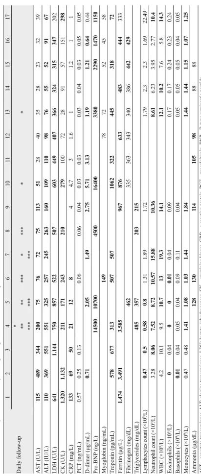
Daily follow-up 1 2 3 4 5 6 7 8 9 10 11 12 13 14 15 16 17 * ** *** ** *** *** * *** *** * * AST (U/L) 11 5 489 344 200 75 76 72 75 11 3 51 28 40 35 28 23 32 39 AL T (IU/L) 11 0 369 551 551 325 257 245 263 160 109 11 0 98 76 55 52 91 67 LDH (U/L) 641 1.144 750 857 522 507 603 449 407 366 324 315 347 202 CK (U/L) 1.320 1.132 21 1 171 243 210 279 100 72 28 91 57 151 298 CRP (mg/L) 133 69 50 21 12 8 4 4.7 3 1.6 1 1.2 1 1 PCT (ng/mL) 0.57 0.25 0.13 0.06 0.06 0.04 0.03 0.03 0.03 0.04 0.03 0.05 0.05 D-dimer ( μg/mL) 0.71 2.05 1.49 2.75 5.71 3.13 1.19 1.21 0.64 0.44 Pro-BNP (ng/L) 14500 10700 4500 16400 3380 2290 1470 1150 Myoglobin (ng/mL) 149 78 72 52 45 58 T roponin (pg/mL) 578 677 313 507 507 1062 322 445 318 72 Ferritin ( μg/L) 1.474 3.491 3.585 967 876 633 483 444 333 Fibrinogen (mg/dL) 462 335 363 343 340 386 442 429 T riglycerides (mg/dL) 485 357 203 215 Lymphocytes count (×10 9/L) 0.47 0.5 0.58 0.8 1.31 1.89 1.72 1.79 2.3 2.3 1.69 22.49 Neutrophil count (×10 9/L) 3.28 8.06 7.52 8.72 10.57 15.88 10.36 8.61 6.23 3.95 2.77 10.4 WBC (×10 9/L ) 4.2 10.1 9.5 10.7 13 19.3 14.1 12.1 10.2 7.6 5.8 14.3 Eosinophils (×10 9/L) 0 0.04 0 0 0.01 0.04 0.09 0.17 0.17 0.24 0.23 0.24 Basophils (×10 9/L) 0.01 0.04 0.05 0.04 0.09 0.1 1 0.04 0.05 0.05 0.05 0.04 0.05 Monocytes (×10 9/L ) 0.47 0.48 1.41 1.08 1.03 1.44 1.84 1.44 1.44 1.15 1.07 1.25 Ammonia ( μg/dL) 128 130 11 4 105 98 88 88 AST ‒ aspartate transaminase, AL T
: alanine transaminase, LDH: lactate dehydrogenase, CK
‒ creatinine kinase, CRP ‒ c-reactive protein, PCT ‒ procalcitonin, BNP ‒
B-type natriuretic peptide
T
ab. 1.
T
reatment days and laboratory
fi
ndings ar
e given; * stem cell transplantation, ** tocilizumab administration, *** ster
oid tr
of both lungs were commonly held, especially in the second tho-rax CT. In our patient, the lesions seen in both lungs had ground-glass appearance, and areas of consolidation were compatible with COVID-19 infection (21, 22). After the MSC transplantation for the last time in our patient, bilateral lung symptoms regressed on control thorax CT. After the patient had been discharged, there was no problem in his medical checkups.
After the fi rst MSC transplantation in our patient, the values of AST, ALT, LDH, CK, pro-bnp, ferritin, triglyceride, fi brinogen, ammonia, and myoglobin began to decrease. The second time the MSCs had been given, CRP reached normal values (Tab. 1). We thought that MSCs were related to the immunomodulatory ef-fect on cytokine storm. On day 2 after MSC transplantation, the lymphocyte count reached the normal level. What is important here, is that reaching the effi cacy of lymphocyte count on day 2 was achieved by the administration of MSCs and tocilizumab at the same time. In the literature, it is seen that the normal level of the number of lymphocytes was reached on day 5 in the study of Xiaoling Xu et al (23). In our study, the increase in lymphocytes was thought to be related to MSCs transplantation. It was observed that the number of TH-2 cells increased, and the number of TH-1 cells decreased in the immune modulation after MSC transplan-tation. After the fi rst MSC transplant, the proportions of CD4+T cells and CD8+T cells were 66 % and 26.7 %, respectively. After 3 MSC transplantation, the proportion of CD4+T cells was 42.9 % and 39.1 %, while that of CD8+T cells was 18.7 % and 22 %, re-spectively. After the second and third MSC transplantations, it was found that the proportion of CD4+T cells was 42.9 % and 39.1 % while that of CD8+ cells was 18.7 % and 22 %, respectively. In previous studies, the decrease in T lymphocytes has been shown to be due to co-death in T cells infected with the virus (6, 24).
Learning points
The transplantations of MSCs, both systematically and in-trathecally, were effective in the treatment of the central nervous system. This activity is related to the fact that MSCs administered intrathecally can easily cross the blood brain barrier. Combining both ways of MSC treatment in multi-organ and brain involvement
due to COVID-19 infection will increase the effectiveness of treatment.
References
1. Kruse LR. Therapeutic strategies in an
out-break scenario to treat the novel coronavirus orig-inating in Wuhan, China (version 2; peer review: 2 approved) Research 2020.
2. Sun T, Guan J. Novel coronavirus and
cen-tral nervous system. Eur J Neurol 2020. DOI: 10.1111/ene.14227.
3. Ozturk S, Elcin EA, Elcin MY. Mesenchymal
stem cells for coronavirus (COVID-19) induced pneumonia: Revisiting the paracrine hypothesis with new hopes. Aging Dis 2020. DOI: 10.14336/ AD.2020.0403.
4. Li YC, Bai WZ, Hashikawa T. Response to Commentary on “The
neu-roinvasive potential of SARS-CoV-2 may play a role in the respiratory fail-ure of COVID-19 patients”. J Med Virol 2020. DOI: 10.1002/jmv.25824.
5. Atluri S, Manchikanti L, Hirsch AJ. Expanded umblical cord
mes-enchymal stem cells (UC-MSCs) as a therapeutic strategy in managing critically ill COVID-19 patients: the case for compassionate use. Pain Physician 2020: 71‒83.
6. Behnke J, Kremer S, Shahzad T et al. MSC based therapies-new
per-spectives for the injured lung. J Clin Med 2020; 9 (3): 682. https://DOI. org/10.3390/jcm9030682.
7. Kabatas S, Demir CS, Civelek E, Yilmaz I, Kircelli A, Yilmaz C, Akyuva Y, Karaoz E. Neuronal regeneration in injured rat spinal cord
after human dental pulp derived neural crest stem cell transplantation. Bratisl Lek Listy 2018; 119 (3): 143‒151. DOI: 10.4149/BLL_2018_028.
8. Kabataş S, Civelek E, Inci C, Yalçınkaya EY, Gunel G, Kır G, Al-bayrak E, Ozturk E, Adas G, Karaoz E. Wharton’s Jelly-Derived
Mes-enchymal Stem Cell Transplantation in a Patient with Hypoxic-Ischemic Encephalopathy: A Pilot Study. Cell Transplant 2018; 27 (10): 1425‒1433. DOI: 10.1177/0963689718786692.
9. Dai A, Baspinar O, Yeşilyurt A, Sun E, Aydemir Çİ, Öztel ON, Cap-kan DU, Pinarli F, Agar A, Karaöz E. Effi cacy of Stem Cell Therapy in
Ambulatory and Nonambulatory Children with Duchenne Muscular Dys-trophy- Phase I-II. Degenerative Neurol Neuromusc Dis 2018; 8: 63‒77. eCollection 2018. https://www.ncbi.nlm.nih.gov/pubmed/30498389.
10. Okur SÇ, Erdoğan S, Demir CS, Günel G, Karaöz E. The Effect
of Umbilical Cord-Derived Mesenchymal Stem Cell Transplantation in a Patient with Cerebral Palsy: A Case Report. Internat J Stem Cells 2018; 11 (1): 141‒147. https://www.ncbi.nlm.nih.gov/pubmed/29699386.
11. Desforges M, Le Coupanec A, Dubeau P, Bourgouin A, Lajoie L, Dubé M, Talbot PJ. Human Coronaviruses and Other Respiratory Viruses:
Underestimated Opportunistic Pathogens of the Central Nervous System? Viruses 2019; 12 (1). pii: E14. DOI: 10.3390/v12010014.
12. Desforges M, Le Coupanec A, Stodola JK, Meessen-Pinard M, Talbot PJ. Human coronaviruses: viral and cellular factors involved in
neuroinvasiveness and neuropathogenesis. Virus Res 2014; 194: 145‒158.
13. Desforges M, Le Coupanec A, Brison E, Meessen-Pinard M, Tal-bot PJ. Neuroinvasive and neurotropic human respiratory coronaviruses:
potential neurovirulent agents in humans. Adv Exp Med Biol 2014; 807: 75‒96. DOI: 10.1007/978-81-322-1777-0_6.
Route of administration Drug Ritonavir + Lopinavir 2X400mg
Oral Oseltamivir 2X75mg Azithromycin 1X500mg Favipiravir 2X1600mg/2X600mg Hydroxychloroquine 2X200mg/ Methylprednisolone 2X100mg Intravenous Tocilizumab 1X400mg MSC 1st application/ Day 1 3X10⁶/kg Intravenous 2nd application/ Day 3 3X10⁶/kg 3rd application/ Day 6 3X10⁶/kg
4th application/ Day 9 2X10⁶/kg + 1X10⁶/kg Intravenous + intrathecal
14. Sun T, Guan J. Novel coronavirus and central nervous system. Eur J
Neurol 2020. DOI: 10.1111/ene.14227.
15. Helms J, Kremer S, Merdji H, Clere-Jehl R, Schenck M, Kummer-len C et al. Neurologic Features in Severe SARS-CoV-2 Infection. New
Engl J Med 2020. DOI: 10.1056/NEJMc2008597.
16. Mao L, Jin H, Wang M. Neurologic manifestations of hospitalized
pa-tients with coronavirus disease 2019 in Wuhan, China. JAMA Neurol. 2020
17. Han J, Li Y, Li Y. Strategies to enhance Mesenchymal Stem
Cell-Based therapies for acute respiratory distress syndrome. Stem Cells In-ternational 2019: 1‒12.
18. Adas G, Koc B, Adas M, Duruksu G, Subasi C, Kemik O et al.
Effects of mesenchymal stem cells and VEGF on liver regeneration fol-lowing major resection. Langenbecks Arch Surg 2016; 401 (5): 725‒740. DOI: 10.1007/s00423-016-1380-9.
19. Shinmura D1, Togashi I, Miyoshi S, Nishiyama N, Hida N, Tsuji H et al. Pretreatment of human mesenchymal stem cells with pioglitazone
improved effi ciency of cardiomyogenic transdifferentiation and cardiac function. Stem Cells 2011; 29 (2): 357‒366. DOI: 10.1002/stem.574.
20. Gojo S, Gojo N, Takeda Y et al. In vivo cardiovasculogenesis by
direct injection of isolated adult mesenchymal stem cells. Exp Cell Res 2003; 288: 51–59.
21. Shi H, Han X, Jiang N et al. Radiological fi ndings from 81 patients
with COVID-19 pneumonia in Wuhan, China: a descriptive study. Lan-cet Infect Dis 2020.
22. Salehi S, Abedi A, Balakrishnan S, Gholamrezanezhad A.
Corona-virus Disease 2019 (COVID-19): A Systematic Review of Imaging Find-ings in 919 Patients. AJR: 215, July 2020.
23. Xiaoling X, Mingfeng H, Tiantian L et al. Effective Treatment of
Severe COVID-19 Patients with Tocilizumab. chinaXiv:202003.00026v1.
24. Wang X, Xu W, Hu G, Xia S, Sun Z, Liu Z et al. SARS-CoV-2
in-fects T lymphocytes through its spike protein-mediated membrane fusion. Cell Mol Immunol 2020. DOI: 10.1038/s41423-020-0424-9.
Received May 19, 2020. Accepted May 27, 2020.