ARAŞTIRMA YAZISI / RESEARCH ARTICLE
FONDAPARİNUKSUN KIRIK İYİLEŞMESİNE GERÇEKTEN POZİTİF ETKİSİ VAR MIDIR?
SIÇANLAR ÜZERİNDE YAPILAN DENEYSEL BİR ÇALIŞMA
DOES FONDAPARINUX HAVE REALLY POSITIVE EFFECT ON FRACTURE HEALING? AN EXPERIMENTAL STUDY IN RATS
Ahmet Şükrü MERCAN1, Bahattin Kerem AYDIN2, Mehmet Nuri KONYA3, Hakan SOFU⁴, Ertan YILMAZ1, Vedat ŞAHİN⁴
1Özel Avrupa Şafak Hastanesi Ortopedi ve Travmatoloji Kliniği 2Selçuk Üniversitesi Tıp Fakültesi Ortopedi ve Travmatoloji AD. 3Afyon Kocatepe Üniversitesi Tıp Fakültesi Ortopedi ve Travmatoloji AD.
⁴Erzincan Üniversitesi Tıp Fakültesi Ortopedi ve Travmatoloji AD.
Yazışma Adresi / Correspondence: Bahattin Kerem AYDIN
Selcuk University, Faculty of Medicine, Department of Orthopaedics and Traumatology, Selcuklu, Konya 42100 [email protected]
ÖZ
AMAÇ: Antiembolik ajanlar ortopedi ve travmatoloji
kliniklerin-de özellikle artroplasti, tümör ve travma cerrahilerinkliniklerin-de embolizm problemlerini azaltmak için rutin olarak kullanılmaktadır. Fonda-parinuks’un kırık iyileşmesi üzerine etkisi belirsizdir. Bu çalışmanın amacı sıçan modeli kullanarak fondaparinuksun kırık iyileşmesi üzerine etkisinin incelenmesi ve enoksaparinin etkisi ile karşılaş-tırılmasıdır.
GEREÇ VE YÖNTEM: 64 adet Wistar-Albino sekiz gruba randomize
olarak ayrıldı. Genel anestezi altında sol femur kapalı standart kırıkları oluşturuldu. Kontrol grupları (A, B), heparin grupları (C, D), enoksaparin grupları (E, F), ve fondaparinuks grupları (G, H), sırasıyla izotonik NaCl (1cc/gün), heparin (1000 anti Xa IU/kg/gün), enoksaparin (100 anti Xa IU/kg/gün) ve fondaparinuks (0.2mg/kg/ gün) olacak şekilde 14 gün süre ile uygulandı. A, C, E, G gruplarındaki sıçanlar postoperatif 14. günün, B, D, F, H gruplarındakiler ise 28. günün sonunda sakrifiye edildiler. Tüm femurların radyolojik incelemesi standart ön-arka ve yan grafiler kullanılarak Goldberg sınıflamasına göre yapıldı. Histolojik inceleme ise Huo histolojik iyileşme sınıflamasına göre yapıldı. Bu çalışmanın istatiksel analizleri GraphPad Prisma V.3 paket programı kullanılarak yapıldı. Sonuçlarda anlamlılık p<0.05 düzeyi olarak belirlendi.
BULGULAR: Radyolojik incelemede, ikinci ve dördüncü hafta
so-nundaki sonuçlar incelendiğinde, gruplar arasında istatiksel olarak anlamlı fark saptanmadı. Histolojik incelemede ise heparin almış olan H grubundaki iyileşme sonuçları diğer gruplarla karşılaştırıldı-ğında istatiksel olarak anlamlı şekilde kötü olarak saptandı. Diğer gruplar arasında histolojik açıdan, heparin grubu hariç, istatiksel olarak anlamlı bir fark saptanmadı. Ayrıca histolojik ve radyolojik olarak fondaparinuks ve enoksaparin grupları arasında kırık iyileş-mesi üzerine etkileri arasında istatiksel olarak anlamlı fark saptan-madı (p>0.05).
SONUÇ: Çalışmamızda fondaparinuksun enoksaparin ile
karşılaş-tırıldığında kırık iyileşmesi üzerine herhangi ekstra olumlu etkisini saptamadık. Fondaparinuks uygulamasının kırık iyileşmesi üzerine negatif bir etkisi saptanmamış olması nedeniyle travma vakaların-da embolizm problemlerini önlemede kullanılabileceği kanaatin-deyiz.
ANAHTAR KELİMELER: Kırık iyileşmesi, Fondaparinuks,
Enoksapa-rin, HepaEnoksapa-rin, Sıçan
ABSTRACT
OBJECTIVE: Antiembolic agents are routinely used in
orthopaedics and traumatology clinics especially in arthrop-lasty, tumor and trauma surgery to decrease the embolism prob-lems. The effect of fondaparinux on fracture healing is unclear. The aim of this study is to find out the effect of fondaparinux on frac-ture healing and to compare with the effect of enoxaparin using a rat model.
MATERIALS AND METHODS: 64 Wistar-Albino rats were
randomi-zed into eight groups. Standard closed left femur fractures crea-ted under general anaesthesia. The control groups (A, B), heparin groups (C, D), enoxaparin groups (E, F), and fondaparinux groups (G, H), which administered isotonic NaCl solution (1cc/day), hepa-rin (1000 anti Xa IU/kg/day), enoxaparin (100 anti Xa IU/kg/day) and fondaparinux (0.2mg/ kg/day) respectively for 14 days. The rats in groups A, C, E, G were sacrificed at the end of day14 and the rats in groups B, D, F, H were scarified at the end of day 28 postopera-tively. All the femurs were radiologically evaluated with standard AP and lateral X-rays of the sacrificed femurs were rated according to the Goldberg classification system. Histological classification of healing was done according to Huo’s histological healing sca-le. Statistical analysis in this study was performed with GraphPad Prisma V.3 package software. Significance in the results were eva-luated at the level of p<0.05.
RESULTS: Radiological evaluation did not reveal any significant
dif-ference between the groups in the second and the fourth weeks. Histological callus formation was found to be significantly poorer in the heparine group compared to other groups at the end of the fourth week. No significant differences were found between the groups histologically except the heparin group. Besides that, there was no significant difference on fracture healing radiologically and histologically between the enoxaparine and fondaparinux groups.
CONCLUSION: We did not detect any extra positive effect of
fon-daparinux on fracture healing compared to enoxaparin. But fonda-parinux can be used to prevent embolism problems in traumatic cases as the application of fondaparinux has no negative effect bone healing.
KEYWORDS: Fracture healing, Fondaparinux, Enoxaparin,
Hepa-rin, Rat 18:13-18/Ocak/2017
Geliş Tarihi / Received: 12.02.2016 Kabul Tarihi / Accepted: 13.05.2016
INTRODUCTION
Fondaparinux, warfarin, low-molecular-wei-ght-heparins (LMWH) and unfractionated hepa-rin reduce the risk of deep vein thrombosis (1). One of the major problems in trauma surgery is thromboembolic complications (2). Deep vein thrombosis can occur up to 65% of cases wit-hout perioperative anticoagulation (3). Patients with pelvis, lower extremity or vertebral frac-tures cannot be mobilized preoperatively and some time after the surgery. This perioperative period without mobilization can increase the thrombosis risk up to 60%. As because of this, prophylaxis for thrombosis in trauma patients is indisputable. Mechanic and pharmacologic methods are commonly used together to redu-ce the risk. But anticoagulant pharmacotherapy is the gold standard thromboprophylaxis for patients undergoing surgery except for the pa-tients who have high risk for bleeding. The effi-ciency and reliability of pharmacological agents are used for thromboprophylaxis in orthopa-edic surgery have been compared and shown LMWHs are superior to heparins and warfarins in preventing DVT and related pulmonary em-bolism (4, 5).
LMWH effects on bone metabolism and fractu-re healing afractu-re controversial. Most studies have reported adverse effects on bone cycle; howe-ver, there are other studies suggesting in the opposite (6-8). Recently, fondaparinux, is one of the popular LMWH reported that it has no negative effects on bone cycle and osteoblasts (9,10) and also fondaparinux is considered to be cost effective and more efficacious than LMWH (1).
As there is limited number of studies about the effects of LMWH on fracture healing; the effect of fondaparinux is not clear (4-12). Up to date, there is not any publication of comparing the effects of heparin, enoxaparin and also fonda-parinux in the same study. The aim of this study to compare the effects of heparin, enoxaparin and fondaparinux on fracture healing using a rat femur fracture model. Our hypothesis is fon-daparinux would have positive effect on fractu-re healing compafractu-red to enoxaparin.
MATERIALS AND METHOD
Sixty-four male Wistar-Albino rats were used with approval from the Experimental Animals Ethics Committee of the Istanbul University Cerrahpasa Medical School. The study was con-ducted at the Experimental Animals Research Laboratory of the Istanbul University Cerrah-pasa Medical School. The mean age of the rats included in the study was 2.9 months (2.6-3.1 months) and their mean body weight was 195 grams (175-215 grams). The animals were ran-domly divided into eight groups and 8 animals were placed in each cage named A, B, C, D, E, F, G, and H.
The combination of Ketamine (Ketalar, Pfizer, Istanbul, Turkey) 50mg/kg and Xylazinne (Rom-pun, Bayer, Istanbul, Turkey) was used intrape-ritoneally for anaesthesia of the rats. The tech-nique which Bonnarens and Einhorn described was used for the animal fracture model (13). Radiographs were taken immediately postope-ratively to verify the fracture configuration and the wire placement. Any rats with comminuted fractures excluded from the study. The animals in group A and B were given 1 cc isotonic NaCl solution (I.E Ulugay, Istanbul, Turkey) sc, group C and D were given 100 anti Xa IU/kg of enoxa-parin (Clexane, Sanofi-Aventis, Istanbul, Turkey) (14) sc once daily starting from the operation day for two weeks. Also starting from the opera-tion day, 0.2mg/kg fondaparinux (Arixtra, Glaxo Smith Kline’s, Istanbul, Turkey) (15-17) sc in group E, F and heparin (Nevparin, Mustafa Nev-zat Ilac San, Istanbul, Turkey) 1000 anti Xa IU/kg (18) sc once daily administered for two weeks. The same person did all the injections.
The rats in group A, C, E and G were scarified at the end of day 14 after the operation. The rema-ining rats were scarified at the end of day 28. Cervical dislocation technique under high dose ether vapour was used for euthanasia. After the rats were killed their left femurs were disarticu-lated from their hip and knee joints. Soft tissues on femoral bone were peeled off without har-ming callus tissue. All the left femurs were eva-luated for radiological and histological aspects. All the femurs were radiologically evaluated with standard AP and Lateral X-rays of the
sacrificed femurs were rated according to the Goldberg classification system (0= non healing, 1=callus formation but non complete union, 2= complete union) for radiological evaluation (19). Two blind orthopaedic surgeons perfor-med scoring.
For histological evaluation all the femurs were fixed in 10% formalin solution for two weeks time and then fixed in Bouin’s solution for another two days. The specimens were embedded in paraffin blocks and cut for 3 micron of 4 sections for each animal. The sections were stained with haematoxylin and eosin. Histological classification of healing was done according to the histological healing scale published by Huo et al. (20) (Table1). All the
histological and radiological results evaluated statistically as the drug treated groups and the control group both in themselves and with each other as well as mutually.
Statistical analysis in this study was perfor-med with GraphPad Prisma Version 3 package software (GraphPad Software, Inc. San Diego, CA, USA). Data evaluation used the Kruskal Wallis test in intergroup comparisons, Dunn’s multiple comparison test in subgroup compa-risons, the Mann-Whitney U-test in pair group comparisons, and chi-square and Fisher’s exact tests in the comparison of qualitative data. Sig-nificance in the results were evaluated at the level of p<0.05.
RESULTS
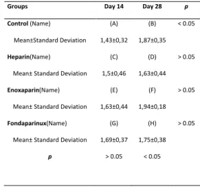
During the postoperative period no complication or death detected. Radiological results according to the mean Goldberg scores and also the results of statistical analyses were
presented in Table 2. Statistically significant correlation was found between the blind orthopaedic surgeons who made the radiologic evaluation. According to Goldberg classification system there was only significant difference in control groups (A (day14) and B (day 28)) results, the other drug groups showed no significant difference according to their results at day 14 and 28 (Table 2).
The mean histologic results according to Huo classification were presented in Table 3. According to the histologic results there was significant difference between the day 14 and Table 2: The mean scores and statistical findings of the
groups radiologically.
Table 1: Scoring system for the histological evaluation
Table 2: The mean scores and statistical findings of the groups radiologically. The Goldberg clasification system was used for radiological evaluation of fracture healing as (0): non healing, (1): callus formation but non complete union, (2): complete union.
Groups Day 14 Day 28 p
Control (Name) Mean±Standard Deviation (A) 1,43±0,32 (B) 1,87±0,35 < 0.05 Heparin(Name) Mean± Standard Deviation (C) 1,5±0,46 (D) 1,63±0,44 > 0.05 Enoxaparin(Name) Mean± Standard Deviation (E) 1,63±0,44 (F) 1,94±0,18 > 0.05 Fondaparinux(Name) Mean± Standard Deviation (G) 1,69±0,37 (H) 1,75±0,38 > 0.05 p > 0.05 < 0.05 Table 1.Scoring system for the histological evaluation Score Histologic Findings 1 Fibrous tissue 2 Predominantly fibrous tissue with little cartilage 3 Equal amounts of fibrous tissue and cartilage tissue 4 Only cartilage tissue 5 Predominatly cartilage tissue with little immature (woven) bone 6 Equal amounts of cartilage and immature bone tissue 7 Predominatly immature bone with little cartilage tissue 8 Healing with immature (woven) bone 9 Immature bone with little mature bone 10 Healing with mature (lamellar) bone
Table 3: The mean scores and statistical findings of the
groups histologically.
Groups Day 14 Day 28 p
Control (Name) Mean±Standard Deviation (A) 3,56±1,24 (B) 7±2,56 <0.05 Heparin(Name) Mean±Standard Deviation (C) 5,19±2,2 (D) 3,37±1,58 < 0.05 Enoxaparin(Name) Mean±Standard Deviation (E) 3,88±2,57 (F) 7,75±2,6 < 0.05 Fondaparinux(Name) Mean±Standard Deviation (G) 3,81±1,03 (H) 6,69±2,69 < 0.05 p > 0.05 < 0.05 The scoring system published by Huo et al (23) was used for histological evaluation. This scale is presented in Table 1.
day 28 scores in each drug groups separately. When the groups were compared according to their results at day 14, there was no significant difference. But there was significant difference among the groups for their day 28 results. The poorest results were in heparin group (group D) which showed significant difference compared to the other drugs and control groups. There was no significant difference among the other groups’ (B, F, H) results (Table 4).
DISCUSSION
According to our results fondaparinux has no positive effect on bone healing radiologically and histologically. We also found out that enoxaparin and fondaparinux have no negative effect on fracture healing histologically and radiologically. The negative effect of heparin was detected only histologically when the 4 weeks results were evaluated. Despite the hypothesis of our study; fondaparinux would have positive effect on fracture healing compared to enoxaparin; there was no significant difference between the control, enoxaparin and fondaparinux except heparin that has significantly poorest results.
At the beginning of this study, we thought that heparin and the enoxaparin would affect fracture healing negatively but fondaparinux that has positive effects on osteoblasts according to the previous papers would effect positively on fracture healing (6-12). Hereby, fondaparinux would be used in traumatic patients with fractures safely as an antiembolic agent was a part of our hypothesis. Actually we detected negative effects of heparin on fracture healing but we did not detect the negative effects of enoxaparin on healing. Especially fondaparinux also did not affect the fracture healing in this experimental study.
Thromboembolic complications are still prob-lematic in trauma surgeries both preoperati-ve and especially in the postoperatipreoperati-ve period. The use of antiembolic agents is absolutely ne-cessary. The major agents that used today are LMWH, fondaparinux and just in case heparin. Heparin, one of the first used agents used for thromboembolism prophylaxis has negative effects on bone healing and also bone metabolism, which was published in many papers (21-25). Today the use of this agent is limited with the patients who have cardiovascular or neurovascular problems that needs active monitoring. We also found out that heparin has negative effects on fracture healing process especially. The result of this study is consistent with the previous studies in the literature.
LMWH agents are the most popular drugs to prevent thromboembolic events. But according to some publications LMWH have negative effects on fracture healing as heparin (8, 26, 27). There some papers regarding the LMWH have no negative effects on fracture healing (9,27). Also there are some papers concluded that the LMWH can effect bone turnover and healing process negatively when they used for a long time period with high doses (28-30). In our study we found that enoxaparin has no negative effects both radiologically and histologically. This result is similar with previous studies (8, 11, 27). We think that our study follow up is for 4 weeks, which is average for fracture healing process in rats.
Fondaparinux is a synthetic anti-thromboem-bolic agent that has a selective inhibitor effect on Factor Xa. As it has no effect on thrombin it has no effect on platelet function also (31). There are limited numbers of papers about its activity on fracture healing. Mainly it is belie-ved that fondaparinux has no adverse effect on bone cycle and fracture healing (11). In a study about the effects of dalteparin, enoxaparin, standard heparin and fondaparinux on human osteoblast cells, it was found that fondapari-nux has more positive effects according to the Table 4: Comparision of each groups’ histological results
at day 28. Table 4: Comparision of each groups’ histological results at day 28. Groups p Control Group (B) / Heparin Group (D) P < 0.05 Control Group (B) / Enoxaparin Group (F) P > 0.05 Control Group (B) / Fondaparinux Group (H) P > 0.05 Heparin Group (D) / Enoxaparin Group (F) P < 0.05 Heparin Group (D) / Fondaparinux Group (H) P < 0.05 Enoxaparin Group (F) / Fondaparinux Group (H) P > 0.05 Dunn's Multiple Comparision Test was used to compare each groups results.
mitochondrial activity and osteoblast protein synthesis. They also concluded that fondapari-nux could be used to prevent heparin induced negative effects on fracture healing implant in-tegration (9). According to our study, our results are consistent with that fondaparinux has no negative effect on bone healing. But we could not detect any positive effects on bone healing as they mentioned in their study.
This study also has some limitations. First, lack of biomechanical evaluation. One of the criteria for fracture healing is biomechanical analysis of united fracture. We could not analyse this due to the lack of availability in the laboratory where we conducted our study. The second limitation is the limited kinds of drugs that used in this study. As there were 4 groups in this study the other drugs could be studied such as rivaroxaban, dalteparin, nadroparin. The third limitation is the number of rats and follow up time could be longer for more reliable results. Up to now, we could not find any study compa-ring heparin, enoxaparin and fondaparinux in the same experimental fracture model. Radio-logical evaluations were performed by two ort-hopaedist independent from the study. It was observed that the evaluation findings of both were in accord with each other.
In conclusion, according to our results showed no significant positive effect of fondaparinux on fracture healing histologically and radiologically comparing the enoxaparin and control groups. We did not detect any extra positive effect of fondaparinux on fracture healing compared to enoxaparin. We think that fondaparinux can be used to prevent embolism problems in traumatic cases as the application of fondaparinux has no negative effect bone healing.
ACKNOWLEDGEMENTS
We want to thank to Rana KONYALIOGLU who is a biostatistics expert for the statistical analyses of this manuscript. Also we want to thank to Dr. Rahime TANRITANIR for the histological evaluation.
REFERENCES
1. Marsland D, Mears SC, Kates SL. Venous thromboembolic
prophylaxis for hip fractures. Osteoporos Int 2010; 21: 593-604.
2. Shackford SR, Davis JW, Hollingsworth-Fridlund P, et al.
Venous thromboembolism in major trauma. Am J Surg 1990; 159:365– 369.
3. Leclerc JR, Geerts WH, Desjardins L, et al. Prevention of deep
vein thrombosis after major knee surgery– a randomized, double-blind trial comparing a low molecular weight heparin fragment (enoxaparin) to placebo. Thromb Haemost 1992; 67: 417- 423.
4. Lindner T, Cockbain AJ, El Masry MA, et al. The effect of
anticoagulant pharmacotherapy on fracture healing. Expert Opin Pharmacother 2008; 9(7):1169–1187.
5. Palmer AJ, Koppenhagen K, Kirchhof B, et al. Efficacy and
safety of low molecular weight heparin, unfractionated heparin and warfarin for thromboembolism prophylaxis in orthopedic surgery: A meta-analysis of randomized clinical trials. Haemostasis 1997; 27: 75– 84.
6. Shaughnessy SG, Young E, Deschamps P, et al. The effects
of low molecular weight and standard heparin on calcium loss from fetal rat calvaria. Blood 1995; 15:86 (4): 1368-1373.
7. Muir JM, Andrew M, Hirsh J, et al. Histomorphometric analysis
of the effects of standard heparin on trabecular bone in vivo. Blood 1996; 15: 88 (4): 1314-1320.
8. Street JT, McGrath M, O’Regan K, et al. Thromboprophylaxis
using a low molecular weight heparin delays fracture repair. Clin Orthop2000; 381: 278-289.
9. Matziolis G, Perka C, Disch A, et al. Effects of fondaparinux
compared with dalteparin, enoxaparin and unfractionated heparin on human osteoblasts. Calcif Tissue Int. 2003; 73(4): 370-379.
10. Hanschin AE, Trentz OA, Hoerstrup SP, et al. Effect of low
molecular weight heparin (dalteparin) and fondaparinux
(Arixtra) on human osteoblasts in vitro. Br J Surg. 2005; 92 (2): 177-183.
11. Demirtas A, Azboy I, Bulut M, et al. Investigation of the
effects of Enoxaparin, Fondaparinux and Rivaroxaban used in thromboembolism prophylaxis on fracture healing in rats. Eur Rev Med Pharmacol Sci. 2013; Jul (14): 1850-1856.
12. Say F, Iltar S, Alemdaroglu KB, et al. The effect of various
types of low molecular weight heparins on fracture healing. Thromb Res 2013; 131(3):114-119.
13. Bonnarens F, Einhorn TA. Production of a standard closed
fracture in laboratory animal bone. J Orthop Res 1984; 2(1): 97-101.
14. Folwarczna J, Janiec W, Gawor M, et al. Effects of enoxaparin
on histomorphometric parameters of bones in rats. Pol J Pharmacol 2004; 56(4):451-457.
15. Schlitt A, Buerke M, Hauroeder B, et al. Fondaparinux
and enoxaparin in comparison to unfractionated heparin in preventing thrombus formation on mechanical heart valves in an ex vivo rabbit model. Thromb Haemost; 2003; 90:245-251.
16. Chung LT, Holton HL, Silverman PR. The effect of
fondaparinux versus enoxaparin in the survival of a congested skin flap in a rabbit model. Ann Plast Surg 2006; 56: 312-15.
17. Olanders K, Börjesson A, Zhao X, et al. Effects of anticoagulant
treatment on intestinal ischaemia and reperfusion injury in rats. Acta Anaesthesiol Scand 2005; 49: 517-524.
18. Folwarczna J, Janiec W, Sliwinski L. Effects of heparin and
low-molecular weight heparin on bone mechanical properties in rats. Thromb Haemost. 2004; 92 (5): 940-946.
19. Goldberg VM, Powell A, Shaffer JW, et al. Bone grafting: role
of histocompatibility in transplantation. J Orthop Res 1985; 3:389-404.
20. Huo MH, Troiano NW, Pelker RR, et al. The influence of
ibuprofen on fracture repair: biomechanical, biochemical, histologic, and histomorphometric parameters in rats. J Orthop Res 1991; 9:383-390.
21. Douketis JD, Ginsberg JS, Burrows RF, et al. The effects of
long-term heparin therapy during pregnancy on bone density. A prospective matched cohort study. Thromb Haemost.1996; 75(2): 254-257.
22. Von Mandach U, Aebersold F, Huch R, et al. Short-term
low-dose heparin plus bedrest impairs bone metabolism in pregnant women. Eur J Obstet Gynecol Reprod Biol 2003; 10; 106(1): 25-30.
23. Simmons HA, Raisz LG. Effects of acid and basic fibroblast
growth factor and heparin on rezorption of cultured fetal rat long bones. J Bone Miner Res 1991; 6 (12):1301-1305.
24. Chigot P, De Gennes C, Samama MM. Osteoporosis induced
either by unfractionated heparin or low molecular weight heparin. J Mal Vasc 1996; 21(3): 121-125.
25. Barbour DA, Kick SD, Steiner JF, et al. A prospective study
of heparin-induced osteoporosis in pregnancy using bone densitometry. Am J Obstet Gynecol 1994; 170(3): 862-869.
26. Kock HJ, Werther S, Uhlenkott H, et al. Influence of
unfractionated and low molecular weight heparin on bone healing: an animal model. Unfallchirurg 2002; 105 (9): 791-796.
27. Hak DJ, Stewart RL, Hazelwood SJ. Effect of low molecular
weight heparin on fracture healing in a stabilized rat femur fracture model. J Orthop Res 2006; 24(4): 645-652.
28. Muir JM, Hirsh J, Weitz JI, et al. A histomorphometric
comparison of the effects of heparin and low-molecular
weight heparin on cancellous bone in rats. Blood 1997;1; 89(9): 3236-3242.
29. Rajgopal R, Bear M, Butcher MK, et al. The effect of heparin
and low molecular weight heparins on bone. Thromb Res 2008; 122 (3): 293-298.
30. Casele HL, Laifer SA. Prospective evaluation of bone density
in pregnant women receiving the low molecular weight heparin enoxaparin sodium. J Matern Fetal Med 2000; 9(2): 122-125.
31. Walenga MJ, Jeske P W, Bara L, et al. Biochemical and
pharmacologic rationale for the development of a synthetic heparin pentasaccharide Thromb Res 1997; 86:1:1-36.