The Immunostimulatory Effect of Lactic
Acid Bacteria in a Rat Model
Murat Karamese*1, Hakan Aydin2, Emin Sengul3, Volkan Gelen4, Cigdem Sevim5, Duran Ustek6, Emre Karakus5
1Department of Medical Microbiology, Faculty of Medicine, Kafkas University, Kars, 2Department of
Virology, Faculty of Veterinary Medicine, 3Department of Physiology, Faculty of Veterinary Medicine,
Ataturk University, Erzurum, 4Department of Physiology, Faculty of Veterinary Medicine, Kafkas University,
Kars, 5Department of Pharmacology and Toxicology, Faculty of Veterinary Medicine, Ataturk University,
Erzurum, 6Department of Medical Genetics, Faculty of Medicine, Medipol University, Istanbul, Turkey
ABSTRACT
Background: Probiotics are “live”, beneficial microbes that provide important health benefits in their hosts. There is significant interest in the modulation and regulation of the immune function by probiotics. Objective: To investigate the immunomodulatory effects of a probiotic mixture, including Lactobacillus and Bifidobacterium species, by detecting serum cytokine and immunoglobulin levels. Methods: The rats were randomly divided into 4 groups. The first group was “Control group” and other 3 groups were probiotic application groups who received different doses of probiotics. The probiotic mixture included 12 probiotic bacteria, mostly Lactobacillus and Bifidobacterium strains. Probiotic mixture was administered to rats for 12 consecutive days. TNF-α, TGF-β, IL-1-β, IL-6, and IL-10 levels as well as serum IgG and IgA concentrations were detected in the sera after 12 days. Results: Probiotics led to a decrease in the levels of TNF-α, IL-6 and TGF-β; however, they led to increase in the serum levels of IL-10, IgG and IgA. There were significant differences between control group and probiotic application groups (p<0.05). Conclusion: These data suggest that the commensal microbiota are important for stimulating both proinflammatory and regulatory responses in order to rapidly clear infections and minimize inflammation-associated tissue damage.
Karamese M, et al. Iran J Immunol. 2016; 13(2):220-228.
Keywords: Bifidobacterium, Cytokines, Immunomodulation, Lactobacillus, Probiotics
--- *Corresponding author: Dr Murat Karamese, Department of Medical Microbiology, Faculty of Medicine, Kafkas University, Kars, Turkey, Tel: (+) 905548638853, Fax: (+) 904742251193, e-mail: [email protected]
INTRODUCTION
Probiotics are “live”, beneficial microbes that provide important health benefits in their hosts (1). Several bacterial species can be used as probiotics, especially Lactobacillus, Bifidobacterium, Pediococcus, Lactococcus and Leuconostoc species; however the most critically important species are believed to be Lactobacillus casei, Lactobacillus acidophilus, and Bifidobacterium species. These bacteria are part of the gastrointestinal microflora, and are especially useful in the digestion of dairy products (2-4). Many scientific studies have reported that the regular intake of probiotics or their related products, especially Lactobacillus and Bifidobacterium, significantly improves human health through a range of effects, including the biosynthesis of vitamins (e.g. vitamin K) (5), detoxification of xenobiotics (6), metabolic fermentation of indigestible dietary fiber (7), competition with pathogenic microbes for binding sites on mucosal epithelial cells (8), and the modulation, regulation, and improvement of the host’s immune response (1).
There is significant interest in the modulation and regulation of the immune function by probiotics, and some studies have examined the relationship between probiotics and the immune response; however, the exact mechanisms of immune modulation by these probiotic organisms are not entirely known. Although there have been some suggestions, such as production of bacteriocin, colonization site interference, lowering of colonic pH status, competition for available nutrients in the colon, competition for binding sites on the epithelial cells, and nonspecific stimulation of the immune system (2). The beneficial effects of probiotics are based on their capability to differentially regulate the production of anti-inflammatory and pro-inflammatory cytokines (9). The smooth functioning of the immune system is crucial for the prevention of infectious diseases; but the strategies currently employed for the modulation of immune responses are quite limited. The improvement of immune system functions, or certain related parameters, can be achieved through the application of probiotics. Moreover, several probiotic strains have drawn special attention in recent years for their encouraging abilities to increase animal and human health, especially through the prevention of chronic diseases and the improvement of natural immune protection (10).
The immunomodulatory effects could contribute to cytokines being released from immune cells, further regulating the innate and adaptive immune responses, and evidence shows immune responses directed toward both of the T helper cell types (11). Cytokines include interleukins (ILs), tumor necrosis factors (TNFs), transforming growth factor (TGF), interferons (IFNs) and chemokines produced and released by immune cells, such as lymphocytes, granulocytes, macrophages, mast cells, epithelial cells, and dendritic cells (DCs) (12). It is well-known that cytokines play crucial roles in the immune system by binding to specific receptors on the cell membrane, triggering one or more of the cellular cascades that lead to the induction, improvement, or inhibition of a number of cytokine-regulated genes in the nucleus. They increase the capability of host defense response to invasion by bacterial, viral, and/or parasitic pathogenic components (13,14).
The main goal of this study was to investigate the immunomodulatory effects of a probiotic mixture, including mostly Lactobacillus and Bifidobacterium species, by detecting specific immune parameters, such as serum cytokine and immunoglobulin levels. The predominant and most important bacteria that reside in the small intestine
are the Lactobacillus and Bifidobacterium species. For this reason, a mixture of lactic acid bacteria which mostly contained these 2 bacterial strains was used in this study. MATERIALS AND METHODS
Preparation of Probiotic Mixture. To study the effect of probiotic mixture on immune modulation of rats were divided into two experimental groups. Controls animals were fed with regular food and experimental groups were fed with a mixture of live probiotics (Enzibody®, KenzBioTech, USA). Enzibody® was provided by Kenz Biotech, Houston, TX, USA. The probiotic mixture is shown in Table 1. Bacteria were grown on MRS BROTH (Cat: CM0359, Thermo Scientific, USA) overnight at 37°C. Bacteria were separated from the culture supernatant by centrifugation (15 min at 3000×g), washed three times with ice cold phosphate saline buffer (PBS) (pH=7.2) and resuspended in PBS. All bacteria were mixed in a 1:1 ratio. The final concentration of mixture contained 1x10¹³cfu lactic acid bacteria. The mixtures were stored at 4oC after the preparation till the day of experiment.
Table 1. Lactic acid bacteria strains in the experimental mixture.
Bacteria Strain
Bifidobacterium infantis ATCC-15697
Bifidobacterium thermophilum ATCC-25867
Bifidobacterium longum ATCC-15707
Lactobacillus acidophilus ATCC-43121
Lactobacillus casei ATCC-393
Lactobacillus paracasei ATCC-25302
Lactobacillus helveticus ATCC-15009
Lactobacillus plantarum ATCC-14917
Lactobacillus bifidus ATCC-11863
Lactobacillus brevis ATCC-14869
Leuconostoc mesenteroides ATCC-8293
Lactococcus lactis ATCC-19435
Animals and Administration of Probiotic Mixture. This project was approved by the Local Ethics Committee of Animal Experiments of Ataturk University, Veterinary Faculty with the number of 36643897-484. Then, twenty female Wistar albino rats, 12 week old and weighing 220 ± 10 g, were purchased from Ataturk University Medical Experimental Research and Application Center with the ethical approval form. Animals were kept in polycarbonate boxes at temperatures ranging between 19ºC and 22ºC, with a standard 12-hour light-dark cycle.
The rats were randomly divided into 4 groups, called the Control (Group 1), 1x10¹¹ CFU/day application group (Group 2), 1x10¹⁰ CFU/day application group (Group 3), 1x10⁹ CFU/day application group (Group 4). Probiotic mixtures were administered to rats for 12 consecutive days using the mentioned dosages. Probiotics were given by oral gavage (0.3 mL).
Sample Collection and ELISA. The rats used in this study were euthanized with high dose of thiopental sodium anesthesia. Blood was drawn from the apex of the cardiac ventricle, and collected into sterile blood collection tubes containing EDTA. Then, the blood samples were centrifuged at 4000 rpm for 10 minutes. After centrifugation, the serum supernatant to be used for further analysis was aliquoted into microcentrifuge tubes, and stored at -80°C. To test the probiotic immunomodulation, the cytokine and immunoglobulin levels were detected by commercially available ELISA kits: Rat IL-1-beta ELISA Kit (Elabscience Biotechnology Co.,Ltd, USA), Rat IL-6 ELISA Kit (Elabscience Biotechnology Co.,Ltd, USA), Rat IL-10 ELISA Kit (Elabscience Biotechnology Co.,Ltd, USA), Rat TNF-alpha ELISA Kit (Elabscience Biotechnology Co.,Ltd, USA), Rat TGF-beta ELISA Kit (Elabscience Biotechnology Co.,Ltd, USA), Rat IgA ELISA Kit (Elabscience Biotechnology Co.,Ltd, USA), and Rat IgG ELISA Kit (Elabscience Biotechnology Co.,Ltd, USA).
All of the chemicals and 96-well ELISA microplates were brought to room temperature before use. The first 7 wells were used for the standards; the 8th was used for the blank, and 100 μl of serum was added to each of the other wells. After incubation (at 37°C, for 90 min), a 100 μl Biotinylated Detection Antibody was added to each well, then the plate was replaced in the incubator (at 37°C, for 60 min). After a 3 step-washing, 100 μl of HRP conjugate was added to each well, placing the plate in the incubator again (at 37°C, for 30 min). After the final 5 step-washing, 90 μl of substrate reagent was added, with 15 min incubation under darkened conditions. Finally, a 50 μl stop solution was added to the wells and the plate was read at a wavelength of 450 nm.
Statistical Analysis. The data were analyzed using the Statistical Package for the Social Sciences (SPSS) version 20.0 statistical software (IBM, SPSS, Inc., Chicago, IL, USA). Those continuous variables with parametric distributions were then analyzed using the one-way ANOVA test. The differences between the groups were defined as statistically significant when the p-value was less than 0.05.
RESULTS
Based on their functions, 7 parameters (cytokines and immunoglobulins) were categorized into 4 groups: pro-inflammatory cytokines, anti-inflammatory cytokines, lymphocyte agonist cytokines, and immunoglobulins the results are explained accordingly.
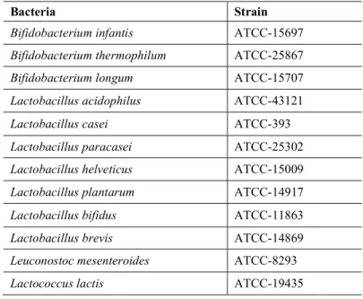
Pro-Inflammatory Cytokines. The pro-inflammatory IL-1-β, IL-6 and TNF-α levels were detected in the serum samples. It was found that the IL-1-β levels were significantly higher in all of the probiotic application groups when compared with the control group (p<0.05). Although the increase in the IL-1-β did not occur in a dose dependent manner in the application group, a nearly 3-fold increase was detected (Figure 1).
Figure 1. Pro-inflammatory cytokines levels of all experimental groups A: IL-1-β levels; B: IL-6 levels and C: TNF-α levels *p <0.05 difference within probiotic application groups and from control group.
In contrast with the IL-1-beta levels, significant decreases were detected in both the IL-6 and TNF-α levels of the probiotic application groups. In all probiotic application groups IL-6 levels were significantly different from control group (p<0.05) (Figure 1). IL-IL-6 level, the 1x10⁹, 1x10¹⁰, and 1x10¹¹ CFU application groups were 125.1 pg/ml, 103.6 pg/ml, and 106.5 pg/ml, respectively, while its level in the control group was 254.6 pg/ml.
Figure 2. Anti-inflammatory cytokines levels of all experimental groups A: IL-10 levels and B: TGF-β levels *p <0.05 difference within probiotic application groups and from control group.
A similar result can be seen in the levels of TNF-α in the probiotic application groups in which there were statistically significant differences between the experimental groups and the control group (p<0.05) (Figure 1). The TNF-α levels were lower in the probiotic application groups and, as with the IL-6 levels, a greater decrease was detected in the 1x10⁹ and 1x10¹⁰ CFU application groups.
Anti-inflammatory Cytokines. The IL-10 levels were found to be increased in the high dose (1x10¹¹ CFU) probiotic application group (p<0.05). Other two doses led to an increase in IL-10 level; however, this increase is not significantly different when compared to the controls (p>0.05).
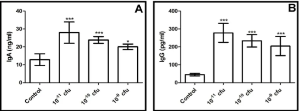
Figure 3. Immunoglobulin levels of all experimental groups A: IgA levels and B:IgG levels *p <0.05 difference within probiotic application groups and from control group.
Moreover, the levels of IL-10 in the 1x10⁹, 1x10¹⁰, and 1x10¹¹ CFU application groups were 289.5 pg/ml, 279 pg/ml, 371.1 pg/ml, respectively, and 226.5 pg/ml in the control group (Figure 2).
Lymphocyte Agonist. In the present study, the TGF-β-1 levels were detected as lymphocyte agonist, and a decrease was detected in all of the probiotic application groups (Figure 2). 1x10¹⁰ and 1x10¹¹ CFU application groups were significantly different from control (p<0.05) while, no significant difference was detected between the 1x10⁹ CFU probiotic application groups and control group (p>0.05). As seen in Figure 5, probiotic treatment led to an important change in the TGF-β levels.
Immunoglobulins. The serum IgA and IgG concentrations were measured in all of the experimental groups, and the results were as expected, with the immunoglobulin concentrations of all of the probiotic treatment groups being significantly different from the control group (p<0.05). In terms of the IgA concentrations, probiotics led to an increase in the serum IgA concentrations in the rats in a dose-dependent manner (Figure 3). The high dose probiotic application (1x10¹¹ CFU) led to the greatest increase in the IgA concentrations. In addition, significant differences were detected within the probiotic application groups (p<0.05). On the other hand, when the serum IgG concentrations were evaluated in our study, it was seen that the probiotic application caused a dramatic increase in the rats in a dose-dependent manner (Figure 3). Statistical differences were detected between the experimental groups and the control (p<0.05). DISCUSSION
There are some realistic evidences suggesting that an important mechanism by which probiotics provide health benefits is through the modulation of immune functions. However, in order to understand how these bacteria improve human health remains largely unknown (15). Actually, probiotics do not have the same effects in every individual, due to the environmental and epigenetic interactions with the genetic make up of the host. The currently used probiotic products usually focus on the healthy
It is generally believed that the inflammation process is regulated by pro-inflammatory and anti-inflammatory cytokines. One, anti-inflammatory cytokine, IL-10, is produced mainly by monocytes, T cells, B cells, natural killer cells, macrophages, and dendritic cells, and regulates the inflammatory process by inhibiting the over expression of many proinflammatory cytokines, chemokines, and chemokine receptors. IL-10 is an important cytokine for commensal intestinal bacteria and in its absence, severe intestinal inflammation may occur. One previous study reported that the administration L. reuteri to IL-10-deficient mice reduced the development of colitis, suggesting that probiotics show anti-inflammatory actions in the intestine. Similar results were seen when IL-10 knockout mice were administered L. salivarius and B. infantis.
Recently, Bifidobacterium strains have been shown to induce IL-10-producing regulatory T cells in the colon. Probiotic application to modulate immune mechanisms can stimulate the production of IL-10 and TGF-β, which are critical mediators of intestinal homeostasis. Most scientific studies support the fact that probiotic applications, especially Lactobacillus and Bifidobacterium strains, stimulate IL-10 production. Similar to the current literature, our probiotic mixture application, especially in high dose (1x10¹¹ CFU) led to a significant increase in the IL-10 levels in rats.
Wide ranges of both Bifidobacterium and Lactobacillus have induced the expression of anti-inflammatory/regulatory cytokines, especially TGF-β, associated with Treg suppressive function/tolerance. The level of a potent regulatory cytokine, TGF-β-1, was decreased in the present study, when compared with the control group. Currently, there is contradictory evidence about this issue. For instance, some studies showed that certain probiotic strains increase the TGF-β-1 level, while others reported no significant changes in the TGF-beta-1 levels. In addition, Biswas et al. reported that the TGF-β-1 production was up-regulated in the rat spleen after probiotic treatment. However, the same study pointed out that the TGF-β-1 expression was down-regulated when the animals were infected with A. salmonicida.
An imbalance between the Th1 (IFN, IL-2, and TNF-α) and Th2 responses (IL-4, IL-5, and IL-10) has been detected in both human and animal models. This may lead to a chronic inflammatory response from the production of pro-inflammatory cytokines, such as IL-1, IL-6, and TNF-α. It has recently been shown that the gut microbiota induces macrophages and dendritic cells (DCs) to produce IL-6 and IL-1-β, which both trigger Th17 differentiation. The same cytokines also trigger the differentiation of IL-10-producing B regulatory cells.
TNF-α and IL-6 play important roles in some immune functions and metabolic disorders. As in TGF-β-1, some studies have claimed that probiotic application can lead to an increase in the TNF-α and IL-6 levels, while others state otherwise. For example, Morita et al. performed a study testing several probiotic bacteria, and they reported that all of the tested bacteria increased the production of IL-6, IL-12, and TNF-α. Moreover, Rangavajhyala et al. reported that Lactobacillus and Bifidobacterium induced the production of TNF-α and one study reported that the application of L. casei led to decreases in the TNF-α and IL-6 levels. On the other hand, in another study, the probiotics reduced the expression of TNF-α, IL-6, beta-defensin 2, and Toll-Like Receptor-2. The differences between the scientific studies with regard to the levels of the cytokines could be explained by the fact that the effects/activities of probiotics are strain specific, and that a combination of probiotics could be beneficial. In the present study, TNF-α and IL-6 levels decreased after the application of probiotics.
Many probiotic strains are obviously able to trigger the production of IgA via B cells, which help maintain humoral immunity by binding to antigens. Serum IgA is produced in the bone marrow, and is also regulated from the mucosal IgA system with an independent mechanism. In the current literature, two studies were performed which examined the serum IgA levels which reported an approximately 10% increase in the serum IgA concentrations after the consumption of the probiotic bacterial strain (Lactobacillus johnsonii).
In other studies no increase in the serum IgA concentration after the intake of yogurt products was detected however the different probiotic strains used in these studies may be the reason for the differences in the serum IgA levels. For example, probiotic strains such as Lactobacillus spp and Bifidobacterium spp have been demonstrated to increase IgA secretion. Probiotic bacteria have also been shown to trigger the epithelial cell expression of IL-10, TGF-β, and IL-6 which stimulate IgA production and class-switching. Finally, probiotics can stimulate the expression of immunoglobulin receptors on the surface of intestinal epithelial cells (17). Parallel with the current information, our results showed that there were increases in the serum IgA levels in all of the probiotic application groups.
In the present study, it was determined that the IgG concentrations of the rat serum samples were increased in the probiotic application groups. This data shows similarities with the current literature. For example, Paineau et al. used 1x10¹⁰ CFU Bifidobacterium lactis and L.acidophilus strains in their study, and reported that these bacteria significantly decreased the serum IgG levels. Another study determined that the growth phase of orally administered Lactobacillus strains affected the IgG response differently.
Finally, probiotics, especially Bifidobacterium and Lactobacillus, could be further tested in experimental models of a low grade inflammation state, such as in obesity. Additionally, the capability of probiotics to reduce the systemic levels of IL-6 could also be researched, with regard to the link in the reduction of glucose homeostasis. Studies clearly demonstrate the capability of probiotics to modulate/regulate the immune response via the down-regulation of pro-inflammatory cytokines, such as TNF-α, and IL-6, and the up-regulation of the anti-inflammatory cytokine IL-10. These effects appear to present a health benefit, with evidence of increased epithelial barrier function and decreased tissue inflammation. These data suggest that the commensal microbiota are important for stimulating both proinflammatory and regulatory responses in order to rapidly clear infections and minimize inflammation-associated tissue damage.
ACKNOWLEDGEMENTS
Thanks for Dr. Ismail CAN for invaluable support to some steps of the experiments and methods. The current study was supported by Kafkas University, Scientific Research Project Council under the project number: 2016-TS-43.
Karamese M, et al. REFERENCES
1. Hardy, H., et al., Probiotics, prebiotics and immunomodulation of gut mucosal defences: homeostasis and immunopathology. Nutrients, 2013; 5: 1869-912.
2. Ashraf, R. and N.P. Shah, Immune system stimulation by probiotic microorganisms. Crit Rev Food Sci Nutr, 2014; 54: 938-56.
3. DuPont, A.W. and H.L. DuPont, The intestinal microbiota and chronic disorders of the gut. Nat Rev Gastroenterol Hepatol, 2011; 8: 523-31.
4. Gareau, M.G., P.M. Sherman, and W.A. Walker, Probiotics and the gut microbiota in intestinal health and disease. Nat Rev Gastroenterol Hepatol, 2010; 7: 503-14.
5. Bentley, R. and R. Meganathan, Biosynthesis of vitamin K (menaquinone) in bacteria. Microbiol Rev, 1982; 46: 241-80.
6. Maurice, C.F., H.J. Haiser, and P.J. Turnbaugh, Xenobiotics shape the physiology and gene expression of the active human gut microbiome. Cell, 2013; 152: 39-50.
7. Nilsson, A.C., et al., Including indigestible carbohydrates in the evening meal of healthy subjects improves glucose tolerance, lowers inflammatory markers, and increases satiety after a subsequent standardized breakfast. J Nutr, 2008; 138: 732-9.
8. Candela, M., et al., Interaction of probiotic Lactobacillus and Bifidobacterium strains with human intestinal epithelial cells: adhesion properties, competition against enteropathogens and modulation of IL-8 production. Int J Food Microbiol, 2008; 125: 286-92.
9. Dong, H., I. Rowland, and P. Yaqoob, Comparative effects of six probiotic strains on immune function in vitro. Br J Nutr, 2012; 108: 459-70.
10. Ben Salah, R., et al., Lactobacillus plantarum TN8 exhibits protective effects on lipid, hepatic and renal profiles in obese rat. Anaerobe, 2013; 23: 55-61.
11. Foligne, B., et al., Probiotic properties of non-conventional lactic acid bacteria: immunomodulation by Oenococcus oeni. Int J Food Microbiol, 2010; 140: 136-45.
12. Savan, R. and M. Sakai, Genomics of fish cytokines. Comp Biochem Physiol Part D Genomics Proteomics, 2006; 1: 89-101.
13. Biswas, G., et al., Elevated cytokine responses to Vibrio harveyi infection in the Japanese pufferfish (Takifugu rubripes) treated with Lactobacillus paracasei spp. paracasei (06TCa22) isolated from the Mongolian dairy product. Fish Shellfish Immunol, 2013; 35: 756-65.
14. Mulder, I.E., S. Wadsworth, and C.J. Secombes, Cytokine expression in the intestine of rainbow trout (Oncorhynchus mykiss) during infection with Aeromonas salmonicida. Fish Shellfish Immunol, 2007; 23: 747-59.
15. van Hemert, S., et al., Identification of Lactobacillus plantarum genes modulating the cytokine response of human peripheral blood mononuclear cells. BMC Microbiol, 2010; 10: 293.
16. van Baarlen, P., J.M. Wells, and M. Kleerebezem, Regulation of intestinal homeostasis and immunity with probiotic lactobacilli. Trends Immunol, 2013; 34: 208-15.
17. Resendiz-Albor, A.A., et al., Regionalization of pIgR expression in the mucosa of mouse small intestine. Immunol Lett, 2010; 128: 59-67.