182
ÖZ
GİRİŞ ve AMAÇ: Geleneksel saptırıcı kolostomi tekniği ile laparotomisiz kolostomi tekniklerini karşılaştırmak ve laparotomisiz teknikte ameliyat öncesi görüntüleme tetkiklerinin faydasını göstermek amaçlanmıştır.
YÖNTEM ve GEREÇLER: Laparotomisiz hasta grubunda doğru stoma yerinin tespiti için 3 boyutlu bilgisayarlı tomografi (BT) ve düz grafi tetkikleri kullanıldı. Bu hastaların sonuçları ile başka bir merkezde laparotomi ile saptırıcı kolostomi açılan hastaların sonuçları karşılaştırıldı. BULGULAR: Laparotomisiz kolostomi uygulanan 18 hasta ve laparotomili kolostomi uygulanan 16 hasta çalışmaya dahil edildi. Demografik veriler ile birçok hasta ve işlem ilişkili parametre benzer bulundu. İnsizyon boyutu (4.8±0.8 karşın 13.3±1.9cm, p<0.001) operasyon süresi (31.4±13.0 karşın 46.7±7.9dk, p<0.001) cerrahi alan enfeksiyonu (0 vs 4 [25%], p=0.039) ve yatış süresi (4 [3-30] karşın 5 [4-34]gün, p=0.01) laparotomisiz grupta anlamlı olarak daha az idi.
TARTIŞMA ve SONUÇ: Saptırıcı kolostomi oluşturulmasında laparotomisiz teknik güvenli ve yararlı olabilir. Laparotomide sakınılarak insizyon boyutu, operasyon süresi ve yatış süreleri kısaltılıp cerrahi alan enfeksiyonları azaltılabilir. Mevcut çalışma, laparotomisiz kolostomi planlanan hastalarda görüntüleme yöntemlerinden yararlanılmasını önermektedir.
Anahtar Kelimeler: kolostomi, laparotomisiz, tomografi, fekal diversiyon, cerrahi
ABSTRACT
INTRODUCTION: We aimed to compare the outcomes of no-laparotomy and conventional diverting colostomy techniques and to describe the process and benefits of using preoperative imaging tools in no-laparotomy procedure. METHODS: Patients intended to receive no-laparotomy diverting colostomy, have preoperative imaging tools of 3D computerized tomography and X-ray examinations in order to predict the best location for the stoma construction. The perioperative outcomes in these cases were compared with those obtained from the patients operated with conventional diverting colostomy with laparotomy at another institution. RESULTS: Eighteen and 16 patients had a diverting colostomy with no-laparotomy technique after preoperative assessment, and conventional procedure. Demographics and most of the patient- and procedure-related factors were similar. Length of incision, (4.8±0.8 vs. 13.3±1.9cms, p<0.001) operation time (31.4±13.0 vs 46.7±7.9mins, p<0.001) and the rate of surgical site infection (0 vs 4 [25%], p=0.039) and hospitalization period (4 [3-30] vs 5 [4-34]days, p=0.01) were significantly less in no-laparotomy group.
DISCUSSION and CONCLUSION: No-laparotomy technique may be safe and beneficial while performing a diverting colostomy. Length of incision, operation time and
hospitalization period are shortened if a laparotomy is avoided, and the rate of surgical site infection decreases. Current study recommends preoperative imaging tools when a no-laparotomy technique is intended.
Keywords: colostomy, fecal diversion, image guided surgery, no-laparotomy, 3D tomography
İletişim / Correspondence: Dr. Mustafa Celalettin Haksal
İstanbul Medipol Üniversitesi, Genel Cerrahi Ana Bilim Dalı, Kolorektal&onkolojik Cerrahi Bilim Dalı, İstanbul, Türkiye E-mail: [email protected] Başvuru Tarihi: 02.12.2018 Kabul Tarihi: 06.02.2019
P
P
r
r
e
e
o
o
p
p
e
e
r
r
a
a
t
t
i
i
f
f
G
G
ö
ö
r
r
ü
ü
n
n
t
t
ü
ü
l
l
e
e
m
m
e
e
K
K
ı
ı
l
l
a
a
v
v
u
u
z
z
l
l
u
u
ğ
ğ
u
u
n
n
d
d
a
a
L
L
a
a
p
p
a
a
r
r
o
o
t
t
o
o
m
m
i
i
s
s
i
i
z
z
v
v
e
e
K
K
l
l
a
a
s
s
i
i
k
k
S
S
a
a
p
p
t
t
ı
ı
r
r
ı
ı
c
c
ı
ı
K
K
o
o
l
l
o
o
s
s
t
t
o
o
m
m
i
i
:
:
Ç
Ç
o
o
k
k
M
M
e
e
r
r
k
k
e
e
z
z
l
l
i
i
V
V
a
a
k
k
a
a
K
K
o
o
n
n
t
t
r
r
o
o
l
l
Ç
Ç
a
a
l
l
ı
ı
ş
ş
m
m
a
a
s
s
P
P
r
r
e
e
o
o
p
p
e
e
r
r
a
a
t
t
i
i
v
v
e
e
I
I
m
m
a
a
g
g
i
i
n
n
g
g
G
G
u
u
i
i
d
d
e
e
d
d
N
N
o
o
-
-
L
L
a
a
p
p
a
a
r
r
o
o
t
t
o
o
m
m
y
y
v
v
s
s
C
C
o
o
n
n
v
v
e
e
n
n
t
t
i
i
o
o
n
n
a
a
l
l
D
D
i
i
v
v
e
e
r
r
t
t
i
i
n
n
g
g
C
C
o
o
l
l
o
o
s
s
t
t
o
o
m
m
y
y
:
:
A
A
M
M
u
u
l
l
t
t
i
i
-
-
I
I
n
n
s
s
t
t
i
i
t
t
u
u
t
t
i
i
o
o
n
n
a
a
l
l
C
C
a
a
s
s
e
e
-
-
C
C
o
o
n
n
t
t
r
r
o
o
l
l
S
S
t
t
u
u
d
d
y
y
Mustafa Celalettin Haksal1, Nuri Okkabaz2, Mehmet Seker3, Nuri Emrah Göret2, Yunus Emre Altuntas2, Cengiz Erol3, Mustafa Öncel1
1 İstanbul Medipol Üniversitesi, Genel Cerrahi Ana Bilim Dalı, Kolorektal&onkolojik Cerrahi Bilim Dalı, İstanbul, Türkiye 2Kartal Eğitim ve Araştırma Hastanesi, Genel Cerrahi Kliniği, İstanbul, Türkiye
183 INTRODUCTION
Fecal diversion is necessitated for various benign or malignant conditions. Although stoma creation has traditionally required laparotomy, various minimal invasive techniques have been proposed to decrease the physiological burden of surgery (1-5). Laparoscopic stoma creation has been used for over two decades as an alternative technique to the conventional open stoma construction, since it has shown to be associated with shorter hospital stay, less postoperative analgesia requirement, and shorter time to flatus, bowel movement and tolerance of a solid diet (6-10).
Some other techniques have proposed to use the stoma incision for catching and pulling the planned bowel segment through for stoma creation without an additional incision or trocar site for the exploration. Trephine colostomy has been shown to be associated with improved cosmesis, less pain, quicker recovery compared with laparotomy (11,12). Case series and reports have described the variations of this technique, and have used further attempts including blunt finger dissection or insertion of camera through incision for further exploration (4,5). Single incision laparoscopic surgery (SILS) trocars have been used through planned stoma site for the same purpose (3,13). Some have also used colonoscopy for assisting the stoma creation (2). However, complications have been reported related to limited visualization in no-laparotomy including the stapling of wrong limbs and inadvertent small bowel injury (12).
Current study describes two different preoperative imaging techniques on defining the most suitable location when a stoma creation has been intended without a laparotomy. In addition, the outcomes of no-laparotomy colostomy creation technique are compared with the results of conventional stoma construction procedure with laparotomy.
METHODS
A novel diverting colostomy technique has been initiated in 2015 at Department of Colorectal Surgery at XXXXX Medical School. This practice has aimed to create a diverting transvers colostomy
through the stoma site (no-laparotomy group), where an experienced radiologist preoperatively marked using computerized tomography images with/without special abdominal x-ray scans. The details of the imaging techniques are described below.
The patients operated with no-laparotomy transvers colostomy creation technique have been compared with those operated with a conventional stoma creation method with laparotomy (laparotomy group) at Department of General Surgery at YYYYYYY. A retrospective analyze has been completed including all consecutive patients necessitated a diverting colostomy for benign or malignant problems between 2015 and 2017 in both departments.
The inclusion criterion is the procedure of diverting colostomy as the sole operation, thus patients have been excluded if they received a diverting colostomy as the consequence of a resection-anastomosis procedure. In addition, trauma patients necessitating other repairs or presented with intraabdominal sepsis such as those presenting with an iatrogenic colon perforation at the time of colonoscopy have not been included to the study in order to homogenize the information. Prior to the initiation of the data retrieval the local ethics committee has approved the content of the study (No:10840098-604.01.01-E.21331) and the
study was registered at
http://www.researchregistry.com (researchregistry3679).
Preoperative Imaging Techniques
The aims of the technique are to exclude the necessity of a laparotomy or laparoscopy during the stoma creation and to minimize the size of the stoma in order to prevent stoma related complications. 3D computerized tomography guided diverting transverse colostomy technique necessitates close co-operation of the surgeon with an experienced radiologist, who defines the projection of transverse colon on the skin using the 3-D coating technique (Figure 1). Then the distances between the target location for the stoma and umbilicus and xiphoid are measured and noted. In some patients, clinicians may decide to continue with an additional measure. In these cases three different size coins are vertically attached on the skin between the umbilicus and xiphoid. Then a
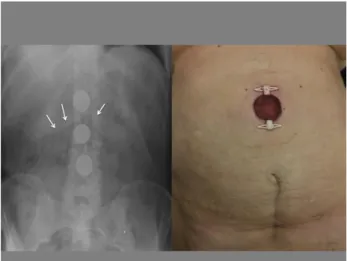
184 standard abdominal x-ray is taken while the patient is in a supine position. Transverse colon is recognized with the gas formation in it, and the most suitable location for stoma is estimated while considering the images of the coins (Figure 2).
Figure 1. Computerized tomography is used for the assessment of appropriate stoma location. The exact location of targeted bowel segment (on the right) is calculated using 2D and 3D images. Black star (*) refers to xiphoid. Umbilicus (black arrow) is considered as the reference point. Then the estimated stoma site (red arrow) is calculated and marked while measuring its distancefrom the umbilicus. The images are taken with 256-slice array computerized tomography (Phillips Brilliance ICT 256-slice scanner, Netherlands). 3D images are created with CT viewer application and Volume Rendering Programme at the work station (Phillips Intellispace Portal Workspace, Netherlands) At the beginning period of the study, surgeons have preferred to use X-ray examinations, however consequently the preoperative evaluation has evolved to use of computerized tomography as the single technique imaging in years. Transvers colon does not have attachments to the retroperitoneum, which makes it highly mobile. Thus, this portion of the colon can be easily taken to the skin without tension, so this bowel segment has chosen for stoma construction. The most appropriate location for stoma is marked with the cooperation of the surgeon and radiologist. Finally, stoma-therapy nurse has examined the patient, questioned the intended stoma site for patients’ postoperative comfort, and then has had the option of relocating it a nearby location, 2-3 cm far from the initial site. After the decision of accurate stoma location, a 3 to 5 cm-long midline facial incision is completed beneath the circular skin incision under the general anesthesia. The transverse colon is detected through
this incision and taken out of the abdomen with meticulous mobilization from its attachments particularly omental connections. The stoma is created over of loop ostomy rod. No additional incision or trocar insertion has been planned for these cases (Figure 2). If an additional incision or trocar insertion is necessitated in no-laparotomy group, it is defined as the failure of the technique.
Figure 2.Plain abdominal graph shows the shadows of the coins, and gas in the transvers colon (white arrows) (on the left). The stoma is created where the second coin is located (on the right). The patient has an infra-umbilical midline incision
The patients have operated at YYYYYY Hospital received a conventional diverting colostomy procedure through a midline incision. Details of postoperative care have been individualized related to the nature of the primary disease.
These measures have been abstracted and compared between the two groups: patients’ demographics, American Society of Anesthesiology (ASA) score, body mass index (BMI), and concomitant diseases, previous abdominal operation and type of incision, preoperative radiation therapy or chemotherapy for the primary disease, number of on-chemotherapy patients, the indication for stoma, timing of the operation, length of incision, operation time, bowel segment for the stoma, time to the beginning of stoma function, length of stay, stoma and general complications, re-operation rate and reason, and rates and reasons of re-operation and 30-day mortality reasons. Finally, stoma size and the presence of a possible parastomal hernia have been specifically questioned with the re-evaluation of the computerized tomography scans in patients for whom this
185 diagnostic tool has been used for the evaluation of ongoing disease. The latest scan has been used for this evaluation, and the interval between the operation and time for the CT has been also noted as follow-up period. If the patient has been using chemotherapy during the last 30 days before operation, the condition is defined as ‘on-chemotherapy’.
Statistical Analysis
Data were analyzed by using SPSS 21.0 for Windows (Armonk, NY, IBM Corp). Results were given as percentages, mean and standard deviations or median and ranges. Quantitative and qualitative variables were compared with Student’s t-test or Mann-Whitney U test and Chi-square (Pearson’s or Fischer’s Exact) tests, respectively. A p value less than 0.05 was considered to be significant.
RESULTS
During the study period, a diverting transvers colostomy has been performed in 18 consecutive patients at XXXXX Colorectal Surgery Department. For assessing the stoma site, 3D tomography guidance was preferred for all patients, and in 9 (50%) cases additional assistance was necessitated for marking with abdominal X-rays and attached coins on the bully. Accordingly the stoma site was marked. There were not any patients, who necessitated an additional incision or trocar insertion in no-laparotomy group, so the success rate of the technique was 100%. During the same period a total of 27 cases received a diverting colostomy at YYYYYY General Surgery Department. In order to homogenize the information, trauma patients (n=9) and those has necessitated a colostomy because of an iatrogenic perforation during colonoscopy (n=2) have been excluded leaving 16 cases in laparotomy group. Thus a total number of 34 patients (20 [58.8%] women with a median [±SD] 53.3±16.4 years) are the subjects of the current study.
Demographics and other patient related features were similar within the groups, however there were significantly more diabetic patients in the laparotomy group (p=0.039) (Table 1). The indications for stoma creation and operation timing were not statistically different between the
groups. Although the rate of a history of a radiation therapy was similar, there were significantly more patients in no-laparotomy group who had received chemotherapy (p=0.009). The incision size, operation time and length of stay were significantly shorter in no-laparotomy group (p<0.001 for all). However, in contrast to the no-laparotomy group, where transvers colon was used for stoma construction, sigmoid colon was preferred as stoma site for 62.5% of patients in laparotomy group (p<0.001) (Table 2). The rates of stoma related problems were similar between the groups. Besides surgical site infection rate was significantly more in laparotomy group (Table 3). Two patients necessitated a reoperation; stoma was revised in one (5.6%) in no-laparotomy group because of prolapse, and another case (6.2%) in laparotomy group necessitated wound closure because of evisceration (p=0.999). Two (11.1%) cases both in no-laparotomy group deceased because of sepsis and respiratory failure on postoperative days 6 and 30, respectively.
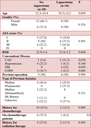
Table 1. Demographics and Other Patient Related Features No-laparotomy (n=18) Laparotomy (n=16) P Age 52.3±18.4 54.5±14.2 0.699 Gender (%) Female Male 12 (66.7) 6 (33.3) 8 (50) 8 (50) 0.324 ASA score (%) I II III IV 5 (27.8) 9 (50) 4 (22.2) 0 3 (18.8) 6 (37.5) 7 (43.8) 0 0.501 BMI 22.9±3.6 23.4±3.2 0.696 Concomitant Disease CAD Hypertension DM COPD 1 (5.6) 4 (22.2) 0 3 (16.7) 5 (31.2) 1 (6.2) 4 (25) 1 (6.2) 0.078 0.340 0.039 0.604 Previous operation 9 (50) 8 (50) 0.999
Type of Previous Incision
Midline Pfannenstiel Midline (Infraumblical) Mc Burney Unknown 4 (44.4) 0 2 (22.2) 1 (11.1) 2 (22.2) 2 (25.0) 3 (37.5) 0 0 3 (37.5) 0.151 History for chemotherapy 10 (55.6) 2 (12.5) 0.009 On-chemotherapy patients 6 (33.3) 1 (6.2) 0.09 History for radiation therapy 5 (27.8) 2 (12.5) 0.405
ASA: American Society of Anesthesiology, CAD: Coronary artery disease, COPD: Chronic obstructive pulmonary disease, DM: Diabetes mellitus
186
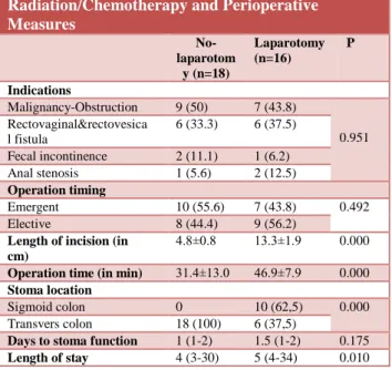
Table 2. The Indications for Stoma Creation, Timing for Operation and History for Radiation/Chemotherapy and Perioperative Measures No-laparotom y (n=18) Laparotomy (n=16) P Indications Malignancy-Obstruction 9 (50) 7 (43.8) 0.951 Rectovaginal&rectovesica l fistula 6 (33.3) 6 (37.5) Fecal incontinence 2 (11.1) 1 (6.2) Anal stenosis 1 (5.6) 2 (12.5) Operation timing Emergent 10 (55.6) 7 (43.8) 0.492 Elective 8 (44.4) 9 (56.2)
Length of incision (in cm)
4.8±0.8 13.3±1.9 0.000
Operation time (in min) 31.4±13.0 46.9±7.9 0.000
Stoma location
Sigmoid colon 0 10 (62,5) 0.000 Transvers colon 18 (100) 6 (37,5)
Days to stoma function 1 (1-2) 1.5 (1-2) 0.175
Length of stay 4 (3-30) 5 (4-34) 0.010
Table 3. Complications and 30-Day Mortality
No-laparotomy (n=18) Laparotomy (n=16) P Early Stoma Complications Retraction 0 3 (18.8) 0.094 Prolapse 1 (5.6) 0 0.999 Mucosal dehiscence 1 (5.6) 0 0.999 Total 2 (11.1) 3 (18.8) 0.648 Surgical Site Infection Wound infection 0 2 (12.5) 0.214 Evisceration 0 2 (12.5) 0.214 Total 0 4 (25) 0.039 30-day mortality 2 (11.1) 0 0.487
Current study retrospectively has evaluated postoperative computerized tomography scans in order to question a parastomal hernia and final size of the stoma. Eleven (61.1%) and 5 (31.3%) patients have had CT scans for various reasons mostly for the evaluation of primary disease in no-laparotomy and no-laparotomy groups, respectively; and the interval between the stoma creation and CT scan has been significantly longer in no-laparotomy group (8.6±7.2 vs 3.2±2 months, p=0.04). These examinations have revealed that the rates of parastomal hernia (2 [18.2%] out of 11 vs 1 [20%] out of 5 in no-laparotomy and laparotomy groups, respectively, p=0.999) and the sizes of the fascia defects have been similar (39.7±25.5 vs 44.4±5.7 mms in no-laparotomy and laparotomy groups, respectively, p=0.697).
DISCUSSION
Fecal diversion may be required for a various number of conditions, and is sometimes the sole practice in the abdominal cavity, particularly when a resection-anastomosis or repair procedure is not the case. For these instances, different techniques have been described for stoma creation without a laparotomy or insertion of additional trocars (1-5,12-14). Trephine stoma has been proposed years ago, and different variations have been described (2,5,11,12). The introduction of the SILS port from the stoma site have been shown to be feasible, since the exploration is practicable and the most suitable bowel segment is easy to detect with the laparoscopic examination of the abdominal cavity (3,13). Direct positioning of the camera through the stoma site without the insertion of a trocar has been also proposed as gasless laparoscopic-assisted intestinal stoma creation technique (5). It has been also advocated that mobilization of the colon with finger dissection is practical through the stoma site (12). However, it has been reported that limited visualization may lead complications including the stapling of wrong limbs and inadvertent small bowel injury (12). In our opinion, another major concern of these techniques is the fact that surgeon does not know the best location for stoma creation on the skin at the beginning of the operation, where the bowel segment is easily taken out for a tension-free stoma construction, since a preoperative work-up has never been considered during the preoperative period. Preoperative imaging may increase safety and success rate of no-laparotomy stoma creation. In a previous study, it was noted that 4 patients out of 36 (11%) necessitated a laparotomy particularly because the bowel segment could not be taken to the wound (12). However, after a proper preoperative work-up, the success rate was 100% in our series. A previous case report has also shown that a no-laparotomy technique may be suitable in even a multiply scarred abdomen, when a preoperative plan for the patient is considered with the use of computerized tomography (15). Thus, we believe that the technique has been described in this study, has a new point of view, since preoperative imaging gives the surgeon to make a preoperative individualized plan for the patient to find the best location for stoma.
187 Since it has been aimed to analyze the outcomes of no-laparotomy technique in constructing a diverting colostomy without a laparotomy, current study includes the patients, who have had a conventional colostomy creation with laparotomy in another institution as a control group. There have been more diabetic cases in laparotomy group and there have been more patients who had chemotherapy in no-laparotomy group. In addition, sigmoid colon has been the location for colostomy in significantly more patients in laparoscopy group. Besides these differences, we believe that the patients in no-laparotomy and laparotomy groups seem to be homogenous and comparable.
Previous studies have shown that the outcomes may improve including operation time, postoperative pain, length of stay and time to chemotherapy or radiation therapy, if the stoma creation is achieved without necessitating a laparotomy (16,17). Current study has also revealed the benefits of no-laparotomy technique. Regarding the perioperative outcomes, operation time is longer in laparotomy group, probably because entering to the abdomen and closing the incision have required extra time. As expected, the length of incision has been longer in this group. However, although number of patients in the study is limited, the rate of surgical site infection is significantly lower, and the length of stay is significantly shorter in no-laparotomy group. This is an important point, since 4 patients (25%) have experienced surgical site infection, and a laparotomy has necessitated due to evisceration in a case. Although high frequency of surgical site infection may be questioned, it is probably related to the poor condition of the patients and/or the underlying disease in our series. In our opinion, these results are exhibiting how important is to avoid laparotomy in these patients. Some may also question the safety of no-laparotomy technique particularly in those who has previously had an abdominal surgery. In a previous study by Stephenson et al (12) 12 out of 36 (37%) patients has undertaken a no-laparotomy technique, had had previous abdominal surgery. However, same series has also shown that all 4 patients who necessitated a conversion to laparotomy had had previous laparotomy, which was the only significant factor for conversion. In another study, no-laparotomy technique was achieved in 4 (71%)
out of 7 cases (18). In our study half of the patients in the no-laparotomy group had the history for an abdominal operation, and of those 4 (44%) had an upper midline incision, where is possibly interfering with stoma site. Preoperative operative planning may be beneficial in these patients. A case report has shown that this technique may also be practical in a multiple scarred abdomen (15). Similarly, the conversion to conventional or laparoscopic surgery has been necessitated in no-laparotomy group in the current study. Thus, in our opinion, current study shows that no-laparotomy technique may still be feasible and safe in cases, who have previous laparotomy.
Current study has aimed to evaluate the parastomal hernia rate in long term after colostomy creation. Besides the limited the number of patients, who have scanned with computerized tomography during the follow-up period, no statistical difference has been shown between the risks of hernia in two groups. We believe that regarding this long-term complication; both techniques may be similar, although the number of cases in the study is restricted.
Current study has some limitations. The retrospective design of the study and the limited number of patients particularly while analyzing the long-term parastomal hernia rate are the two major concerns of the study. In addition, since the patients in two groups are operated at different institutions, the outcomes may be biased with the surgeon and hospital related aspects. For example, more than half of the patients in laparotomy group have had a sigmoid colostomy, while all patients in the no-laparotomy group have had a transvers colostomy, which may be probably related to the routine practice and experience of the surgeons. However, we still believe that this comparative study describes a considerable technique, which may be used in patients requiring a diverting stoma.
CONCLUSION
Current study suggests preoperative imaging in all cases for whom a diverting colostomy without laparotomy is intended. Length of incision, operation time and hospitalization period are shortened if a laparotomy is avoided, and the rate of surgical site infection decreases. In our opinion,
no-188 laparotomy diverting colostomy technique is a safe and rapid procedure, and may have better results compared to conventional technique.
REFERENCES
1. Hasegawa J, Hirota M, Kim HM, Mikata S, Shimizu J, Soma Y, et al. Single-incision laparoscopic stoma creation: experience with 31 consecutive cases. Asian J Endosc Surg 2013; 6: 181-5.
2. MacCormack B. Colonoscopic assisted trephine stoma in children with chronic constipation. Ann R Coll Surg Engl 2014; 96: 387.
3. Tanaka E, Fukunaga Y, Nakashima M, Nagasue Y, Akiyoshi T, Konishi T, et al. Laparoscopic incisionless stoma creation for patients with colorectal malignant stricture. Surg Laparosc Endosc Percutan Tech 2015; 25: 37-9.
4. Jacobsen HL, Harvald TB, Rosenberg J. No-trocar laparoscopic stoma creation. Surg Laparosc Endosc Percutan Tech 2006; 16: 104-5.
5. Hellinger MD, Martinez SA, Parra-Davila E, Yeguez J, Sands LR. Gasless laparoscopic-assisted intestinal stoma creation through a single incision. Dis Colon Rectum 1999; 42: 1228-31.
6. Swain BT, Ellis CN. Laparoscopy-assisted loop ileostomy: an acceptable option for temporary fecal diversion after anorectal surgery. Dis Colon Rectum 2002; 45: 705-7. 7. Hollyoak MA, Lumley J, Stitz RW.
Laparoscopic stoma formation for faecal divertion. Br J Surg 1998; 41: 226-8.
8. Young CJ, Eyers AA, Solomon MJ. Defunctioning of the anorectum: historical controlled study of laparoscopic vs open procedures. Dis Colon Rectum 1998; 41: 190-4.
9. Young MT, Hwang GS, Menon G, Feldmann TF, Jafari MD, Jafari F, et al. Laparoscopic versus open loop ileostomy reversal: is there an advantage to a minimally invasive approach? World J Surg 2015; 39: 2805-11.
10. Shah A, Moftah M, Hadi Nahar Al-Furaji H, Cahill RA. Standardized technique for single port laparoscopic ileostomy and colostomy. Colorect Dis 2014; 16: 248-52.
11. Senepati A, Philips RK. The trephine colostomy: a permanent left iliac end colostomy without recourse to laparotomy Ann R Coll Surg 1991; 73: 305-6.
12. Stephenson ER Jr, Ilahi O, Koltun WA. Stoma creation through the stoma site: a rapid safe technique. Dis Colon Rectum 1997; 40: 112-5.
13. Zagniyan KN, Murrell Z, Fleshner PR. Scarless single-incision laparoscopic loop ileostomy: a novel technique. Dis Colon Rectum 2011; 54: 1542-6.
14. Atallah S, Albert M, larach S. Technique for constructing an incisionless laparoscopic stoma. Technique Coloproctol 2011; 15: 345-7.
15. Craig E, Garner J, Amin S. Stoma sitting using CT guidance in multiply scarred abdomen. Tech Coloproctol 2007; 11: 281-2. 16. Nylung G, Oresland T, Hulten L. The
trephine colostomy: formation of a stoma without laparotomy. Eur J Surg 1997; 163: 627-9.
17. Anderson ID, Hill J, Vohra R, Schofield PF, Kiff ES. An improved means of faecal diversion: the trephine stoma. Br J Surg 1992; 79: 1080-1.
18. Patel P, Wright A, Messersmith R, Palmer J. Does trephine colostomy procedure a satisfactory stoma? Colorectal Dis 2001; 3: 270-1.