case report 140 Le Infezioni in Medicina, n. 2, 140-143, 2016 Corresponding author Ergenekon Karagöz E-mail: [email protected] n INTRODUCTION
T
ularemia is a serious infectious zoonosis caused by a small, pleomorphic, non-motile, nonsporulating aerobic, gram-negative bacillus, Francisella tularensis.The infection can develop after contact with in-fected animals or inin-fected arthropod vectors (es-pecially, ticks). Tularemia is endemic in many re-gions in North America, Scandinavia and Japan where the route of infection depends mostly on contact with infected animals and ticks, howev-er, in Turkey the most prominent and main route of infection is by consuming non-chlorinated drinking water [1, 2]. The most frequent form is the ulceroglandular form which is characterized by a papuloerythrematous ulcer, with a
necro-sis in the middle portion of the bite site, fever and regional painful lymphadenopathy, which is usually induced by means of a vector. Significant cellulitis or lymphangitis may not be observed if a bacterial superinfection does not accompany the disease.
In the present case, our aim was to report a pa-tient with facial cellulitis that developed due to tularemia where a regional lymphadenopathy was not observed, and which was accompanied with severe facial edema, rash and increased tem-perature after a tick-bite on hairy skin. Our pa-tient presented with one of the very rare forms of tularemia in which the definitive diagnosis was made possible only via serology. The condition induced by a tick bite was considered a valuable case due to the presence of atypical dermal find-ings such as lymphocytic cellulitis and its route of transmission.
Our patient is one of the rare tularemia cases in medical literature with an atypical dermatologi-cal finding such as facial cellulitis.
Tick-related facial cellulitis caused
by Francisella tularensis
Ferhat Arslan1, Ergenekon Karagöz2, Ebru Zemheri3,
Haluk Vahabog˘ lu4, Ali Mert1
1Department of Infectious Diseases and Clinical Microbiology, Istanbul Medipol University Hospital, Istanbul, Turkey; 2Department of Infectious Diseases and Clinical Microbiology, Van Military Hospital, Van, Turkey;
3Department of Pathology, Istanbul Medeniyet University Hospital, Istanbul, Turkey;
4Department of Infectious Diseases and Clinical Microbiology, Istanbul Medeniyet University Hospital, Istanbul,
Turkey
Tick-borne illnesses have diverse biological and clin-ical features that make recognition and appropriate treatment challenging. Arthropod-transmitted (ticks, fleas and deer flies) tularaemia remains a concern worldwide.
Generally, two kinds of tularaemia manifestations, namely ulceroglandular and glandular infections, can arise from the bite of an infected arthropod vector. If the ulceroglandular or glandular form is not treated, suppuration can arise from the gland. In addition,
SUMMARY
cellulitis is rarely observed around the ulcers. In our case, with the knowledge of tick exposure to the scalp, tularaemia was not initially considered for facial cel-lulitis without regional lymphadenopathy and also due to apparent failure to respond to doxycycline and gentamicin therapy. Serological confirmation in the late stages of the disease suggests the importance of clinical suspicion in such rare conditions.
141
Tick-related facial cellulitis caused by Francisella tularensis
n CASE REPORT
A 49-year-old female patient referred to our hos-pital due to complaints, including a rash on the forehead, slight facial swelling and headache. The patient’s medical background revealed that a tick was extracted from her scalp at a state hos-pital approximately 6 days before and that the patient experienced headache, swelling on the forehead and fatigue a day after the procedure. A course of amoxicillin clavulanate and gentami-cin had been administered.
Physical examination of the patient demonstrat-ed a rash with a diameter of approximately 10 cm on the forehead; however there were no other significant findings, except for a fever that con-tinued for two days. Laboratory studies were otherwise normal, stated as follows: WBC 4.1/ mm3, Hb 12,6 g/L, PLT 97.000 mm3, C-reactive
protein (CRP) 1,58 (0-5). The patient was prelim-inarily diagnosed with early stage Lyme disease (erythema chronicum migrans) and cellulitis (Fig-ure 1) and therapy with oral doxycycline 2x100 mg and intravenous ceftriaxone 2x1 g was initi-ated. The serology of Lyme Disease (ELISA) and Francisella tularensis (Tube agglutination), and a buffy coat study carried out for Ehrlichia chaffeen-sis were found negative.
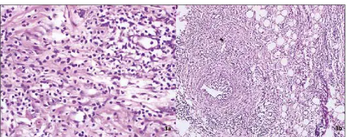
An excisional biopsy sample was taken from the scalp where the tick adhered to the hairy skin and from the dermal region where a rash and an ulcerated lesion were identified. The result of the biopsy indicated an ulceration within the en-tire dermis at the center of the epithelium, a vast number of interstitial perivascular lymphocytes that extended down to the deep layer of the der-mis and a small number of eosinophil-sporadic giant cells (Figures 2 and 3). Under therapy the patient’s fever subsided a day later and vanco-mycin 2 x 1g was added because of the devel-opment of a diffused facial edema and rash on the forehead. Unfortunately, the patient’s com-plaints did not abate, and therefore the medica-tions were withdrawn on the tenth day. Eventu-ally, empiric streptomycin was initiated due to the preliminary diagnosis of tularemia. The pa-tient’s complaints including headache and facial edema began to wane in the first week of therapy and Francisella tularensis IgM 18 U/mL (>10 U/ mL) was determined three weeks after the tick bite while tube agglutination remained negative.
Figure 1 - Facial cellulitis associated with Francisella
tularensis.
Figure 2 - Excisional biopsy taken from hairy areas of
the skin revealed ulceration in the epithelium, inflam-matory cell infiltration extending from the entire der-mis to the fatty tissue.
142 F. Arslan, et al.
Streptomycin administration was interrupted at week 2 due to vertigo which may be a sign of drug toxicity. However, no relapse was observed in the state of the patient after a month’s fol-low-up.
n DISCUSSION
Tularemia, a rare zoonotic infectious disease, can be seen in Turkey and sometimes may lead to idemic outbreaks. Despite previous reports of ep-idemics seen in the Marmara and Western Black Sea regions, many tularemia cases have been re-ported from other regions of the country as well [2]. The clinical form of all the known cases was induced by the consumption of infected drinking water; with all the patients being diagnosed with oropharyngeal tularemia [3]. The vectorial fea-tures of ticks are well known, yet the only cases reported in our country have been two tick-re-lated, ulcero-glandular tularemia patients diag-nosed in the Yozgat Province [2]. Our patient’s di-agnosis was confirmed by serological and/or mo-lecular studies even though the clinical condition had developed after a tick contact. Consequently, in addition to tularemia cases associated with the consumption of contaminated drinking water, it is vital to consider that tick-induced tularemia can also be detected, particularly in areas where the condition is endemic.
Diagnosis is challenging not only because the causative agent cannot be readily cultured for growth on a medium but also due to late results of serological tests. In patients with tularemia, specific antibodies develop within 2-3 weeks af-ter inoculation and may persist for several years. Monitoring of antibody titers in serum during acute and convalescent phases is thus necessary to identify a tularemia infection. In our case, the ELISA test eventually proved to be positive after the preliminary and concomitant tube agglutina-tion were reported to be negative. Recent studies showed that the sensitivity of ELISA was better than tube agglutination [4]. We suggest that the ELISA test is the most useful test in such highly suspicious conditions.
Although lymphadenomegaly was not part of the clinical presentation, the disease could still be categorized as ulceroglandular-type tularemia. Lymphadenopathy may not have been an enti-ty due to previous use of gentamicin and dox-ycycline, in addition to the low virulence of the pathogen in question. Secondary dermal lesions, as part of systemic involvement and which can be observed in patients with tularemia, are de-pendent primarily on its route of entry into the host. Approximately 35-43% of tularemia cases present with secondary dermal lesions and these are more frequent in females than males [2]. Rash and dermal eruptions such as pustule, erythema nodosum, erythema multiform, acneiform lesions
Figure 3 - Inflammatory cells consist of lymphocytes that display perivascular and interstitial distribution as well
as eosinophils (Figure 3a). The lymphoid cells tend to show vascular wall infiltration without signs of fibriniod necrosis in focal areas (Figure 3b). These findings with the clinical history in this case can be evaluated as an ul-ceroglandular type tularemia.
143
Tick-related facial cellulitis caused by Francisella tularensis
or hives (urticaria) may develop in all clinical forms of tularemia, especially in forms related to diffused maculopapular or vesiculopapular erup-tions [(5]. A small number of rare tularemia cas-es where cellulitis developed after contact with a tick are reported in literature [6, 7]. Our patient is one of these rare “cellulitis” cases in literature, induced by previous contact with a tick and di-agnosed in accordance with clinical-pathological correlation.
This case was presented with the aim to empha-size the possibility of a rare combination of cellu-litis and tularemia. We should be aware that the presentation of tularemia may have atypical fea-tures presenting with atypical dermal lesions; ad-ditionally we must bear in mind that cases may be associated with the consumption of contaminated drinking water, whereas this condition may show up in endemic regions where tick bite is the main source of disease.
Conflict of interest: The authors state that they have no conflicts of interest.
n REFERENCES
[1] Sjostedt A. Tularemia: history, epidemiology, patho-gen physiology, and clinical manifestations. Ann. N. Y.
Acad. Sci. 1105, 1-29, 2007.
[2] Yesilyurt M., Kilic S., Cagasar O., Celebi B., Gul S. Two cases of tick-borne tularemia in Yozgat province, Turkey. Mikrobiyol. bul. 45, 746-754, 2011.
[3] Ulu Kilic A., Kilic S., Sencan I., et al. A water-borne tularemia outbreak caused by Francisella tularensis sub-species holarctica in Central Anatolia region.
Mikrobiy-ol. Bul. 45, 234-247, 2011.
[4] Sharma N., Hotta A., Yamamoto Y., et al. Detection of
Francisella tularensis-specific antibodies in patients with tularemia by a novel competitive enzyme-linked immu-nosorbent assay. Clin. Vaccine Immunol. 20, 9-16, 2013. [5] McGinley-Smith D.E., Tsao S.S. Dermatoses from ticks. J. Am. Acad. Dermatol. 49, 363-392; quiz 93-96, 2003. [6] Junkins A.D., Snyder J.W. Cellulitis, headache, and fever following tick bites. J. Clin Microbiol. 49, 6, 2085, 2387, 2011.
[7] Senel E., Satilmis O., Acar B. Dermatologic man-ifestations of tularemia: a study of 151 cases in the mid-Anatolian region of Turkey. Int. J. Dermatol. 54e, 33-37, 2015.