Corresponding author Bahadır Ceylan
E-mail: [email protected], [email protected]
n INTRODUCTION
A
spergillus is a mould frequently found in na-ture and inhalation of infectious conidias is common [1]. Inhaled conidia can cause invasive diseases in immunosuppressed patients. Dissem-inated disease may develop in patients with inva-sive pulmonary aspergillosis (IPA) due to the abil-ity of the fungus to invade vascular structures [2]. The most common risk factors of invasive asper-gillosis (IA) are haematologic malignancies, hae-matopoietic stem cell transplantation (HSCT), sol-id organ transplantation, and long-term sterosol-id treatment [3-5].Although the first entry point of the infectious agent into the body is the respiratory system, it may rarely enter through the gastrointestinal system [1]. Primary gastrointestinal aspergillosis (PGA) is the least common form of IA and knowl-edge is limited to case reports and autopsy find-ings [6-25].Here, we report a patient with PGA and review the general characteristics of cases of PGA in the literature.
n CASE REPORT
A 65-year-old man developed a febrile neutropen-ic episode (FNE) on the third day following an au-tologous HSCT for plasmocytoid variant diffuse large B-cell gastric non-Hodgkin’s lymphoma. He had been treated with 6 cycles of R-CHOP (ritux-imab-cyclophosphamide, doxorubicin, vincris-tine, prednisolone) and a complete response had been obtained. The autologous stem cell
trans-Primary gastrointestinal aspergillosis:
a case report and literature review
Bahadır Ceylan1, Mesut Yılmaz1, Hüseyin Saffet Beköz2, Saime Ramadan3, Gülhan Ertan Akan4, Ali Mert1
1Medipol University, Faculty of Medicine, Department of Infectious Diseases and Clinical Microbiology,
Istanbul, Turkey;
2Medipol University, Faculty of Medicine, Department of Hematology, Istanbul, Turkey; 3Medipol University, Faculty of Medicine, Department of Pathology, Istanbul, Turkey; 4Medipol University, Faculty of Medicine, Department of Radiology, Istanbul, Turkey
Invasive aspergillosis is a severe infection that generally involves the lungs. Primary gastrointestinal aspergillo-sis is the least common form of invasive aspergilloaspergillo-sis. A patient aged 65 years developed a febrile neutropen-ic episode following an autologous stem cell transplant for plasmacytoid variant diffuse large B-cell gastric non-Hodgkin’s lymphoma. He had abdominal pain on the second day of the febrile neutropenic episode and ileus occurred on the sixth day. His general condition de-teriorated despite broad spectrum antibiotics and caspo-fungin treatment, and intestinal perforation occurred on
SUMMARY
the nineteenth day of the febrile neutropenic episode. Pathological examination of the resected jejunum and ileum revealed mould hyphae compatible with asper-gillus. The patient died due to massive gastrointestinal bleeding on the fifth post-operative day. Although a rare condition, primary gastrointestinal aspergillosis should be kept in mind while treating neutropenic patients with gastrointestinal symptoms.
Keywords: aspergillosis, gastrointestinal tract, ileum, jeju-num.
plantation had been performed upon early recur-rence of the disease 10 months after initial treat-ment. He was living in an urban area and was not working. The patient was given 2 cycles of ICE (ifosfamide, carboplatin, etoposide) protocol to provide remission before HSCT. Neutropenia occurred on the tenth day after ICE protocol and lasted 4 days. Lymphopenia occurred on the third day after ICE protocol and lasted 10 days. HSCT was performed by applying BEAM (carmustine, etoposide, cytosine arabinosid, melfalan) proto-col. Neutropenia occurred on the ninth day after BEAM protocol and FNE developed at the same time. Fluconazole had been given to the patient at a dose of 200 mg once a day for prophylaxis against the yeast infection. Chest X-ray was nor-mal and physical examination was nornor-mal other than a body temperature of 38.4oC. Biochemical
examination revealed C-reactive protein (CRP) 8 mg/L (normal: 0-5) and procalcitonin 4.19 ng/ mL(normal <0.5 ng/mL). Piperacillin/tazobac-tam was initiated empirically. A slight abdominal pain developed on the second day of treatment. There was slight tenderness in the abdomen of the patient during palpation. Abdominal ultra-sonography showed fine perisplenic fluid. Ab-dominal computed tomography (CT) revealed an increased thickness of the jejunal wall (thickness 10 mm) (Figure 1). Thorax tomography was nor-mal. Treatment was switched to a combination of colistin, tigecyclin and meropenem on the fourth day of FNE because carbapenem resistant
Kleb-siella pneumoniae was isolated from blood cul-tures. Procalcitonin was 5.11 ng/mL, and serum CRP level was 366 mg/L on the first day of this treatment. Ultrasonography, that was done on the sixth day of FNE because of worsened abdominal pain and new-onset vomiting, revealed a dilata-tion in the small intestines. Abdominal tomogra-phy showed an increased thickness of the jejunal wall (at the same size as before), dilatation of the whole small intestines except the terminal ileum, misty mesentery, and a large number of mesen-teric lymphatic ganglion, the largest being 14x10 mm. Thorax tomography was normal. A nasogas-tric tube was inserted, and 2000 mL of gasnasogas-tric flu-id was removed from the stomach. Neutropenia lasted 8 days. Caspofungin was added empiri-cally due to the gradual deterioration of clinical and laboratory parameters on the eleventh day of FNE. At this time, abdominal pain was worse,
Figure 1 - Abdominal computerized tomography of the
patient on the second day of febrile neutropenic ep-isode [Two-pointed black arrows show the increased thickness of the jejunal wall (thickness 10 mm)]. body temperature was high, serum CRP level was 405 mg/L, and serum procalcitonin level was 4.98 ng/mL. The patient’s body temperature re-turned to normal after the third day of treatment with caspofungin, and abdominal pain contin-ued with decreasing severity. Procalcitonin was 1.89 ng/mL and serum CRP level was 89 mg/L on the eighteenth day of FNE. An acute abdomen developed on the following day and abdominal CT revealed sub-diaphragmatic free air and flu-id in the abdomen consistent with intestinal per-foration. At this time, serum CRP level was 132
mg/L and procalcitonin level was 2.39 ng/mL. Two nodules, one with a dimension of 17x13 mm in the superior segment of the left lower lobe and the other with a dimension of 6 mm in the anteri-or segment of the right upper lobe were seen on thorax tomography. The patient was operated on, and necrotic and perforated areas were observed on the surface of the small intestine during the
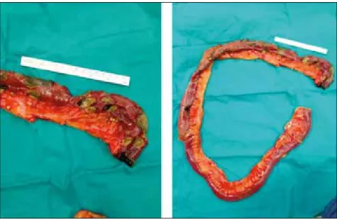
operation. Twenty five cm of ileum and 100 cm of jejunum were resected and end-to-end anastomo-sis was carried out. Ulcers and necroanastomo-sis were seen in the mucosa of the resected intestinal segments (Figure 2). The patient was admitted to the inten-sive care unit in the post-operative period and received mechanical ventilation. Upper gastroin-testinal bleeding developed on the fifth day after
Figure 2 - Macroscopic
ap-pearance of resected jejunum in the patient with primary gastrointestinal aspergillosis (Scale is 15 cm; A: Proximal part of the resected jejunum, B: All resected jejunum; ne-crotic and perforated areas are remarkable)
Figure 3 - Images on the
pathological examination of the resected jejunum. A: Peri-odic acid Schiff (x400 magnifi-cation), septate mould hypha branching at 45-degree angle; B: Gomori Methenamine-Sil-ver (x400 magnification), sep-tate mould hypha branching at 45-degree angle; C: Hema-toxylin-eeosin (x20 magnifica-tion), transmural necrosis and perforation in the jejunum; D: Hematoxylin&eosin (x40 mag-nification), submucosal gran-ulomatous inflammation con-taining giant cells
the operation. Upper gastrointestinal endoscopic examination revealed that the mucosa of the es-ophagus and stomach and the anastomotic line were normal. However, the mucosa of the small intestine after the anastomosis line could not be evaluated because there was too much blood in the lumen.
The patient died due to massive gastrointestinal bleeding on the fifth post-operative day.
Asper-gillus spp was isolated from the sputum culture taken the day before the death. Pathological ex-amination of the resected intestinal segments re-vealed granulomas containing Langerhans-type giant cell and mould hypha branching at a
45-de-gree angle infiltrating all intestinal layers (Figure 3). There was no angioinvasion of hypha.
n LITERATURE REVIEW
We searched the PubMed database system using the key words (“aspergillosis”, “invasive asper-gillosis”, “primary gastrointestinal asperasper-gillosis”, “digestive tract aspergillosis”, “colonic aspergil-losis”, “small bowel aspergilaspergil-losis”, “jejunal gillosis”, “ileal aspergillosis”, “esophageal asper-gillosis” and “aspergillus appendicitis”) for cases recorded between 1980 and May 2017. The
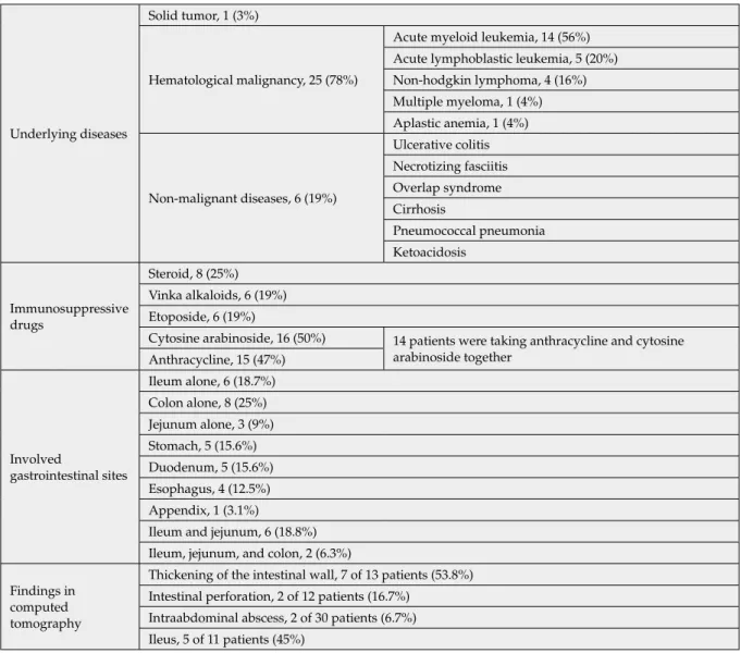
refer-Table 1 - General characteristics of patients with primary gastrointestinal aspergillosis.
Underlying diseases
Solid tumor, 1 (3%)
Hematological malignancy, 25 (78%)
Acute myeloid leukemia, 14 (56%) Acute lymphoblastic leukemia, 5 (20%) Non-hodgkin lymphoma, 4 (16%) Multiple myeloma, 1 (4%) Aplastic anemia, 1 (4%) Non-malignant diseases, 6 (19%) Ulcerative colitis Necrotizing fasciitis Overlap syndrome Cirrhosis Pneumococcal pneumonia Ketoacidosis Immunosuppressive drugs Steroid, 8 (25%) Vinka alkaloids, 6 (19%) Etoposide, 6 (19%)
Cytosine arabinoside, 16 (50%) 14 patients were taking anthracycline and cytosine arabinoside together Anthracycline, 15 (47%) Involved gastrointestinal sites Ileum alone, 6 (18.7%) Colon alone, 8 (25%) Jejunum alone, 3 (9%) Stomach, 5 (15.6%) Duodenum, 5 (15.6%) Esophagus, 4 (12.5%) Appendix, 1 (3.1%)
Ileum and jejunum, 6 (18.8%) Ileum, jejunum, and colon, 2 (6.3%) Findings in
computed tomography
Thickening of the intestinal wall, 7 of 13 patients (53.8%) Intestinal perforation, 2 of 12 patients (16.7%)
Intraabdominal abscess, 2 of 30 patients (6.7%) Ileus, 5 of 11 patients (45%)
ences of each manuscript were checked to prevent duplicated cases.
Patients with PGA proven by histo-pathological and/or culture methods were included, while ones with gastrointestinal involvement due to disseminated disease and oral or pharyngeal as-pergillosis were excluded from our study.
A total of 20 manuscripts and 31 cases were found in the literature [6-25].
General characteristics of the patients
Thirty-one patients (19 male, 12 female, mean age between 18 and 49 years) with PGA were found in the literature [6-25].Details of the characteristics of the patients are presented in Table 1.
Underlying diseases and immunosuppressive drugs used prior to development of PGA
The most common underlying disease was hema-tologic malignancy (25 cases, 78.1%). Five of these patients had also undergone allogenic HSCT and one patient had undergone otologous HSCT due to Wilms’ tumor. The most commonly used drugs prior to development of PGA were cytosine arab-inoside (16 cases, 50%) and steroids (8 cases, 25%). Four of the eight patients using steroids were neu-tropenic at the time of diagnosis of PGA. Nine-teen patients (67.9%) were neutropenic at the time of diagnosis of PGA. PGA-associated symptoms developed after a median of 11 (min-max: 3-303) days of immunosuppressive treatment. PGA de-veloped after 30 days (at 77, 303, and 145 days) in 3 patients. Seven patients (30.4%) had bacteremia concomitantly or before the development of PGA.
Symptoms
The patients were suffering from abdominal pain, (17 of 23 patients, 72%), diarrhoea, (6 of 23 pa-tients, 26.1%), vomiting (8 of 23 patients 34.8%), and disphagia (2 of 23 patients 8%).
Clinical findings
Ileus (5 of 22 patients, 22.7%), acute peritonitis (3 of 23 patients, 13%), and gastrointestinal bleeding (9 of 23 patients, 39.1%) were discovered in the patients.
Aspergillus dissemination from the gastrointestinal system
Aspergillus infection spread to the lung and brain in 1 patient and only to the lung in 6 patients.
Diagnostic methods
Fungus culture (13 of 18 patients, 72%), patholog-ic examination (27 of 28 patients 96.4%), fungus culture + pathologic examination (8 of 32 patients, 23%), high serum galactomannan antigen level (7 of 11 patients, 63%) were used as diagnostic meth-ods.
Aspergillus fumigatus (9 of 10 patients, 90%) and
Aspergillus flavus (1 of 10 patients, 10%) were iso-lated by fungus culture. Transmural necrosis (15 of 23 patients, 65.2%) and vessel invasion (14 of 20 patients, 60%) were seen in pathologic exam-ination.
Diagnostic interventional procedures
Surgical resection (n=18, 56.3%), autopsy (n=4,12.5%) and, endoscopy, (n=9, 29.1%) were used in patients.
Macroscopic appearance of intestine
Mucosal ulcer (14 of 21 patients, 66.7%), mass in the gastrointestinal system (3 of 22 patients, 13.6%), and intestinal necrosis (14 of 22 patients, 63.6%) were seen in the intestine.
Antifungal therapy
Antifungal drugs were used for a mean of 13±6 days (range 4-20). The delay of antifungal treat-ment was 8±4.6 days (range 4-30). Voriconazole (6 of 17 patients, 35%), amphotericin (10 of 17 pa-tients, 59%), and anidulafungin (1 of 17 papa-tients, 6%) were used. In 9 patients, the antifungal drug’s name was not known.
Mortality
Nineteen patients (59.4%) died within 16±9.7 days (range 7-30).
n DISCUSSIONS
In patients with PGA, the first entry site of the as-pergillus spores into the body is the gastrointesti-nal tract and patients usually have predominant gastrointestinal symptoms [26]. Our case did not have any respiratory symptoms or any radiologic findings of aspergillosis in the lung at the begin-ning. However, pulmonary nodules were seen on the thorax CT on the nineteenth day of febrile neutropenia when intestinal perforation
devel-oped. Aspergillus fumigatus was isolated from tra-cheal aspirate culture during the post-operative period. With these findings, we believe that our case was PGA and that pulmonary involvement developed through a secondary spread from the gastrointestinal tract. We also believe that the presence of gastric lymphoma in our case might have facilitated the colonization and subsequent-ly invasion of aspergillus spores into the wall of the intestines.
In the past, the most important risk factor for as-pergillus infection was neutropenia [27]. Howev-er, only one third of the patients had neutropenia at the time of development of invasive aspergillo-sis in recent publications. This is due to increased use of other immunosuppressive drugs such as steroids [28, 29]. Our study showed that only half of the patients with PGA were neutropenic and one third of the patients were using steroids. It is known that aspergillus spores cannot invade the intact mucosa as opposed to Candida spp. [1]. The half of the patients with PGA were using cytosine arabinoside that often causes mucositis and dis-ruption of mucosal integrity [30]. We think that mucosal problems due to cytosine arabinoside and other immunosuppressive drugs may have great significance in the pathogenesis of PGA. Invasive aspergillosis in allogeneic stem cell transplant cases may exceed 20%, but this ratio is reported to be as low as 2-6% in otology HSCT cases [31, 32]. The most important risk factor for invasive aspergillosis is known as neutropenia lasting over 21 days [27]. The risk of aspergillo-sis in autologous HSCT cases is low due to their neutropenia period lasting less than 14 days. Our patient is an interesting case if we consider that invasive aspergillosis rarely occurs after au-tologous HSCT. We think that neutropenia and lymphopenia that had occurred after two cycles of ICE protocol might have contributed to the de-velopment of PGA. Our case had also received 6 cycles of R-CHOP treatment 1 year before HSCT. It is known that rituximab in this regimen reduc-es the number of B cells and this decrease usual-ly resolves within 1 year [33].It is known that B lymphocytes do not only contribute to immunity by secretion of antibodies, but also affect the func-tions of Th lymphocytes with produced cytokines [33]. Rituximab used a year before may have con-tributed to the development of the PGA.
As in invasive pulmonary aspergillosis, the most
common underlying disease was haematological malignancies in the patients with PGA [1].
PGA often manifests itself with non-specific clin-ical and radiologclin-ical findings. It is quite natural that these nonspecific clinical and radiological findings may be confused with other diseases such as necrotizing enterocolitis, gastrointesti-nal GVHD, and Clostridium difficile enterocolitis which are more common than PGA in neutropen-ic patients. We excluded the clostridium colitis in the differential diagnosis of our case because our patient had no diarrhoea. GVHD could not develop in a patient that has undergone autolo-gous stem cell transplantation. But we could not exclude necrotizing enterocolitis caused by fac-ultative anaerobe bacteria. However, despite the effective antibiotic treatment for the microorgan-ism isolated from blood and the improvement of neutropenia on the eighth day, the persistence of gastrointestinal symptoms and ileus should be a warning sign of atypical issues such as PGA. The abdominal CT of the present case showed the thickening of the jejunum wall and this was a non specific finding. In our case, an endoscopy accessing the jejunum could allow us to diagnose the PGA early. Because of the rare occurrence of PGA and its non specific clinical and radiological findings, a vast majority of patients may have a delayed or no antifungal treatment at all. Inade-quate or delayed antifungal treatment may have a role in high mortality in patients with PGA. The most frequently involved sites reported in literature are the ileum, jejunum, and colon in pa-tients with PGA, but this finding may be mislead-ing since the entire gastrointestinal tract has not been evaluated in terms of aspergillus infection in all cases. Colon, stomach, and oesophagus asper-gillosis are easier to diagnose than ileum and je-junum aspergillosis because they are easily acces-sible by endoscopy. Therefore, most patients with PGA were diagnosed through surgical resection and autopsy.
In the literature, serum galactomannan levels were found to be high in some patients with PGA [34]. In immunodepressed patients with gastro-intestinal symptoms and high body temperature who do not have pulmonary or paranasal sinus aspergillosis, we think that high serum galacto-mannan levels may be helpful in suggesting PGA if we take into consideration the cases in the liter-ature. We did not measure serum galactomannan
levels in our patient because of the lack of lung-re-lated symptoms and radiological findings.
In the majority of patients with PGA, macroscopic examination revealed mucosal ulcers and necrosis and histopathological examination revealed trans-mural necrosis and vascular invasion of mould hypha branching at a 45-degree angle. PGA was diagnosed in 72% of cases by microbiological iden-tification of microorganism and pathological ex-amination revealed hyphae structures compatible with aspergillus in almost all cases. Microorgan-isms can be observed in tissue samples as narrow as 3-6 microns wide, septated hypha with dichoto-mous acute angle (45°C) branching in the setting of invasive aspergillosis [35]. However, some hyaline mould, including Scedosporium spp and Fusarium
spp, have the same appearances as Aspergillus spp in histopathologic examination. Thus, it is impor-tant to confirm Aspergillus spp by methods such as culture or polymerase chain reaction. We observed mould hypha which is compatible with aspergillus in tissue section in our case, but we did not use mi-crobiologic methods in the diagnosis because we did not consider PGA in the preliminary diagnosis. Therefore, it is difficult to say that the fungus that invades the intestines is definitely Aspergillus spp. Our patient had no symptoms related to the lungs, and thorax tomography was normal at the begin-ning and on the sixth day of FNE attack. However, radiological findings compatible with aspergillus infection appeared and Aspergillus spp. was isolat-ed from tracheal aspirate samples when neutrope-nia improved and intestinal perforation occurred. For this reason, pulmonary infection was thought to be caused by secondary spreading of the infec-tion from the gastrointestinal tract. Based on the results of the isolation of Aspergillus spp. from the tracheal aspirate sample and fungal hyphae seen in the pathological examination of the intestinal biopsy, we thought that Aspergillus spp. was the causative agent of the intestinal lesion.
One of the main characteristics in the pathogen-esis of aspergillus infection is tissue necrosis due to vascular invasion [1]. It is the reason for serious complications such as ileus and intestinal perfo-ration and for a high rate of surgical intervention in such patients.
Similarly to invasive pulmonary aspergillosis, the most commonly isolated aspergillus species in PGA is Aspergillus fumigatus [1].
To conclude, PGA is a rare condition with
non-specific clinical and radiological findings, and it should be suspected in immunosuppressive pa-tients with gastrointestinal symptoms who are non-responsive to antibacterial treatment.
Disclosure of potential conflict of interest: All the authors declare that they have no conflict of interest.
n REFERENCES
[1] Patterson T.F. Aspergillus species, In Principles and Practice of Infectious Diseases (Mandell GL., Bennett JE. and Dolin R., Eds) 2010, pp 3241-53. Churchill Living-stone, Philadelphia.
[2] Barnes P.D., Marr K.A. Aspergillosis: spectrum of disease, diagnosis, and treatment. Infect. Dis. Clin. North Am. 20, 3, 545-561, 2016.
[3] Gavalda J., Len O., San Juan R., et al. Risk factors for invasive aspergillosis in solid-organ transplant recipi-ents: a case-control study. Clin. Infect. Dis. 41, 1, 52-59, 2005.
[4] Marr K.A., Carter R.A., Crippa F., Wald A., Corey L. Epidemiology and outcome of mould infections in he-matopoietic stem cell transplant recipients. Clin. Infect. Dis. 34, 7, 909-917, 2002.
[5] Pagano L., Fianchi L., Leone G. Fungal pneumonia due to molds in patients with hematological malignan-cies. J. Chemother. 18, 4, 339-352, 2006.
[6] Akyol Erikci A., Ozyurt M., Terekeci H., Ozturk A., Karabudak O., Oncu K. Oesophageal aspergillosis in a case of acute lymphoblastic leukaemia successfully treated with caspofungin alone due to liposomal am-photericin B induced severe hepatotoxicity. Mycoses. 52, 1, 84-86, 2009.
[7] Bizet J., Cooper C.J., Zuckerman M.J., Torabi A., Mendoza-Ladd A. A bleeding colonic ulcer from inva-sive Aspergillus infection in an immunocompromised patient: a case report. J. Med. Case Rep. 8, 407, 2014. [8] Catalano L., Picardi M., Anzivino D., Insabato L., Notaro R., Rotoli B. Small bowel infarction by Aspergil-lus. Haematologica. 82, 2, 182-183, 1997.
[9] Cha S.A., Kim M.H., Lim T.S., et al. Invasive Prima-ry colonic aspergillosis in the immunocompetent host without classical risk factors. Yonsei Med. J. 56, 5, 1453-1456, 2015.
[10] Choi J.H., Yoo J.H., Chung I.J., et al. Esophageal as-pergillosis after bone marrow transplant. Bone Marrow Transplant. 19, 3, 293-294, 1997.
[11] Choi S.H., Chung J.W., Cho S.Y., Kim B.J., Kwon G.Y. A case of isolated invasive Aspergillus colitis pre-senting with hematochezia in a nonneutropenic patient with colon cancer. Gut Liver. 4, 2, 274-277, 2010.
ini-tial manifestation of disseminated aspergillosis. Chest. 101, 3, 877-879, 1992.
[13] Eggimann P., Chevrolet J.C., Starobinski M., et al. Primary invasive aspergillosis of the digestive tract: re-port of two cases and review of the literature. Infection. 34, 6, 333-338, 2006.
[14] Fieber J.H., Atladottir J., Solomon D.G., et al. Dis-seminated enteroinvasive aspergillosis in a critically ill patient without severe immunocompromise. J. Surg. Case Rep. 11, 4, 2013.
[15] Gonzalez-Vicent M., Diaz M.A., Colmenero I., Se-villa J., Madero L. Primary gastrointestinal aspergillosis after autologous peripheral blood progenitor cell trans-plantation: an unusual presentation of invasive asper-gillosis. Transpl. Infect. Dis. 10, 3, 193-196, 2008. [16] Kazan E., Maertens J., Herbrecht R., et al. A retro-spective series of gut aspergillosis in haematology pa-tients. Clin. Microbiol. Infect. 17, 4, 588-594, 2011. [17] Marchesi F., Lepanto D., Annibali O., et al. Sytemic strongyloidiasis and primary aspergillosis of digestive tract in a patient with T-cell acute lymphoblastic leu-kemia. Leuk. Res. 35, 7, 978-980, 2011.
[18] Mora D., Barba P., Ruiz I., et al. Primary gastroin-testinal aspergillosis 6 months after allogeneic hemato-poietic cell transplantation: a case report. Transpl Infect. Dis. 15, 3, 107-110, 2013.
[19] Prescott R.J., Harris M., Banerjee S.S. Fungal infec-tions of the small and large intestine. J. Clin. Pathol. 45, 9, 806-811, 1992.
[20] Shah S.S., Birnbaum B.A., Jacobs J.E. Disseminated aspergillosis inciting intestinal ischaemia and obstruc-tion. Br. J. Radiol. 74, 888, 1145-1147, 2001.
[21] Sousa A.B., Ferreira G., Veiga J., Carvalho A. Clin-ical picture: Bowel infarction due to aspergillosis. Lan-cet. 359, 9302, 210, 2002.
[22] Sulik-Tyszka B., Figiel W., Krawczyk M., Wrob-lewska M. Invasive aspergillosis of the stomach and co-infection with candida krusei in a patient with ter-minal liver failure: a case report. Transplant Proc. 48, 9, 3149-3152, 2016.
[23] Tresallet C., Nguyen-Thanh Q., Aubriot-Lorton M.H., et al. Small-bowel infarction from disseminated aspergillosis. Dis. Colon Rectum. 47, 9, 1515-1518, 2004 [24] Tresallet C., Seman M., Hoang C., Menegaux F. Gastric perforation from potential primary digestive aspergillosis. Surgery. 148, 1, 158-159, 2010.
[25] Weingrad D.N., Knapper W.H., Gold J.,
Mertels-mann R. Aspergillus peritonitis complicating perforat-ed appendicitis in adult acute leukemia. J. Surg. Oncol. 19, 1, 5-8, 1982.
[26] Patterson T.F., Thompson G.R., Denning D.W., et al. Practice Guidelines for the diagnosis and manage-ment of Aspergillosis: 2016 Update by the Infectious Diseases Society of America. Clin. Infect. Dis. 63, 4, e1-e60, 2016.
[27] Gerson S.L., Talbot G.H., Hurwitz S., Strom B.L., Lusk E.J., Cassileth P.A. Prolonged granulocytopenia: the major risk factor for invasive pulmonary aspergil-losis in patients with acute leukemia. Ann. Intern. Med. 100, 3, 345-351, 1984.
[28] Marr K.A., Carter R.A., Boeckh M., Martin P., Co-rey L. Invasive aspergillosis in allogeneic stem cell transplant recipients: changes in epidemiology and risk factors. Blood. 100, 13, 4358-4366, 2002.
[29] Wald A., Leisenring W., van Burik J.A., Bowden R.A. Epidemiology of Aspergillus infections in a large cohort of patients undergoing bone marrow transplan-tation. J. Infect. Dis. 175, 6, 1459-1466, 1997.[30] Naidu M.U., Ramana G.V., Rani P.U., Mohan I.K., Suman A., Roy P. Chemotherapy-induced and/or radiation thera-py-induced oral mucositis--complicating the treatment of cancer. Neoplasia 6, 5, 423-431, 2004.
[31] Marr K.A., Carter R.A., Boeckh M., Martin P., Co-rey L. Invasive aspergillosis in allogeneic stem cell transplant recipients: changes in epidemiology and risk factors. Blood 100, 4358-4366, 2002.
[32] Gil L., Styczynski J., Komarnicki M. Infectious complication in 314 patients after high-dose therapy and autologous hematopoietic stem cell transplanta-tion: risk factors analysis and outcome. Infection 35, 421-427, 2007.
[33] Kelesidis T., Daikos G., Boumpas D., Tsiodras S. Does rituximab increase the incidence of infectious complications? A narrative review. Int. J. Infect Dis. 15, 1, e2-e16, 2011.
[34] Nucci M., Nouér S.A., Cappone D., Anaissie E. Early diagnosis of invasive pulmonary aspergillosis in hematologic patients: an opportunity to improve the outcome. Haematologica 98, 11, 1657-1660, 2013.
[35] Verweij P.E., Brandt M.E. Aspergillus, Fusarium, and other opportunistic moniliaceous fungi, In Manual of Clinical Microbiology (Murray PR, Baron EJ, Landry ML, Jorgenesen JH, Eds), 2007, pp 1802. ASM Press, Washington DC.
![Figure 1 - Abdominal computerized tomography of the patient on the second day of febrile neutropenic ep-isode [Two-pointed black arrows show the increased thickness of the jejunal wall (thickness 10 mm)].](https://thumb-eu.123doks.com/thumbv2/9libnet/5413828.102661/2.850.436.767.402.983/figure-abdominal-computerized-tomography-neutropenic-increased-thickness-thickness.webp)